Cardio Images Flashcards
Name the pathologic process and how it results/examples.

Concentric hypertrophy
Increased systolic loads (increased afterloads)
Examples: aortic stenosis, pulmonic stenosis, pulmonary hypertension in PDA, cats with hyperthyroidism (systemic hypertension)
Name the pathologic process and how it results/examples.

Eccentric hypertrophy & dilation
Increase in diastolic load (increased preload)
Examples: AV or semilunar insufficiencies, arteriovenous shunts
Mass is usually increased, but walls are thin
Name the pathologic process and what does this indicate.
What can this be seen with?

Subendocardial fibrosis
Best indicator or dilation in atria
Seen with either type of cardiac hypertrophy
This image shows three sequelae to what pathologic process?
Name the sequelae.

LHF
Sequelae: pulmonary congestion & edema, LA enlargement
This image shows a sequelae to what pathologic process?
Name the sequelae.

LHF
Sequelae: pulmonary edema
This image shows a sequelae to what pathologic process?
Name the sequelae.

LHF
Sequelae: hemosiderosis “heart failure cells”
This image shows a sequelae to what pathologic process?
Name the sequelae.

RHF
Sequelae: passive hepatic congestion “nutmeg liver”
This image shows a sequelae to what pathologic process?
Name the sequelae.

RHF
Sequelae: hydrothorax
This image shows what pathologic process?
Define the pathologic process.

RHF secondary to pulmonary disease (dirofilariasis) = “Cor pulmonale”
(other causes - chronic obstructive pulmonary dz, PTE, neoplasia)
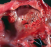
Name the congenital abnormality indicated by the arrow.
What are the consequences?
Name the congenital abnormality indicated by the star.

Atrial septal defect
Consequences: excessive flow from LA to RA in neonate → RV dilation → elevated CVP & blood in lungs → pulmonary hypertension → RV hypertrophy → reversal of flow through defect → cyanosis
High ventricular septal defect

Name the congenital abnormality, where is it located?
What are the consequences?

Ventricular septal defect in membranous portion
Consequences: pulmonary vascular resistance drops after birth → L to R shunt → RV/LV pressures equalize → eccentric hypertrophy of both ventricles → pulmonary hypertension → shunt reversal R to L → cyanosis → death
Name the congenital abnormality, where is it located?
What are the consequences?

Ventricular septal defect in muscular portion
Consequences: pulmonary vascular resistance drops after birth → L to R shunt → RV/LV pressures equalize → eccentric hypertrophy of both ventricles → pulmonary hypertension → shunt reversal R to L → cyanosis → death
(none in neonate)

Name the congenital abnormality.
How long after birth is this considered normal?

PDA
Normally closes within 5 days after birth

Name the congenital abnormality.
Which arrow indicates the abnormality (top, middle or bottom)?

PDA
Middle
Top = aorta
Bottom = pulmonary artery

Name the pathologic process.
What diseases does this commonly accompany?

Subendocardial hemorrhage
Associated diseases: septicemia, endotoxemia, anoxia, electrocution, trauma, agonal change
Name the pathologic process.
What are 6 causes of this process?

Subendocardial mineralization
Causes: dystrophic (necrotic), metastatic (hyperCa), vit D toxicity, calcinogenic plant toxicosis in cattle, Ca:P imbalance, Johne’s disease
Name the pathologic process.
What are 6 causes of this process?

Subendocardial mineralization
Causes: dystrophic (necrotic), metastatic (hyperCa), vit D toxicity, calcinogenic plant toxicosis in cattle, Ca:P imbalance, Johne’s disease
Name this pathologic process.
What are 3 causes, in order of decreasing incidence?
What are some gross findings that are characteristic of this process?
Which anatomic location is most commonly affected?

Vegetative valvular and mural endocarditis
Causes: bacteria, parasites, fungi
Characteristics: large yellow/gray friable masses of fibrin on valves “vegetations” (fibrin, leukocytes, bacteria, granulation tissue microscopically)
Mitral > aortic > tricuspid > pulmonary
Name this pathologic process.
Which portion of the heart is affected with this process?
Uremic endocarditis
Affected: left atrium
Ulcerative
Name this pathologic process.
What is it characterized by and which structure is most commonly affected?

Endocardiosis
Myxomatous valvular degeneration
Mitral > tricuspid
Name this pathologic process.
What is it characterized by and which structure is most commonly affected?

Endocardiosis
Myxomatous valvular degeneration
Mitral > tricuspid
This image shows a possible sequalae of endocardiosis, name the sequalae.
What are the other 6 sequelae of endocardiosis?

Left atrial rupture (also shown here)
Others: valvular incompetency, congestive HF (R or L), atrial dilation, jet lesions, hemopericardium, chordae tendinae rupture

Name this pathologic process.
What species is this usually seen in?
Why are these not of concern?

Hematocyst
Ruminants
Do not cause problems, regress within a few months after birth
Name this pathologic process.
What species is this usually seen in?
Why are these not of concern?

Lymphocyst
Ruminants
Do not cause problems, regress within a few months after birth
Name the pathologic process and specific cause.
What species are affected?
What do the arrows indicate?

Myocardial necrosis
Cause: nutritional - vitamin E/selenium deficiency
Species: lambs, calves, swine, horses
LEFT: areas of necrosis
RIGHT: mineralization of necrotic fibers
Name the pathologic process and specific cause.
What is this disease process called?
Myocardial necrosis
Cause: nutritional - vitamin E/selenium deficiency
“Mulberry heart disease”
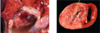
Name the pathologic process and specific cause.
What species is this commonly in?

Myocarditis
Cause: Clostridium chauvoei
Cow
Name the pathologic process and specific cause.
What species is this commonly in?

Myocarditis
Cause: Blastomyces dermatitidis
Dog
Name the pathologic process and specific cause.
What species is this commonly in?

Myocarditis
Cause: canine parvovirus-2
Dog
Name the pathologic process and specific cause.
What species is this commonly in?

Myocarditis
Cause: Trypanosoma cruzi “Chagas disease” (protozoa)
Dog
Name this pathologic process and underlying disease.

Concentric hypertrophy
Feline HCM
Name this pathologic process and underlying disease.

Concentric hypertrophy
Feline HCM
Name this disease process and species.
What is this disease process associated with?

Feline DCM
Associated: taurine deficiency
Name this pathologic process.
What species is commonly affected and what is likely the underlying cause?
What are the consequences?

Excessive moderator bands (false tendons)
Feline - likely congenital, manifests in older cats
Consequences: HF and death
Name the disease process and species/signalment.
What is the underlying cause?
What are some common gross findings and consequences?

DCM
Species/signalment: dogs - young to middle aged giant/large breeds
Cause: genetic, may be x-linked (some breeds)
Gross findings: LHF, biventricular failure, all chambers dilated +/- subendocardial/atrial fibrosis
Consequences: HF, unexpected death
Name the disease process and species/signalment.
What gross finding is notable here?

DCM
Species/signalment: dogs - young to middle aged giant/large breeds
Gross finding: subendocardial fibrosis
Name the disease process and species/breed.
What do the pale areas represent?

Wooly coat cardiomyopathy
Bovine - wooly/curly haired Herefords
Pale areas = necrosis
Name the pathologic process.
Name some underlying causes.

Hydropericardium
Causes: generalized anasarca, hypoalbuminemia, congestive heart failure, neoplasia
Name the pathologic process.
How is this differentiated from the “true form”?
What are the consequences?

Hemopericardium
Clotted blood = “true”
Consequences: rapidly results in death
Name the pathologic process and underlying cause.

Serous atrophy of pericardial fat
Cause: cachexia of any cause
Name the pathologic process, species and underlying causes.

Fibrinous pericarditis
Horse (Foal)
Causes: Mycoplasma felis, mare reproductive loss syndrome
Name the pathologic process, species and underlying cause.

Fibrinous pericarditis
Swine
Causes: Haemophilus suis “Glasser’s disease”
Name the pathologic process, species and underlying cause.

Chronic constrictive pericarditis (purulent pericarditis)
Bovine
Cause: result of traumatic perforation from FB in reticulum → purulent pericarditis → chronic constrictive pericarditis → severe cardiac dysfunction
Name the disease process.

Aortic body tumor “chemodectoma”
This is a primary neoplasm of the heart
Name and describe the disease process
What species can this be seen in?
What is likely the origin?

Rhabdomyoma
Definition: anomalous formations of perinatal cardiac myocytes
Species: pigs >>> cattle, sheep, dogs
Origin: possibly Purkinje cell in swine
Name the disease process and species.

Metastatic lymphoma
Dog