Stroke and TIA Flashcards

1
Q
  1. Definition of Stroke and TIA in general?
  2. What are the causes of brain ischemia? 2
  3. What are the causes of brain hemorrhage?
A
  1. Definition: Alteration of cerebral blood flow
  2. Brain ischemia
    - Thrombosis,
    - embolism or systemic hypoperfusion
  3. Brain hemorrhage
    - Intracerebral hemorrhage
2
Q

Name the vessels in the cerebral arterial circulation (circle of willis)

8

A

See picture

3
Q

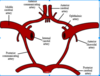
Arterial cerebral circulation

Note the posterior circulation and it’s major blood supply. Why is this interesting?

What is the most common vessel for ischemic stroke?

A

Posterior circulation in the area of the cerebellum and brainstem strokes are not as common but since they involve the brainstem they have terrible outcomes

MIddle cerebral artery

4
Q

What is the most common type of stroke?

What are the cardiac sources of ischemic stroke?6

What are the other causes of ischemic stroke? 4

A

Ischemic stroke is the most common stroke type.

CARDIAC CAUSES

  1. Atrial fibrillation
  2. ASD/VSD
  3. Recent AMI
  4. Endocarditis
  5. Caradiac tumor
  6. Valvular disorder

OTHER CAUSES

  1. Atherosclerotic plaques
  2. Vasculitis
  3. Prothrombotic state
  4. Cerebral hemorrhage

–(20% of strokes)

5
Q

How could atherosclerotic plaques cause stroke?

2

A
  1. Emboli from rupture
  2. Lack of perfusion from stenosis of vessels
6
Q

AFIB cause of stroke:

  1. Commonly from what?
  2. What area does it come from in the heart mostly?
  3. Therapy to decrease the risk of stroke? 2
A
  1. Embolization of intracardiac thrombi
  2. Most commonly from the left atrial appendage

3.

  • Anticoagulation decreases the risk of stroke by up to 70%
  • Aspirin decreases the risk of stroke by 20-25%
7
Q

What are two congenital sources of stroke?

A
  1. Atrial septal defect
  2. Ventricular septal defect
8
Q

If atrial septal defect is associated with what it can cause stroke?2

What about VSD?

1

A

ASD

  1. If associated with a R to L shunt can cause stroke
  2. Patent foramen ovale

Ventricular septal defects

  1. If associated with a R to L shunt can cause a stroke
9
Q
  1. When is stroke most common in MI?
  2. What area of the heart is most responsible for this?
  3. What are the factors of the event that lead to stroke? 3
A
  1. Most common in patients after an anterior wall infarction
  2. Left ventricular wall mural thrombi

3.

  • Large infarctions
  • LV dilation
  • CHF
10
Q
  1. The emboli from endocarditis is from what?
  2. Cardiac tumors can cause strokes how? 3
A
  1. Endocarditis
    - Emboli from vegetations
  2. Cardiac tumor
    - Obstruction of blood flow
    - Can lead to arrhythmias (like afib)
    - Embolization of tumor fragments
11
Q
  1. What is the most common valve disorder that causes stroke?
A

Rheumatic mitral stenosis is the most commonly associated with stroke

12
Q

What are the two kinds of hemorrhagic strokes that equally make up the 20% of strokes?

A
  1. Spontaneous intracerebral hemorrhage (10% of all strokes)
  2. Subarachnoid hemorrhage (the other 10% of hemorrhagic strokes)
13
Q

Subarachnoid hemorrhages can be from what? 2

A
  1. Intracranial aneurysm
  2. Arteriovenous malformations
14
Q

Causes of spontaneous intracerebral hemorrhage

3

A
  1. Associated with poorly controlled hypertension
  2. Bleeding disorders
  3. Amyloid angiopathy
15
Q
  1. Spontaneous intracerebral hemorrhages associated with poorly controlled hypertension are often found where?
  2. Less commonly where?
  3. What are also associated with HTN or DM?
A

Commonly located in the

  1. basal ganglia and
  2. less commonly in the pons, thalmus, cerebellum or cerebral white matter
  3. Lacunar infarcts are associated with HTN or DM
16
Q

How does amyloid angiopathy cause stroke?

A

Amyloid deposits lead to weakening of the cerebral blood vessels resulting in stroke

17
Q

Causes of subarachnoid hemorrhage

6

A
  1. Trauma
  2. Spontaneous SAH is usually related to a ruptured AVM or aneurysm
  3. Abnormal vascular composition (amyloid angiopathy or dissection)
  4. Illicit drug use such as cocaine or amphetamines
  5. Intracranial arterial dissections
  6. 20% may have no identifiable cause
18
Q
  1. Intracranial aneurysm is most commonly located where?
  2. How do aneurysms typically present?
  3. What determines the risk of ruptures? 2
  4. In general, size over ____carries a high risk of rupture
A
  1. Most commonly located in the circle of Willis
  2. Aneurysm is usually asymptomatic until rupture
  3. Size and location determine the risk of rupture
  4. In general, size over 1 cm carries a high risk of rupture
19
Q

Describe the pathophysiology of Arteriovenous malformations?

2

A
  1. Abnormal arterial to venous connection
  2. The venous side will often develop pressures as high as arterial which leads to rupture.
20
Q
  1. Arteriovenous malformation pts are at risk for stroke and what else?
A

seizures

21
Q

Name the subtypes of hemorrhagic stroke? 2

Name the subtypes of ischemic strokes? 3

A
  1. Hemorrhagic

Intracerebral hemorrhage

Subarachnoid hemorrhage

  1. Ischemic

Anterior circulation

Posterior circulation

Lacunar

22
Q
  1. Intracerebral hemorrhage definition?
  2. Major causes? 6
  3. Accumulation of blood over minutes to hours forms what?
  4. As this grows what increases?
A
  1. Arterial bleeding directly into the brain parenchyma
  2. Major causes:
    - HTN,
    - trauma,
    - bleeding disorder,
    - amyloid angiopathy,
    - illicit drug use,
    - AVMs
  3. Accumulation of blood over minutes to hours forming a localized hematoma
  4. Neurologic symptoms increase gradually as the hematoma grows
23
Q

Intracerebral hemorrhage:

  1. What is destroyed as the hematoma enlarges?
  2. What makes this life-threatening?
  3. How would this stroke have a higher morbidity and mortality?
  4. Goal of treatment?
A
  1. Brain tissue is destroyed as the hematoma enlarges.
  2. Pressure created by blood and surrounding brain edema is life-threatening
  3. Large hematomas have a high mortality and morbidity.
  4. The goal of treatment is to contain and limit the bleeding.
24
Q
  1. What are the two major causes of subarachnoid hemorrhages? 2
  2. Bleeding into where? 2
  3. Aneurysm bleeds into the CSF under arterial pressure and increases what?
  4. How long does bleeding last? and what is a common risk?
A
  1. 2 major causes are ruptured aneurysm (most common) or AVM
  2. Bleeding into the CSF and the space surrounding the brain
  3. increases the intracranial pressure
  4. Bleeding lasts a few seconds but rebleeding is common
25
Q

Subarachnoid hemorrhage

Main treatment goal? 2

A
  1. Main treatment goal is identification of source of bleeding and treatment before rebleeding occurs.
  2. The other goal of treatment is to prevent brain damage due to delayed ischemia related to vasoconstriction of intracranial arteries.
26
Q

The other goal of treatment is to prevent brain damage due to delayed ischemia related to vasoconstriction of intracranial arteries.

Blood within the CSF induces what?

A

vasoconstriction, which can be intense and severe

27
Q

2/3 of all ischemic strokes affect the anterior circulation: Which vessels? 2

A

Middle cerebral artery

Anterior cerebral artery

28
Q

MCA is the most commonly involved vessel in ischemic stroke due to what?

A

the direct flow from the internal carotid artery and it’s large size

29
Q

Blood supply to the posterior portion of the brain, including the occipital lobes, cerebellum and brainstem. What are arteries are involved?

A
  1. Vertebral artery
  2. Basilar artery (90% mortality)
  3. Posterior cerebral artery
30
Q

Ischemic strokes: Lacunar infarcts are what?

Where can you see them? 6

A

Small lesions (less than 5mm) that occur in the penetrating arterioles in the

  1. basal ganglia,
  2. pons,
  3. cerebellum,
  4. internal capsule,
  5. thalamus and
  6. deep cerebral white matter
31
Q
  1. How would you describe the prognosis of lacunar strokes?
  2. What are they often seen as on a CT? 2
A

Less morbidity and mortality than other strokes

On CT sometimes seen as

  1. “punched-out hypodense areas” but sometimes
  2. no abnormalities can be seen
32
Q
  1. What is by far the most common subtype of stroke?
  2. What is the culprit vessel in most ischemic strokes?
  3. Anterior strokes occur from what?
  4. Posterior strokes occur from what?
A
  1. By far the most common type of stroke is ischemic
  2. The MCA is the culprit vessel in most ischemic strokes
  3. Anterior strokes occur from occlusion off the internal carotid artery or it’s branches
  4. Posterior strokes occur from occlusion off the vertebral artery or it’s branches
33
Q
  1. What kind of strokes can HTN cause? 2
  2. There is an overlap of ICH and SAH. What does the diagnosis depend on?
A
  1. Hypertension may cause
    - intracerebral hemorrhages (vessel rupture)
    - lacunar infarcts from small vessel occlusion
  2. It all depends on the depth of the vessel that has ruptured.
34
Q

Risk factors for strokes?

A

Hypertension

Diabetes mellitus

Hyperlipidemia

Cigarette smoking

Cardiac disease

Elevated blood homocysteine levels

AIDS

Recreational drug abuse

Heavy alcohol consumption

Family history of stroke

Overweight

Ischemic heart disease

Peripheral vascular disease

Sedentary lifestyle

Men > 45 and women > 55 years old

Oral contraceptives when combined with smoking in women

Hypercoagulopathy

Polycythemia

Prior stroke

Sleep apnea

Bleeding disorders

35
Q
  1. What is aphasia?
  2. What does it impair? 2
  3. Experience difficulty with what?
A
  1. Acquired communication disorder

2.

  • Impairs ability to process language but does not affect intelligence
  • Impairs ability to speak and understand others
    3. Experience difficulty reading and writing
36
Q

What are the types of aphasia?

4

A
  1. Global
  2. Anomic
  3. Broca’s
  4. Wernicke’s
37
Q

What is the most severe from of aphasia?

What are the clinical symptoms? 3

A

Global aphasia

  1. Produce few recognizable words
  2. Understands little or no spoken speech
  3. Can neither read or write
38
Q

What is anomic aphasia?

What are the characteristics of this? 4

A

Persons who are left with a persistent inability to supply words for the things that they want to talk about.

  1. Significant in nouns and verbs
  2. Understand speech well**
  3. Read adequately
  4. Poor writing ability
39
Q
  1. What is the broca’s area supplied by?
  2. What is severely reduced?
  3. What else is affected? 2
  4. What is Broca’s aphasia also called?
A
  1. Broca’s area supplied by superior division L MCA
  2. Speech output severely reduced limited mainly to short utterances of less than four words
  3. Reading, writing are affected also
  4. AKA expressive aphasia
40
Q
  1. What does the speech sound like in Broca’s aphasia?
  2. How is cognition and comprehension affected?
A
  1. Formation of sounds often laborious/clumsy
  2. Comprehension fair, cognitively intact
41
Q

Wernickes aphasia

  1. Wernicke’s area is supplied by what?
  2. How will their speech be affected?
  3. How is the pts comprehension affected? 2
  4. Writing and reading affected?
  5. Also known as?
A
  1. Wernickes area supplied by inferior division of the L MCA

2.

  • Fluent, but meaningless spontaneous speech—jargon of real words and nonwords
  • Individual unaware of language errors unlike in Broca’s aphasia
    3. Comprehension poor
    4. Writing, reading affected the same
    5. AKA receptive aphasia
42
Q
  1. What is dysarthria?
  2. What is dysconjugate gaze?
  3. What is apraxia?
A
  1. Dysarthria- Problem with the muscles that produces speech
  2. Dysconjugate gaze- Failure of the eyes to turn together in the same direction
  3. Apraxia- Difficulty with the motor planning to perform tasks or movements when asked
43
Q
  1. What is dystaxia?
  2. What is agnosia?
A
  1. Dystaxia- Lack of muscle coordination
  2. Agnosia- Inability to process sensory information. Often there is a loss of ability to recognize objects, persons, sounds, shapes, or smells.
44
Q

Describe the fast campaign?

4

A

F Facial droop

A Arm weakness

S Speech difficulties

T Time to call 911

45
Q

Which patient has symptoms of a stroke?

A. Sudden onset severe headache

B. Severe dizziness, nausea and vomiting

C. Acute speech deficit

D. Sudden onset impaired consciousness without focal neurologic deficits

E. Right sided facial droop with weakness of the right arm and leg

A

ALL OF THEM

46
Q

Subarachnoid hemorrhage

  1. Onset of symptoms?
  2. Sudden increase in ICP may cause what?
  3. Sudden severe headache described how? What is this followed by?
  4. Usually what do we NOT see?
  5. They may have signs of what kind of irritation?
  6. Symptoms may be preceded by what? 2
A
  1. Symptoms begin abruptly
  2. cessation of activity (knees may buckle, loss of memory)
  3. Sudden, severe headache “thunderclap” followed by vomiting
  4. Usually no focal neurologic signs****
  5. May have signs of meningeal irritation
  6. Symptoms may be preceded by heavy physical exertion or sex
47
Q

Intracerebral hemorrhage

  1. Onset?
  2. Symptoms increase in correlation with what?
  3. What symptoms occur in half of pts? 2
  4. Symptoms may be preceded by what? 2
A
  1. Symptoms are slower in onset than SAH and increase over minutes to hours
  2. Symptoms worsen as the hematoma enlarges

3.

  • Headache
  • vomiting

occur in about half of patients

  1. Symptoms may be preceded by heavy physical exertion or sex
48
Q
  1. Intracerebral hemorrhage: Neurological symptoms will vary based on what?
  2. With a large hematoma what may you have?
A
  1. depending on location and size of bleed and may be similar to ischemic symptoms
  2. With large hematoma may have decreased LOC
49
Q

The brain damage on one side results in neurologic deficits where?

A

on the opposite (contralateral) side of the body

50
Q

General symptoms of an anterior circulation stroke (ACA and MCA)

May have one or all of the following symptoms:

3

A
  1. Face-hand-arm-leg contralateral hemiparesis
  2. Aphasia
  3. Dysarthria
51
Q

Anterior cerebral artery occlusion may cause what?

3

A
  1. Leg weakness and sensory loss
    - Contralateral side affected
  2. Arm (esp. proximal) weakness and sensory loss
    - Contralateral side affected
  3. Urinary incontinence
52
Q

Middle cerebral artery occlusion may cause:

5

A
  1. Contralateral hemiplegia in the face-arm-leg
  2. Homonymous hemianopsia
  3. If on the L and it is the dominant hemisphere = aphasia (wernickes or brocas aphasia)
  4. Nondominant right hemisphere = confusion, spatial disorientation, sensory and emotional neglect
  5. Apraxia
53
Q

Structures that rely on the posterior circulation blood supply

A
  1. Brainstem
  2. Thalamus
  3. Hippocampus
  4. Cerebellum
  5. Visual cortex
  6. Temporal lobes
  7. Occipital lobe
54
Q

The brainstem has 3 parts and houses cranial nerves III-XII

What nerves are in the midbrain? 2

What nerves are in the pons? 4

What nerves are in the medulla? 4

A

Midbrain

III, IV

Pons

V, VI, VII, VIII

Medulla

IX, X, XI, XII

55
Q

Symptoms of posterior circulation stroke

5

A
  1. Vertigo (must rule this out if someone has vertigo!!!)
  2. Diplopia, dysconjugate gaze, ocular palsy, homonymous hemianopsia
  3. Sensorimotor deficits – ipsilateral face and contralateral limbs (crossed findings), drop attack
  4. Dysarthria
  5. Ataxia
56
Q

5 D’s of posterior stroke symptoms

A
  1. Dizziness
  2. Diplopia
  3. Dysarthria

—Difficulty speaking (loss of motor control for speech)

  1. Dysphagia

–Difficulty swallowing

  1. Dystaxia

–Difficulty controlling voluntary movement

57
Q

Lacunar stroke symptoms

4

A
  1. Pure motor loss (weakness)

—Most common presentation in up to 2/3 of cases

  1. Or pure sensory loss
  2. Sensorimotor stroke (2nd most common)
  3. Ataxic hemiparesis
58
Q

Symptoms that may be unique for the followng stroke locations:

  1. Anterior cerebral artery 3
  2. Middle cerebral artery 4
  3. Brain stem 3
  4. Lacunar 2
A

See picture

59
Q

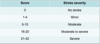
Describe the NIH Stroke Scale

5

A

see picture

60
Q

Acute evaluation

A
  1. Timing of the onset of symptoms is critical
  2. Assessment of stroke risk factors
  3. Physical exam looking for stroke sources
61
Q

Blood work - Labs for strokes?

6

A
  1. Lipid Profile
  2. Blood sugar
  3. CBC
  4. CMP
  5. PT/PTT
  6. Cardiac biomarkers to R/O cardiac ischemia
62
Q

Diagnostic work up for strokes?

5

A
  1. Acute work up = noncontrast CT of the head

–only rules out/in hemorrhagic stroke

  1. Later = MRI ± MRA of the brain
  2. EKG
  3. Ultrasound of carotids
  4. Echocardiogram
63
Q

Window of opportunity to start treating stroke patients is _____hours, but to be evaluated and receive treatment, patients need to get to the hospital within ________?

A

3

60 minutes.

64
Q

Medical Tool bag

4

A
  1. ASA
  2. Heparin or Lovenox
  3. Fibrinolytics
  4. Percutaneous intracerebral intervention in selected cases
65
Q
  1. Treatment for Ischemic stroke? (timeline this must be given)
  2. Hemorrhagic stroke?
A
  1. “Clot-busters”

Must give within 3 – 4.5 hours and within an hour of arrival to the hospital

  1. correct cause of hemorrhage
66
Q

What is tPA (Tissue Plasminogen Activator)? 2

What is the biggest risk with this?

A
  1. Clot busting drug aka fibrinolytic aka thrombolytic
  2. Dissolve the clot in ischemic strokes and the neurologic deficits are more likely to improve
  3. If mistakenly give to hemorrhagic stroke patients = death
67
Q

Inclusion criteria for thrombolytics

3

A
  1. Clinical diagnosis of ischemic stroke causing measurable neurologic deficit
  2. Onset of symptoms less than 4.5 hours before beginning treatment
  3. Age ≥18 years
68
Q

Exclusion criteria (History)

15

A
  1. Significant stroke or head trauma in the previous three months
  2. Previous intracranial hemorrhage
  3. Intracranial neoplasm, arteriovenous malformation, or aneurysm
  4. Recent intracranial or intraspinal surgery
  5. Arterial puncture at a noncompressible site in the previous seven days
  6. Symptoms suggestive of subarachnoid hemorrhage
  7. Persistent blood pressure elevation (systolic ≥185 mmHg or diastolic ≥110 mmHg)
  8. Serum glucose <50 mg/dL (<2.8 mmol/L)
  9. Active internal bleeding
  10. Acute bleeding diathesis, including but not limited to conditions defined in ‘Hematologic‘
  11. Head CT scan

Evidence of hemorrhage

Evidence of a multilobar infarction with hypodensity involving >33 percent of the cerebral hemisphere

  1. Platelet count less than 100,000/mm3
  2. Current anticoagulant use with an INR >1.7 or PT >15 seconds
  3. Heparin use within 48 hours and an abnormally elevated aPTT
  4. Current use of a direct thrombin inhibitor or direct factor Xa inhibitor with evidence of anticoagulant effect by laboratory tests
69
Q

Additional relative exclusion criteria for treatment from 3 to 4.5 hours from symptom onset

4

A
  1. Age >80 years
  2. Oral anticoagulant use regardless of INR
  3. Very severe stroke (NIHSS score >25)

( ≥ 20 NIHSS is severe)

  1. Combination of both previous ischemic stroke and diabetes mellitus
70
Q

Hospital Treatment for Stroke

Who should be involved in the management?

6

A
  1. Dietary
  2. Neurology
  3. Physical & Occupational Therapy
  4. Speech Therapy
    - Swallowing evaluation
    - Speech retraining
  5. Nursing
  6. Physical medicine and rehab Dr. consultation
71
Q

Discharge medications for stroke?

6

A
  1. Aspirin
  2. Statin
  3. Anticoagulation?
  4. Plavix?
  5. Antihypertensives (but only if its really really high, they need the perfusion pressure to help the brain heal)
  6. Assess adequacy of blood sugar control if DM
72
Q

Recovery from Aphasia:

After stroke – If symptoms last longer than __________months, complete recovery is unlikely

A

two or three

73
Q

Some residual effects of strokes

5

A
  1. Emotional lability (mood swings, depression)
  2. Perceptual effects: Difficulty recognizing, understanding familiar objects
  3. Difficulty planning and carrying out simple tasks
  4. Loss of awareness (one-side neglect)
  5. Dysphagia (difficulty swallowing) and aspiration
74
Q

Medical complications of stroke

12

A
  1. Bladder dysfunction
  2. Bowel dysfunction
  3. Pressure ulcers
  4. Malnutrition
  5. Dehydration
  6. Falls and injuries
  7. Recurrent strokes
  8. Venous thromboembolism
  9. Dysphagia
  10. Aspiration pneumonia
  11. Seizures
  12. Spasticity
75
Q
  1. What is a Transient ischemic attack (TIA)?
  2. What about the symptoms makes TIAs different?
  3. What kind of strokes are they?
A
  1. A stroke like event lasting less than 24 hours (usually 20 minutes) that occurs secondary to cerebral ischemia
  2. Symptoms resolve completely
  3. All TIAs are ischemic
76
Q

TIA types

4

A
  1. Amarosis fugax
  2. Low flow
  3. Embolic
  4. Thrombotic
77
Q

TIA workup?

3

A
  1. CT or MRI
  2. Carotid ultrasound
  3. Evaluation for source of emboli or thrombus
78
Q

Treatment for TIAs?

A
  1. Consider admission to the hospital if seen within 72 hours of the symptoms
  2. Risk factor management is the same as if the patient has had a stroke
79
Q
  1. When should we activate stroke team?
  2. What are questions to ask that we need to know? 2
  3. What kind of workup should you do for TIAs?
A
  1. Activate the stroke team ASAP even if you are not 100% sure it is a stroke they are there to help

2.

  • Need to know the onset of symptoms
  • Need to know the patient’s baseline neurologic function
    3. Take all TIA’s serious – they warrant a complete workup
80
Q
  1. What kind of treatment do we need to avoid in ischemic stroke?
  2. Up to how many hours can thrombolytics be given?
A
  1. In ischemic stroke avoid aggressive BP control (want to increase cerebral perfusion)
  2. Thrombolytics can be given up to 4.5 hours from symptom onset (0-3 hours best outcomes)