42: Orthopedics Flashcards
What is the treatment for fibrous dysplasia of bone?
Benign bone tumor treated with curettage +/- bone graft
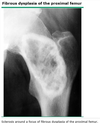
[UpToDate: Fibrous dysplasia is a lesion in which portions of the bone are replaced by fibrous connective tissue and poorly formed trabecular bone. The process originates in the medullary cavity. It is caused by a postzygotic mutation in the guanine nucleotide stimulatory protein (GNAS1) gene. It is more of a skeletal dysplasia than a true neoplasm.
Fibrous dysplasia most commonly presents in the teens or 20s. It may occur in any bone but is most common in the proximal femur, tibia, ribs, and skull . Fibrous dysplasia affects slightly more males than females.
The treatment of fibrous dysplasia depends upon the presence of symptoms. Asymptomatic patients may be observed every six months with serial radiographs. Children with large lesions or lesions in the proximal femur or other weight-bearing bones are observed more frequently.
Curettage, bone grafting, and stabilization may be warranted for fibrous dysplasia that is associated with symptoms (pain, deformity) or fracture; however, there is a high rate of recurrence. Autograft should not be used because it will be resorbed. Bisphosphonate therapy is another alternative for symptomatic patients.
The deformity of fibrous dysplasia may progress with skeletal growth. Fibrous dysplasia usually is static after growth ceases but may be reactivated with pregnancy. Fibrous dysplasia often recurs after curettage and bone grafting.]
What is the treatment for Legg-Calve-Perthes disease?
Maintain range of motion with limited exercise
[Femoral head will remodel without sequelae. Surgery if femoral head is not covered by the acetabulum]
[UpToDate: Children diagnosed with LCP should be made nonweight bearing and referred to an experienced pediatric orthopedist for management. Therapy for LCP is poorly defined, because no large controlled trials are available, and long-term consequences become evident only after decades of follow-up. Treatment focuses on containing the femoral head within the acetabulum through the use of splints or occasionally surgery.
Almost all children do well in the short term. However, long-term outcome depends upon age at time of disease onset and degree of involvement of the femoral head. Children who are younger than six to eight years have a better prognosis, perhaps because more time is permitted for femoral remodeling and because before eight years of age the acetabulum is plastic and can mold to the deformed femoral head, maintaining congruity.]

What is the typical treatment for an ankle fracture?
Cast and immobilization
[UpToDate: Emergent conditions, such as an open fracture or neurovascular impairment, require immediate surgical consultation and treatment. Fracture dislocations must be reduced immediately to prevent severe complications, such as avascular necrosis.
Once emergent conditions are excluded, clinicians should evaluate the fracture more closely, focusing on any malalignment or instability, to determine proper management and follow-up. The ankle should be splinted at 90 degrees (ie, neutral position) to provide support and control pain. Usually, a short-leg posterior splint is sufficient. A sugar-tong (ie, coaptation) splint can be added for additional mediolateral support. If significant swelling or deformity is present, adequate padding should be placed prior to application of the splint to allow for further swelling, while maintaining stability.
Clinicians should instruct the patient to call immediately for:
- Pain that is severe or increasing
- Numbness that is new or worsening
- Skin discoloration (eg, dusky toes) distal to the splint
These complaints may represent vascular compromise or some other serious complication and should be investigated immediately. Any patient complaint of skin irritation, a splint which has become excessively tight or loose, or a splint which has gotten wet should also be assessed. An examination and repeat radiographs to check for acceptable alignment are generally performed during the first follow-up visit at 10 to 14 days.
For stable, nondisplaced, isolated malleolar fractures, the patient should rest, elevate the involved ankle above the level of the heart, and apply ice, while keeping the splint dry. If the injured leg is placed in a prefabricated splint able to withstand ambulation, the patient may bear weight as tolerated. The importance of elevating the leg should be emphasized to patients, as complications with splint treatment often stem from allowing the foot to remain in a dependent position for too long.
Patients awaiting orthopedic consultation or surgery should remain nonweightbearing in a splint (as described above), apply ice while keeping the splint dry, and use pain medication as needed. If surgery is planned in the acute setting, excessive use of narcotic analgesics should be avoided, if possible, until the orthopedic surgeon is able to explain the procedure and obtain informed consent. Management of specific fracture types is discussed immediately below.]

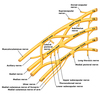
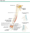
Which nerve roots contribute to the ulnar nerve?
C8-T1

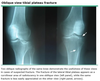
Anterior shoulder dislocation is associated with a risk of injury to what?
Axillary nerve
[UpToDate: Clinicians perform a neurovascular examination paying particular attention to distal pulses and the function of the axillary nerve, which is most commonly injured in anterior shoulder dislocations. Axillary nerve dysfunction manifests as loss of sensation in a “shoulder badge” distribution, although this finding is not reliably present. Deltoid muscle weakness may also be present, but is impractical to assess during the acute injury. Some degree of axillary nerve dysfunction is present in 42% of patients with an anterior dislocation, but most patients recover completely without intervention. In many cases, dysfunction resolves with reduction.
Associated fractures identified on plain radiographs include Hill-Sachs deformities, Bankart lesions, and greater tuberosity fractures. A Hill-Sachs deformity is a cortical depression in the humeral head created by the glenoid rim during dislocation. They occur in 35% to 40% of anterior dislocations and are seen on the AP radiograph with the arm in internal rotation. Bankart lesions occur when the glenoid labrum is disrupted during dislocation and a bone fragment is avulsed. Bony Bankart lesions are present in 5% of patients, while soft tissue Bankart lesions (no bone is avulsed) occur in approximately 90% of patients less than 30 years old with an anterior shoulder dislocation. Greater tuberosity fractures are present in 10% of patients. Indications for orthopedic referral, including selected Bankart and Hill-Sachs lesions, are discussed separately.]

What is the biggest risk factor for non-union following a fracture?
Smoking
[UpToDate: Common reasons for nonunion and malunion include a tenuous blood supply to the fractured bone (eg, scaphoid, proximal fifth metatarsal, talar neck), behaviors that interfere with bone healing (eg, smoking), poor bone fixation (ie, excessive movement at the fracture site), poor apposition of bone fragments (ie, fragment ends too far from one another), and infection.]
Fractures in which 3 areas of the body are associated with avascular necrosis?
- Scaphoid
- Femoral neck
- Talus
[UpToDate: A variety of traumatic and atraumatic factors contribute to the etiology of osteonecrosis. A definitive etiologic role has been established for some of these factors, based upon longitudinal cohort studies or meta-analyses, but not for the majority, which are considered associated risk factors. Use of glucocorticoids and excessive alcohol intake are associated with more than 80% of atraumatic cases.
The pathogenesis of osteonecrosis is an area of controversy. Most experts believe that it is the result of the combined effects of genetic predisposition, metabolic factors, and local factors affecting blood supply, such as vascular damage, increased intraosseous pressure, and mechanical stresses. The early stages of the natural history are unclear, as these stages are largely asymptomatic and the patient does not present until later. It is generally agreed that there is an interruption of the blood circulation within the bone; subsequently, the adjacent area becomes hyperemic, resulting in demineralization, in trabecular thinning, and, later, in collapse.
The histopathologic finding of bone marrow infarction has been noted in marrow samples from patients with some of the same disorders that cause clinically apparent osteonecrosis, but neoplastic disorders, particularly hematologic and lymphoid malignancies and metastatic cancer with associated coagulopathy, are other potential etiologies. The causes of bone marrow infarction (bone marrow necrosis) are discussed elsewhere.]

What is the treatment for a displaced calcaneus fracture?
Open reduction and internal fixation (ORIF)
[UpToDate: Emergent (ie, immediate) surgical referral is required for open fractures, fractures associated with neurovascular injury, fractures associated with dislocation (which must be reduced immediately), and suspicion or diagnosis of acute compartment syndrome. Virtually all intraarticular calcaneus fractures should be assessed and managed by a surgeon, and urgent referral is indicated. In addition, calcaneal fractures that are comminuted or involve noticeable displacement warrant urgent referral. As a general rule, it is best to contact the surgeon at the time of diagnosis. Initial management includes:
- Elevating the affected foot above heart level and applying ice.
- Providing adequate analgesia.
- Assessing the skin and swelling.
- Evaluating for other associated injuries of the feet, ankles, legs, and thoracolumbar spine.
- Possible admission for observation and pain control depending upon the severity of the fracture, and consequent risk of major complications (eg, compartment syndrome), and the presence of concomitant injury.
When classifying calcaneus fractures, the most important step is to distinguish extraarticular from intraarticular injuries. Extraarticular fractures generally have a good prognosis, and if nondisplaced may not require referral. The major types of extraarticular fractures are reviewed in the text.
Intraarticular fractures virtually always require referral. Their management is controversial and surgery may be performed.]

What is the operative treatment for Dupuytren’s contracture?
Transverse carpal ligament release
[UpToDate: Surgery has been the treatment of choice for advanced stages of disease, if function is impaired or if a contracture is progressing. At present, typical interventions are a transection of cords (fasciotomy) or an excision of diseased fascial bands (fasciectomy) with or without excision of the overlying skin. Surgery (limited palmar fasciectomy) should be considered only with functional impairment or in the presence of a contracture that is progressive. The initial results are generally good, but the recurrence rate is high. Flexion deformities >30 to 40 degrees at the metacarpophalangeal (MCP) or >20 degrees at the proximal interphalangeal (PIP) joint have been suggested as indications for surgery. PIP joint contractures are less likely to achieve full correction of the contracture and are less likely to respond to surgery in advanced stages (eg, contractures >60 degrees). The aims of surgery are to reverse digital contractures and to restore hand function. Surgery in younger patients has a much higher recurrence rate than in older patients, but this is likely due to the increased severity of the disease in patients who present with contractures at a younger age.
The likelihood of recurrence after surgery may be related to the degree of cellularity of the lesion. A study of 10 patients found that signal characteristics on magnetic resonance imaging (MRI) predicted cellularity and, thus, may provide prognostic information regarding the likelihood of recurrence after surgery, although further study is warranted before this is used in clinical practice. A second operation can be performed in patients who have a recurrence after the first procedure.
The specific surgical technique used depends upon the individual characteristics of the patient and upon the preferences of the surgeon. More aggressive techniques such as radical fasciectomy or dermofasciectomy do not appear to offer an advantage over limited fasciectomies.]

Which nerve roots contribute to the median nerve?
C6-T1

What is the last clinical sign of compartment syndrome to manifest itself?
Loss of distal pulses
[UpToDate: Several common misconceptions exist pertaining to the clinical diagnosis of ACS. Clinicians should be aware that ACS can occur in the presence of open fractures as these may not necessarily decompress elevated compartment pressures. Moreover, ACS can occur without a fracture (or a crush injury). The diagnosis is often delayed because clinicians fail to consider ACS in patients without a fracture. Arterial pulses and normal capillary refill can persist despite the presence of a prolonged, severe ACS. Pulse oximetry is an insensitive instrument for diagnosis and should not be relied upon.]
What are the non-operative treatments for nerve root compression?
- NSAIDs
- Heat
- Rest
[UpToDate: For patients with acute lumbosacral radiculopathy and painful radicular symptoms caused by disc herniation or foraminal stenosis, the following interventions may be useful:
We suggest short-term treatment with either a nonsteroidal anti-inflammatory drug (NSAID) or acetaminophen (Grade 2C).
We suggest temporary activity modification, including avoidance of provocative activities (Grade 2C).
Physical or manual therapies are often tried for patients with persistent symptoms that are mild to moderate in nature, but there is no convincing evidence that they are effective for this indication. Physical therapy in the first one to two weeks is not recommended because patients with mild symptoms are likely to improve on their own while patients with very severe symptoms cannot participate in exercise therapy.]
What would be the physical manifestation of a nerve root compression of L3 nerve (L2-L3 disc)?
Weak hip flexion
[UpToDate: There is marked overlap of the L2, L3, and L4 innervation of the anterior thigh muscles, making it difficult to differentiate these spinal nerve root levels based on symptoms, neurologic examination, or electrodiagnostic testing. Thus, these radiculopathies are generally considered as a group. These nerve roots are most commonly involved in older patients with symptoms of spinal stenosis.
Acute back pain is the most common presenting complaint, often radiating around the anterior aspect of the leg down into the knee and occasionally down the medial aspect of the lower leg as far as the arch of the foot. On examination, there may be weakness of hip flexion, knee extension, and hip adduction. Higher lesions may result in greater weakness of the hip flexors. Sensation may be reduced over the anterior thigh down to the medial aspect of the lower leg. A reduced knee reflex is common in the presence of moderate weakness.
Electromyography and nerve conduction studies may reveal abnormalities confined to muscles of the affected root(s), including the quadriceps, leg adductors, and iliopsoas, with associated paraspinal abnormalities. Saphenous sensory response remains normal even if sensory loss is prominent in the distal leg.]

How should one treat Salter-Harris I and II fractures?
Closed reduction
[Good prognosis]
[UpToDate:Salter I (Ogden IA-B) — The fracture line extends through the zone of hypertrophic cartilage (zone 3), causing the epiphysis and physeal elements to separate from the metaphysis. Type I injuries can have normal radiographs and the diagnosis is therefore often made clinically when focal tenderness is found over the growth plate.
- A type IB Ogden fracture is characterized by the fracture line extending through the primary spongiosa bone layer resulting in a thin line of bone displaced with the epiphysis. Type IB fractures usually occur in children with systemic diseases such as myeloproliferative disorders. Subsequent growth is usually normal with Type IA and IB fractures.
- A Type IC Ogden fracture has an associated injury to the germinal portion of the physis. Type IC fractures can cause growth arrest and occur rarely after age two to three years.
Salter II (Ogden IIA-D) — The fracture line extends through the physis and then propagates across the physeal-metaphyseal junction into the metaphysis. Type II fractures are the most common physeal fractures. The resultant metaphyseal wedge in a Salter II or Ogden Type IIA fracture is called the Thurston Holland fragment.
- A type IIB involves further extension of the fracture line bidirectionally through the metaphysis creating a free metaphyseal fragment or multiple fragments.
- A type IIC fracture is a transverse physeal fracture that includes a thin layer of metaphysis along with the metaphyseal triangular corner segment.
- A type IID fracture is characterized by the angulation of the two segments resulting in the metaphyseal segment compressing the physis and creating an osseous bridge that leads to permanent growth arrest.]

What test should be ordered for a patient with suspected nerve root compression?
MRI
[UpToDate: For imaging of the lumbar spine, MRI, CT, and CT myelography (CT scan after intrathecal administration of contrast media) are equally sensitive for the diagnosis of disc herniation. For routine initial assessment, an MRI is more informative than CT because it can also identify other intraspinal pathologies, including inflammatory, malignant, and vascular disorders. In addition, MRI is not associated with ionizing radiation and is less invasive than CT myelography.
However, there is a high prevalence of abnormal neuroimaging findings in asymptomatic individuals, including some who have what appears to be frank nerve root compression by MRI. As an example, one study of 98 people without back pain found MRI evidence of disc herniation in 27%. Furthermore, lumbar spine abnormalities on MRI in asymptomatic patients do not appear to be predictive for the future development or duration of low back pain.
Although rarely indicated, CT myelography can visualize spinal nerve roots and their trajectory through the neural foramina. It is useful for patients with intolerance of or contraindications to MRI (eg, implanted electrical devices such as cardiac pacemakers or defibrillators) when standard CT fails to define the anatomic correlates of the clinical presentation. In addition, CT myelography is preferred for patients who have surgically placed spinal hardware that produces magnetic artifacts.
A CT scan can assess osseous structures better than either plain radiography or MRI and is therefore helpful in assessing for bony disease. However, CT alone is unable to visualize nerve roots, so it is not helpful in the direct imaging of a radicular process.]
What is the treatment for Felon?
Incision over the tip of the finger and along the medial and lateral aspects to prevent necrosis of the tip of the finger
[UpToDate: A very early presentation of a pulp space infection without a fluctuant swelling may be treated with warm soaks, rest, elevation, and oral antibiotics. However, most patients with a pulp abscess require surgical intervention. A simple incision and drainage procedure may provide temporary relief; however, it is better to debride the abscess cavity in the operating room because the infection may be more extensive than the symptoms and clinical appearance suggest.]

The posterior tibial artery lies in which leg compartment?
Deep posterior compartment

The anterior tibial artery lies in which leg compartment?
Anterior compartment

Which type of Salter-Harris fracture crosses the epiphysis and the growth plate (physis), but not the metaphysis?
Salter-Harris III
[UpToDate: Salter III (Ogden IIIA-D) — The fracture line extends through the physis and then spreads through the epiphysis into the intraarticular space. If the transverse fracture extends across the complete width of the physis, two epiphyseal segments may be formed.
- A Type IIIB fracture, similar to type IB, courses through the primary spongiosa physeal layer resulting in a thin bony metaphyseal line displaced with the epiphyseal segment.
- Type IIIC injuries involve epiphyses in mostly nonarticular areas.
- Type IIID fractures penetrate the germinal zone and interrupt the blood supply to the avulsed segment. These fractures are difficult to visualize on traditional radiographs.]

What is the treatment for slipped capital femoral epiphysis?
Surgical pinning
[UpToDate: The treatment of SCFE is operative. Children with SCFE, regardless of classification, should be referred promptly to an orthopedic surgeon; they must avoid bearing weight until they have undergone orthopedic evaluation.
Approximately 30% to 60% of patients with unilateral SCFE at presentation eventually have SCFE in the contralateral hip. To prevent delay in diagnosis of the second slip, all patients with unilateral involvement who do not undergo prophylactic repair of the contralateral hip should be followed closely by an orthopedic surgeon until after the child has finished growing. Patients and parents should be instructed to seek medical attention immediately if they experience symptoms of SCFE (eg, nonradiating, dull, aching pain in the hip, groin, thigh, or knee).]

What is the treatment for a clavicle fracture?
Application of a sling due to risk of vascular impingement
[UpToDate: Operative versus nonoperative treatment of displaced midshaft clavicle fractures is individualized and based on factors such as the degree of displacement, shortening, and comminution, as well as functional and cosmetic concerns. The data available to address this important question is limited, but studies do not show a clear benefit to surgery over nonoperative management in many cases.
Patients with nondisplaced or minimally displaced middle third fractures are treated with a sling, analgesics, and regular elbow range of motion exercises. For patients with complete displacement who decline surgery, immobilization using a figure of eight bandage may help to correct or prevent shortening, but a sling is acceptable.
Clinically, fractures of the distal clavicle are easily confused with acromioclavicular separations. Radiographs are necessary to differentiate between the two. Orthopedic referral is recommended for most distal clavicle fractures. An exception is type I fractures confirmed by normal stress views using plain radiographs. Confirmed type I fractures can be managed using a sling and early shoulder range of motion exercises, begun as soon as symptoms allow.
Acute fractures of the proximal clavicle should alert the physician to the possibility of serious internal injury. In most cases, evaluation is performed in the emergency department. If there are no associated injuries and the fracture is nondisplaced, treatment involves sling immobilization. Stress fractures develop insidiously from repetitive stress on the proximal clavicle related to a range of activities, including rowing and gymnastics. Conservative treatment is generally successful.
Among children, 90% of clavicle fractures occur in the middle third. In children 10 and under, the majority are nondisplaced; above age 10, the majority are displaced. Treatment generally does not differ from that recommended for adults, but healing occurs more quickly.]

What is the treatment for a talus fracture?
Closed reduction for most
[Open reduction and internal fixation (ORIF) for severe displacement]
[UpToDate: Most talar head fractures are managed by surgeons, and non-operative treatment of talar neck fractures should be performed only by primary care clinicians experienced in caring for patients with musculoskeletal problems, including the use of braces, casts, and foot orthoses. The treatment of talar head fractures is aimed at maintaining and preserving the articular surfaces of the talus and the stability of the talonavicular joint.
Non-surgical treatment is indicated for isolated, non-displaced impaction or avulsion fractures involving a small portion (<5 mm) of the talonavicular surface without extension into the anterior subtalar joint, as determined by CT. All other talar head fractures are referred for surgical consultation.
Non-displaced talar head fractures involving less than 5 mm of the talonavicular joint surface and not involving the subtalar joint are treated in a short-leg walking cast with a molded arch or in a removable cast boot with arch support for six to eight weeks. Repeat radiographs are obtained every two to three weeks to monitor healing. Casting is continued for six to eight weeks or until signs of healing are present. These signs include the absence of tenderness over the fracture site and radiographic evidence of healing (eg, filling of the fracture line). The patient is then transitioned to a longitudinal arch support for two to three months. There are no long term outcome studies of talar head fractures but these injuries are associated with an increased risk of talonavicular osteoarthritis.]
The sural nerve lies in which leg compartment?
Superficial posterior compartment
[Wikipedia: The sural nerve subserves a purely sensory function, and therefore its removal results in only a relatively trivial deficit. For this reason, it is often used for nerve biopsy, as well as the donor nerve when a nerve graft is performed.]
Buckling of the metaphyseal cortex that is seen in children is called what?
Torus fracture
[UpToDate: A buckle fracture occurs at the distal metaphysis, where the bone is most porous, usually in younger children. This injury is caused by buckling of the cortex due to compression failure. Torus fractures are stable, and treatment is aimed at pain relief, comfort, and protection of the bone from any further injury using a short arm cast or a splint.
Based on several small trials, we suggest that children with a torus (buckle) fracture receive a removable splint for immobilization rather than a below-elbow cast. However, the choice of splint versus cast is dependent on the degree of initial pain, the degree of anticipated activity of the child, and parental preference. A splint can be removed for showering and minor activities, and may be preferable by some caregivers, but a cast offers more protection of the fracture site in active children.
Before treating with a splint, it is crucial to radiographically distinguish a buckle fracture from a nondisplaced greenstick fracture. Patients with greenstick fractures warrant casting as volar splints may not prevent refracture during healing.
Current evidence does not identify the optimal type of removable splint (eg, premolded splint versus molded volar plaster or fiberglass splint). In the authors’ experience, a well-padded, molded volar splint (either plaster or fiberglass) is easy to apply and comfortable.
Taken together, the evidence suggests that children with buckle fractures may be safely treated with a removable splint and that a splint may encourage earlier return to normal activities than a short arm cast. Splinting may initially be more painful than casting and may not be ideal in children with higher baseline pain.]

What would be the physical manifestation of a nerve root compression of L4 nerve (L3-L4 disc)?
- Weak knee extension (quadriceps)
- Weak patellar reflex
[UpToDate: There is marked overlap of the L2, L3, and L4 innervation of the anterior thigh muscles, making it difficult to differentiate these spinal nerve root levels based on symptoms, neurologic examination, or electrodiagnostic testing. Thus, these radiculopathies are generally considered as a group. These nerve roots are most commonly involved in older patients with symptoms of spinal stenosis.
Acute back pain is the most common presenting complaint, often radiating around the anterior aspect of the leg down into the knee and occasionally down the medial aspect of the lower leg as far as the arch of the foot. On examination, there may be weakness of hip flexion, knee extension, and hip adduction. Higher lesions may result in greater weakness of the hip flexors. Sensation may be reduced over the anterior thigh down to the medial aspect of the lower leg. A reduced knee reflex is common in the presence of moderate weakness.
Electromyography and nerve conduction studies may reveal abnormalities confined to muscles of the affected root(s), including the quadriceps, leg adductors, and iliopsoas, with associated paraspinal abnormalities. Saphenous sensory response remains normal even if sensory loss is prominent in the distal leg.]

What is the treatment for for Osgood-Schlatter disease with mild symptoms?
Activity limitation
[UpToDate: Osgood-Schlatter disease usually is a benign and self-limited condition. Symptoms generally resolve once the growth plate is ossified.
Conservative measures are the mainstay of therapy. We suggest the following conservative measures (Grade 2C):
- Application of ice to the involved area after participating in sporting activities.
- Analgesics or nonsteroidal antiinflammatory drugs of limited duration as needed for pain.
- Continued sports participation, provided that the pain can be tolerated and resolves within 24 hours.
- Physical therapy to strengthen the quadriceps and stretch the quadriceps and hamstrings.
Patients with persistent pain that alters their ability to participate in sports for more than three months may benefit from injection of hyperosmolar dextrose (eg, 12.5% dextrose) by a sports medicine specialist or orthopedic surgeon.
Patients who have pain that persists after closure of the proximal tibial growth plate and is related to bony or cartilaginous ossicles may benefit from surgical excision.]

Which 2 muscles lie in the lateral compartment of the leg?
- Fibularis longus
- Fibularis brevis
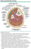
The deep peroneal nerve lies in which leg compartment?
Anterior compartment

What would be the physical manifestation of a nerve root compression of C6 nerve (C5-C6 disc)?
- Weak deltoid
- Weak biceps
- Weak wrist extensors
- Weak biceps reflex
- Weak brachioradialis reflex
What is the treatment for a tibial plateau fracture?
Open reduction and internal fixation (ORIF)
[Open fracture requires external fixator until tissue heals]
[UpToDate: Emergent (ie, immediate) surgical consultation is required for fractures that cause vascular compromise or acute compartment syndrome. Fractures with any degree of displacement or depression, even just a few millimeters, or those associated with suspected or documented meniscal or ligamentous injury merit orthopedic consultation within 48 hours.
Initial treatment — Compression, icing, appropriate analgesics, splinting of the knee in near-full extension, intermittent elevation of the leg above heart level, and strict non-weight bearing are the initial treatments for a tibial plateau fracture. Significant injuries are stabilized and orthopedic consultation is obtained. For fractures without displacement, depression of the tibial plateau, or associated injuries of significance (eg, knee ligament tear), at the first follow-up visit the patient is placed in a hinged brace that is locked in near-full extension and advised to continue non-weight bearing for the affected extremity, and to ambulate with crutches.
Follow-up care — Uncomplicated proximal tibial fractures without any displacement, depression of the tibial plateau, or associated ligamentous or meniscal injury are amenable to non-operative management by clinicians experienced managing such fractures. After brace fitting, the patient returns weekly for the first three weeks following injury. If there is no displacement at two weeks, the patient begins working on knee flexion in the brace with a goal of achieving 90 degrees by four weeks. Plain radiographs are repeated weekly for three weeks and then on a two to three week basis depending on radiographic appearance.
Strict non-weight bearing is the norm for six weeks, but this period may be adjusted based on the injury and clinical progress. Partial weight bearing in the brace can begin once there is adequate radiographic healing (ie, bony callus is present).
Bracing continues until radiographic healing appears complete – this typically requires 8 to 12 weeks. The patient begins exercises to regain lower extremity strength following removal of the brace. Patients rarely regain full function in less than 12 weeks and more often require 16 to 20 weeks.]
What is the treatment for club foot?
Serial casting
[UpToDate: Clubfoot, also referred to as talipes equinovarus, is a complex condition that involves both the foot and lower extremity. It is characterized by the foot being excessively plantar flexed, with the forefoot swung medially and the sole facing inward.
Congenital clubfoot is the most common type. It is usually an isolated anomaly without a well-delineated etiology. Current management is based upon manipulation that includes casting and bracing (referred to as the Ponseti method).]

What is the non-operative treatment for Carpal tunnel syndrome?
- Splint
- NSAIDs
- Steroid injections
[UpToDate: For patients with mild to moderate CTS, effective nonsurgical treatment options for short-term improvement include splinting, glucocorticoids injected into the carpal tunnel, and oral glucocorticoids. Combined therapy may be more effective than the use of any single modality. Referral to an occupational therapist with subspecialty certification in hand therapy may improve outcomes.
For patients with CTS who do not have surgery, we recommend nocturnal wrist splinting in the neutral position as initial therapy in preference to other nonsurgical measures (Grade 1B).
For patients who comply with nocturnal splinting, but remain symptomatic at one month, we suggest continuation of splinting for another one to two months while adding a different nonsurgical modality (ie, a single injection of methylprednisolone or a short course of oral prednisone as discussed below) rather than stopping splinting (Grade 2C).
For patients with CTS not treated with surgery who have an inadequate response to wrist splinting, we suggest a single injection with methylprednisolone (40 mg) as the next therapeutic option rather than oral glucocorticoids (Grade 2B). For patients who decline injection therapy, we suggest treatment with oral glucocorticoids (Grade 2B). We use prednisone 20 mg daily for 10 to 14 days.
A 2003 systematic review found one randomized controlled trial that demonstrated no significant benefit for NSAIDs when compared with placebo for improving CTS symptoms. We recommend not using nonsteroidal anti-inflammatory medication for the treatment of CTS (Grade 1B).]

Subluxation of the radius at the elbow caused by pulling on an extended, pronated arm is called what?
Nursemaid’s elbow
[UpToDate: Radial head subluxation is the most common elbow injury in children. It typically occurs between the ages of one and four years, with a peak incidence between two and three years, although cases have been reported in children younger than six months of age and as old as eight years. Girls are more often affected than boys, and the left arm is more commonly affected than the right
The usual mechanism of injury is axial traction on a pronated forearm with the elbow in extension. With sudden traction on the distal radius, a portion of the annular ligament slips over the head of the radius and slides into the radiohumeral joint, where it becomes trapped. The symptoms that develop are the result of displacement of the annular ligament. By the age of five years, the annular ligament has become thick and strong and is unlikely to tear or be displaced.]

What is the treatment for a metatarsal fracture?
Cast immobilization or brace for 6 weeks
[UpToDate: If the fracture is minimally or nondisplaced and conditions requiring emergent referral have been excluded, initial treatment includes immobilization in a posterior splint and non-weight-bearing with a follow-up visit in three to five days. The injury should be iced and elevated higher than the level of the heart for the first 24 hours.
Metatarsal fractures that are displaced laterally or medially usually do well without correction. Metatarsal fractures displaced more than 3 to 4 mm in a dorsal or plantar direction or with angulation greater than 10 degrees in this plane should probably be reduced or referred. Fractures with lesser degrees of dorsal and plantar displacement can be treated as nondisplaced fractures.
Reduction can frequently be achieved under local anesthesia using either a regional block (particularly useful in patients with multiple metatarsal fractures) or a hematoma block. The latter involves direct injection of local anesthetic into the hematoma that forms at the fracture site.
Once there is adequate anesthesia, reduction can be achieved by placing the toes in Chinese finger traps and allowing gravity to accomplish the reduction. Applying light weights or manual traction to the distal tibia is sometimes helpful. The reduction should be maintained by a molded, bivalved, non-weight-bearing cast, and postreduction radiographs should be obtained to confirm proper alignment. Referral or consultation with an orthopedist is recommended if the clinician is uncertain about the necessity of reduction or uncomfortable about performing the reduction, or if adequate alignment is not maintained by the reduction and immobilization measures described here.
Again, the injury should be iced and elevated higher than the level of the heart for the first 24 hours, while keeping the cast dry.]

Injury to which nerve results in claw hand?
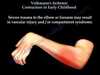
Ulnar nerve
What would be the physical manifestation of a nerve root compression of C7 nerve (C6-C7 disc)?
- Weak triceps
- Weak triceps reflex

Which condition is formed by subluxation or slip of one vertebral body over another?
Spondylolisthesis
[UpToDate: Spondylolysis is a unilateral or bilateral defect (fracture or separation) in the vertebral pars interarticularis, usually in the lower lumbar vertebrae. In young athletes, spondylolysis usually represents a fatigue fracture in the posterior arch of the spine, specifically the bony area of the pars interarticularis (pars) between the zygapophyseal (facet) joints. Although usually an overuse injury, spondylolysis may present following an acute overload. Several observations suggest spondylolysis is primarily a fatigue fracture. First, it has never been reported in a fetus or non-ambulatory person. Second, it occurs most frequently in athletes whose sport involves repetitive increased spinal loads.
Spondylolysis occurs at the fifth lumbar vertebra (L5) approximately 85% to 95% of the time, with an L4 locus in 5% to 15% of cases. Most injuries occur at L5 because the pars interarticularis at this level is subject to a direct pincer-like effect from the inferior articular process of L4 above and the superior articular process of S1 below. Rarely, the injury develops at levels above L4, but it has been reported as high as L1. Multilevel involvement occurs approximately 4% of the time, and bilateral involvement occurs in approximately 80% of cases. When bilateral defects develop, the vertebral body may slip anteriorly relative to the subadjacent vertebra and this is termed spondylolisthesis.]

How does posterior cruciate ligament injury manifest?
Knee pain and joint effusion
[UpToDate: PCL injuries sustained from low-energy trauma (eg, sporting injuries) may present with gross instability, particularly if associated with injuries to posterolateral knee structures, or with more subtle symptoms that can make diagnosis difficult. The presentation of an isolated PCL injury is often subtle, and quite different from that of an injury to the anterior cruciate ligament (ACL), which often involves an acute popping sensation perceived by the athlete at the time of injury, typically while performing a quick pivoting maneuver or landing from a jump, followed by the development of a large knee effusion. Patients with an isolated PCL injury generally do not report feeling or hearing such a “pop.” They may have a mild to moderate knee effusion, a slight limp, pain in the back of the knee (especially with squatting or kneeling), and loss of terminal knee flexion (final 10 to 20 degrees). Complaints of joint instability are more common with multiple knee ligament injuries than an isolated PCL injury. Acutely, many of these athletes may continue to play sports and not seek medical attention. An athlete’s only complaint may be a sensation that “something’s not right” in the knee or of generalized knee pain; the patient may have difficulty being more precise.
Patients suffering from a chronically injured PCL-deficient knee present more often with generalized anterior knee pain that may localize to the medial compartment or patellofemoral joint. According to an observational study of tibiofemoral motion involving open-access magnetic resonance imaging of patients with a PCL-deficient knee, altered kinematics related to PCL deficiency cause a fixed anterior subluxation of the medial femoral condyle in relation to the medial tibial plateau. This appears to increase the risk for degenerative changes in the medial knee. In this study, imaging was performed while patients performed several weight-bearing movements. The lateral compartment of the knee appears to be unaffected by the compressive forces created by PCL deficiency.
Patients with a chronic PCL-deficient knee may complain more of disability than instability that is most noticeable when negotiating inclines such as stairs or ramps. Athletes presenting with a chronic injury may complain of pain when sprinting or decelerating and mild instability. Many individuals with chronic PCL insufficiency are found to have been injured in a previous MVC. Such patients often sustained life-threatening injuries that caused the knee injury to be overlooked.]
Posterior hip dislocation is associated with injury to what?
Sciatic nerve
[Medscape: The sciatic nerve sits just inferoposterior to the hip joint and is injured in approximately 20% of all hip dislocations. These injuries range from nerve contusion to full laceration. The physician must perform a careful neurologic examination at the time of injury to assess sciatic nerve function. Most sciatic nerve injuries do not warrant acute intervention. Neurologic deficits that occur postreduction warrant immediate surgical intervention to decompress or reconstruct the damaged nerve.
Reduction should be attempted as expediently as possible, as prolonged times to reduction are associated with more severe and frequent nerve injuries.]

What causes Volkmann’s contracture?
- Supracondylar humerus fracture resulting in occlusion of anterior interosseous artery
- Closed reduction of humerus leads to reperfusion injury and edema
- This results in forearm compartment syndrome
[UpToDate: Complications of supracondylar fractures include vascular insufficiency, forearm compartment syndrome resulting in Volkmann’s ischemic contracture, nerve injury, and cubitus varus deformity.
Vascular injury and primary swelling from injury can lead to the development of compartment syndrome within 12 to 24 hours. If a compartment syndrome is not treated in a timely manner, the associated ischemia and infarction may progress to Volkmann’s ischemic contracture: fixed flexion of the elbow, pronation of the forearm, flexion at the wrist, and joint extension of the metacarpal-phalangeal joint.]
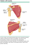
Which 4 muscles make up the rotator cuff
- Supraspinatus
- Infraspinatus
- Teres minor
- Subscapularis
Which type of Salter-Harris fracture is characterized as an epiphysiolysis of the involved growth plate without an associated fracture?
Salter-Harris I
[UpToDate: Salter I (Ogden IA-B) — The fracture line extends through the zone of hypertrophic cartilage (zone 3), causing the epiphysis and physeal elements to separate from the metaphysis. Type I injuries can have normal radiographs and the diagnosis is therefore often made clinically when focal tenderness is found over the growth plate.
- A type IB Ogden fracture is characterized by the fracture line extending through the primary spongiosa bone layer resulting in a thin line of bone displaced with the epiphysis. Type IB fractures usually occur in children with systemic diseases such as myeloproliferative disorders. Subsequent growth is usually normal with Type IA and IB fractures.
- A Type IC Ogden fracture has an associated injury to the germinal portion of the physis. Type IC fractures can cause growth arrest and occur rarely after age two to three years.]

Congenital dislocation of the hip (developmental dysplasia of the hip) is more common in which sex?
Females
UpToDate: Estimates of the incidence of developmental dysplasia of the hip (DDH) are quite variable and depend upon the means of detection, the age of the child, and the diagnostic criteria. It is estimated that dislocatable hips and hips with severe or persistent dysplasia occur in 3 to 5 per 1000 children. Historically, the incidence of DDH with dislocation is 1 to 2 per 1000 children. Mild hip instability is more common in newborns, with reported incidence as high as 40%. However, mild instability and/or mild dysplasia in the newborn period often resolve without treatment. Infants with mild instability and/or mild dysplasia in the newborn period should not be included in estimates of incidence; their inclusion results in overestimation.
In a prospective study, 9030 infants (18,060 hips) were routinely screened for DDH by physical examination and ultrasonography at one to three days of life. Sonographic abnormalities were detected in 995 hips (representing a sonographic incidence of 5.5%). However, on repeat examination at two to six weeks of age with no interval treatment, residual abnormalities were detected in just 90 hips (representing a true DDH incidence of 0.5%), which then proceeded to treatment. In other words, 90% of newborn hips with clinical or sonographic signs of DDH improved spontaneously before two to six weeks of age. It is our opinion that newborns with clinical or sonographic findings of mild laxity or minimal dysplasia have normal immaturity of hip development and should not be diagnosed with or treated for DDH.
The incidence of DDH also varies by race. It is increased in the Lapp and Native American populations (25 to 50 cases per 1000 births), and decreased in populations of African and Asian descent.
Both hips are involved in 20% of patients. Among the unilateral cases, the left hip is affected more often than the right. In a meta-analysis of risk factors for DDH that included ten studies, the relative risk for involvement of the left hip was 1.54 (95% CI 1.25-1.90). The preponderance of left-sided cases may be related to the typical left occiput anterior fetal positioning, in which the left hip is forced into adduction against the mother’s sacrum.
Thorough review of the child’s medical and family history helps to identify risk factors for DDH and exclude other congenital or neuromuscular causes of hip instability. The most important hip-specific risk factors are breech positioning at ≥34 weeks gestation (whether or not external cephalic version is successful), family history of DDH, and female sex. Other factors to be considered include birth order (the risk is increased in first born infants), pregnancy history (eg, the risk is increased in oligohydramnios), and other musculoskeletal abnormalities related to tight intrauterine packaging (eg, torticollis and metatarsus adductus).]

What is the treatment for a scapula fracture WITHOUT glenoid fossa involvement?
Application of a sling
[UpToDate: Scapular fractures account for only 1% of all fractures and less than 5% of fractures to the shoulder complex; they occur in up to 3.7% of blunt trauma patients. As scapular fractures generally require great force, over 90% are associated with other significant injuries, including rib fracture, pneumothorax, and pulmonary contusion. Scapular fractures rarely cause blunt aortic injury. Although the reasons are unclear, in one large prospective study scapula fractures were associated with lower mortality compared with similarly injured patients without them.
We obtain a chest CT in most patients with a scapular fracture following significant blunt chest trauma because of the forces involved and the risk of concomitant injury. We further suggest clinicians obtain consultation with trauma and orthopedic surgery. If the chest CT and workup for extrathoracic trauma reveal no injuries, and no concerns exist about analgesia, comorbidities, or the patient’s social circumstances, patients with scapular fractures may be discharged.]
Which nerve is most commonly injured with lower extremity fasciotomy?
Superficial peroneal nerve
[UpToDate: The superficial peroneal nerve is the most commonly injured nerve during fasciotomy of the leg, and knowledge of its normal and variant anatomy is important to prevent injury during anterior and lateral compartment fasciotomy.
The superficial peroneal nerve branches from the common peroneal nerve at or below the proximal fibular head. The nerve descends in its ‘normal’ course in the lateral compartment adjacent the intermuscular septum of the anterior and lateral compartments. Between 27% and 43% of patients have the superficial peroneal in either the anterior compartment or both the anterior and the lateral compartment of the leg. The superficial peroneal nerve has also been found to run within the septum that separates the anterior from the lateral compartment.]

Which nerve roots contribute to the radial nerve?
C5-C8
Which digits are most commonly affected in Dupuytren’s contracture?
4th and 5th digits (cannot extend fingers)
[UpToDate: Dupuytren’s contracture should be distinguished from diabetic cheiroarthropathy (limited joint mobility), palmar fibromatosis (also referred to as palmar fasciitis), camptodactyly, traumatic scars, Volkmann’s ischemic contracture, and intrinsic joint disease. Limited joint mobility in patients with diabetes involves all fingers except the thumb, whereas Dupuytren’s contracture more commonly affects just the fourth and fifth digits, with the other digits typically spared. In addition, the taut bands or cords and nodules characteristic of Dupuytren’s contracture are not commonly seen with cheiroarthropathy.]

Individuals with a slipped capital femoral epiphysis (SCFE) are at increased risk of what?
Avascular necrosis of the femoral head
[UpToDate: Osteonecrosis (also called aseptic necrosis, avascular necrosis, and ischemic necrosis) of the femoral head is the most serious complication of SCFE and has the worst prognosis. The natural history of osteonecrosis after treatment for SCFE is one of gradual degenerative changes for which reconstructive surgery is often required.
The rate of occurrence of osteonecrosis increases with increasing severity of the slip, occurring in 15% of patients with acute slips. Avascular necrosis rarely occurs in untreated chronic slips since the gradual slipping process permits maintenance of blood supply to the caput through adaptation of the vasculature. However, it can be a complication of operative pinning (if the lateral epiphyseal artery, which supplies the superior weightbearing portion of the femoral head, is injured during surgery).
Unstable SCFE is an important predictor for the development of osteonecrosis, particularly if vascular injury occurs at the time of the slip. Anterior physeal separation at the time of the slip also appears to be associated with subsequent development of osteonecrosis.
Osteonecrosis should be suspected when a patient with a history of SCFE complains of persistent pain and stiffness of the hip. Early in the course of osteonecrosis, the bone scan may show decreased uptake in the femoral head, but later bone scans may show increased uptake as the necrotic bone is replaced with new bone. Osteonecrosis also may be apparent on plain radiographs or MRI. SCFE patients who develop or are suspected to have osteonecrosis should be referred to an orthopedic surgeon.]

Which 3 muscles lie in the superficial posterior compartment of the leg?
- Gastrocnemius
- Soleus
- Plantaris
[Wikipedia: The plantaris is one of the superficial muscles of the superficial posterior compartment of the leg, one of the fascial compartments of the leg.
It is composed of a thin muscle belly and a long thin tendon. While not as thick as the achilles tendon, the plantaris tendon (which tends to be between 30 and 45 cm in length) is the longest tendon in the human body. Not including the tendon, the plantaris muscle is approximately 5–10 cm long and is absent in 8-12% of the population. It is one of the plantar flexors in the posterior compartment of the leg, along with the gastrocnemius and soleus muscles. The plantaris is considered an unimportant muscle and mainly acts with the gastrocnemius.]

What is the treatment for a non-displaced supracondylar humeral fracture in a child?
Closed reduction
[UpToDate: The emergency clinician should promptly identify children with vascular insufficiency and emergently involve an orthopedic surgeon with pediatric expertise. Rarely, these children will require partial closed reduction in the emergency department in an attempt to restore distal circulation. Patients who display a cold, cyanotic hand despite reduction attempts require emergent operative exploration and vascular repair.
Suspected compartment syndrome should prompt measurement of compartment pressure and/or emergent consultation with an orthopedic surgeon with appropriate pediatric expertise. Once confirmed by compartment pressure measurement, immediate management of suspected acute compartment syndrome includes relieving all external pressure on the compartment. Definitive treatment consists of fasciotomy to decompress all involved compartments.
For children with adequate distal circulation and no sign of compartment syndrome, initial therapy consists of pain management and immobilization to prevent further displacement of the fracture.
Emergent orthopedic consultation is indicated for children with an open fracture, neurovascular compromise, or acute compartment syndrome. In addition, prompt involvement of an orthopedic surgeon is necessary for operative care of a child with a Type II or Type III supracondylar fracture. Nondisplaced Type I supracondylar fractures are treated with a long arm splint with orthopedic follow up in one week.]

What is the treatment for a patellar fracture that is not comminuted?
Long leg cast
[UpToDate: Operative management is recommended for displaced or complex fractures in patients able to tolerate both the procedure and postoperative rehabilitation. Nondisplaced patella fractures with an intact extensor mechanism that do not meet operative criteria can be treated nonsurgically. Nondisplaced marginal vertical fractures do not require immobilization. They are treated with activity modification for four to six weeks and progressive range of motion and strengthening exercises.
Acutely, nonsurgical patients should be placed in a knee immobilizer or splint that is placed in full knee extension (not hyperextension). Patients are not to bear weight on the injured leg until a cylinder cast or brace locked in full extension is placed. Compression of the knee using an elastic bandage, ice applied to the patella area, and elevation of the leg above the level of the heart are important for reducing swelling and pain following the initial trauma.
Patients should begin performing strength exercises as soon as possible, including isometric contraction of the quadriceps and straight leg raises, but only while their knee is immobilized.]
What is Felon?
Infection of the terminal joint space of the finger
[UpToDate: The digital pulp, the fleshy mass at the finger tips, is divided into multiple compartments by fibrous septae that provide structural support. Pulp abscesses account for 15% to 20% of all hand infections, and the thumb and index finger are the most commonly affected digits. A severe infection or abscess of the pulp space, called a felon, results in increased pressure and can lead to ischemic necrosis of surrounding tissue, osteomyelitis, flexor tenosynovitis, or septic arthritis of the distal interphalangeal joint (DIPJ). A pulp abscess usually occurs after a puncture wound but may also result from untreated acute paronychia.
Patients with pulp space infections present with pain, cellulitis, and an associated tender fluctuant swelling. The pain is severe and throbbing, worse in the dependent position, and usually does not allow the patient to sleep. The swelling is limited to the soft tissue around the distal phalanx and an area of imminent rupture (pointing) may be obvious. Occasionally, the abscess may spontaneously discharge through the skin, decompressing it and thus reducing symptoms. A radiograph should be obtained to look for any retained foreign bodies and rule out involvement of the distal phalanx.]

Which nerve is responsible for hip adduction?
Obturator nerve
[The obturator nerve arises from the ventral divisions of the L2-L4 in the lumbar plexus; the branch from L3 is the largest, while that from L2 is often very small.]
[UpToDate: The obturator nerve arises from the second, third, and fourth lumbar nerve roots. The fibers then unite posterior to the psoas muscle and pass inferiorly over the sacrum or pelvic brim to the obturator canal. The obturator nerve then bifurcates into anterior and posterior divisions. Both divisions innervate the thigh adductor muscles; the anterior division provides sensory input from the hip joint and anterior medial thigh and the posterior division from the knee.
The obturator nerve may be injured with passage of a trocar through this area (eg, for placement of a transobturator tape or passage of a vascular graft), or during pelvic lymph node dissection in the obturator fossa. During dissection of the obturator fossa, the obturator nerve should be identified. Procedures associated with obturator nerve injury include excision of endometriosis, paravaginal defect repair with dissection in the space of Retzius and obturator bypass.
If unilateral obturator nerve injury occurs, numbness of the inner thigh and minor ambulatory problems will be noted due to weakened adduction of the thigh. Diagnosis of obturator nerve injury is made clinically. Imaging studies do not contribute to the diagnosis. Injection of local anesthetic for a nerve block can be both diagnostic and therapeutic.
Repair of a freshly transected obturator nerve using microsurgical technique and postoperative physiotherapy often results in complete motor recovery.]

Which nerve innervates the intrinsic muscles of the hand (palmar interossei, plamaris brevis, adductor pollicis, hypothenar eminence)?
Ulnar nerve
[UpToDate: At the wrist, the ulnar nerve passes through Guyon’s canal (along with the ulnar artery), the floor of which is formed by the transverse carpal ligament and pisohamate ligament. The roof of Guyon’s canal consists of the palmar fascia and the palmaris brevis muscle. The ulnar nerve then divides into the superficial and deep terminal branches. In the hand, after giving off a branch to the palmaris brevis muscle, the superficial terminal branch supplies the cutaneous ulnar border of the palm and then divides into two digital branches that innervate the palmar or volar surfaces of the fifth and ulnar half of the fourth digit. The deep branch pierces and innervates the opponens digiti muscle and then gives off a branch to the remaining hypothenar muscles just after emerging from Guyon’s canal. In the palm, the deep branch innervates all of the interossei and the third and fourth lumbricals. It then terminates in the thenar eminence where it supplies the adductor pollicis and variable portions of the flexor pollicis brevis muscles.]

What would be the physical manifestation of a nerve root compression of C8 nerve (C7-T1 disc)?
- Weak triceps
- Weak intrinsic muscles of the hand
- Weak wrist flexion

Which nerve roots contribute to the axillary nerve?
C5-C6

Are the radial nerve roots on the superior or inferior portion of the brachial plexus?
Superior portion

Medial collateral ligament injury is caused by what?
Lateral blow to the knee
[UpToDate: Injuries of the medial collateral ligament (MCL), also referred to as the tibial collateral ligament, occur frequently in athletes, particularly those involved in sports that require sudden changes in direction and speed, and in patients struck on the outside of the knee. Most heal well with conservative treatment, but some are associated with other significant injuries and careful evaluation is needed.
According to one systematic review, studies of the epidemiology of knee injuries are deeply flawed and should be interpreted cautiously. Nevertheless, ligament injuries account for up to 40% of all knee injuries, and of these, medial collateral ligament (MCL) injuries appear to be the most common. MCL tears accounted for 7.9% of all injuries in an observational study of 19,530 knee injuries in 17,397 athletes over a 10 year period. The precise incidence of MCL injuries is unlikely to ever be known because many low grade injuries go unreported. While still common, MCL injuries declined in number during the course of an 11-year study of injuries in the Union of European Football Associations (UEFA).]

The peroneal artery lies in which leg compartment?
Deep posterior compartment

What is the treatment for a scapula fracture WITH glenoid fossa involvement?
Internal fixation
[UpToDate: Glenoid neck fractures are relatively rare in the pediatric age group, but have been reported as a skiing injury. On plain radiographs, these fractures will extend from the suprascapular notch to the lateral scapular border inferior of the glenoid rim. When this fracture is associated with a midclavicular fracture, it is considered a “floating shoulder” injury because these structures are essential to providing a foundation to suspend the arm to the axial skeleton. Indications for surgical and nonsurgical management are debated. Displaced fractures may be treated with open or closed reduction, but regardless of treatment, usually have good functional outcomes.
Glenoid fossa fractures (excluding bony Bankart lesions associated with shoulder instability injuries) result from a direct blow to the lateral aspect of the shoulder, driving the humeral head into the glenoid fossa. The fracture can extend inferiorly through the lateral scapular border, medially through the body of the scapula, or produce comminution of the glenoid. In the pediatric population, a fall on the tip of the shoulder will drive the scapula inferiorly while the fracture fragment is held in place by the stronger coracoclavicular ligaments. The fracture will track along the epiphysis of glenoid fossa, located at the upper one-third of the glenoid and inferior to the coracoid process. It is rarely complicated by a suprascapular nerve injury. This injury can mimic an acromioclavicular separation and the Stryker Notch view may help to demonstrate the fracture on radiography. Most of these injuries are treated nonoperatively with immobilization for four to six weeks, but displacement of more than 10 mm or more than 40 degrees of angulation requires surgical management.]
How does an injury to the common peroneal nerve present?
Foot-drop
[Wikipedia: Damage to this nerve typically results in foot drop, where dorsiflexion of the foot is compromised and the foot drags (the toe points) during walking; and in sensory loss to the dorsal surface of the foot and portions of the anterior, lower-lateral leg. A common yoga kneeling exercise, the Vajrasana, has been linked to a variant called yoga foot drop.]

What is the treatment for giant cell tumor of bone?
Total resection +/- XRT
[Benign but 30% risk of recurrence and malignant degeneration risk]
[UpToDate: Surgery is the treatment of choice for a GCTB. For potentially resectable intraosseous lesions (primary or recurrent), we recommend intralesional curettage followed by filling of the cavity with bone cement (polymethylmethacrylate, PMMA) rather than curettage alone (Grade 1B).
Indications for more extensive surgery include extraosseous extension, tumor involving the proximal fibula or distal ulna, a second or later recurrence, a dislocated pathologic fracture, or one in which the articular surface is grossly damaged.
For patients with a GCTB at a site where surgical margins can only be achieved with unacceptable morbidity (as, for example, with a large midline sacral GCTB with no chance of preserving at least one set of sacral nerve roots), or if the patient is a poor surgical candidate, systemic options (ideally through a clinical trial) or radiotherapy are reasonable options.
For sacral tumors, another option is arterial embolization, although published experience is limited.
Local recurrence is accompanied by an increased risk of pulmonary metastases. We suggest screening of such patients for pulmonary metastases with a chest CT (Grade 2B).
Most patients with pulmonary metastases have a favorable outcome and do not die of their disease. Nevertheless, we suggest surgery rather than observation for most patients (Grade 2B). If observation is chosen, close monitoring is needed for early detection of tumor progression.
GCTB is a radiosensitive tumor, and radiotherapy is highly effective, resulting in long-term local control rates ranging from 60% to 84%; rates are lowest for tumors >8.5 cm and for locally recurrent disease.
Radiotherapy is a reasonable option if surgery is contraindicated, or in a situation where negative surgical margins can only be achieved with unacceptable morbidity. One example of where this might be the case is a large midline sacral GCTB. Resection of a small sacral tumor to one side of midline may be possible without unacceptable morbidity if one set of sacral nerves can be preserved, providing reasonable postoperative bladder and bowel function. However, a complete resection of a large midline sacral GCTB would necessitate sacrifice of both sets of sacral nerves, leading to permanent double incontinence.
Radiation has also been used as an adjuvant therapy to reduce local recurrence rates after intralesional surgery for spinal GCTB. There are no trials comparing radiation therapy versus a local adjuvant such as bone cement or burring in this setting.]

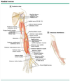
Which nerve is responsible for wrist extension, finger extension, and thumb extension?
Radial nerve
[UpToDate: The radial nerve innervates the extensor muscles of the hand. The radial nerve divides at the proximal forearm into superficial and deep branches. The deep posterior interosseus branch innervates all the extensor muscles in the hand including the extensor digitorum communis, extensor digiti minimi, extensor indicis proprius, extensor pollicis longus, extensor pollicis brevis, and abductor pollicis longus. Radial nerve motor function can be tested by resisting thumb, MCP, or wrist extension.]
Which nerve is responsible for finger abduction (spread fingers)?
Ulnar nerve
[Wikipedia: In human anatomy, the dorsal interossei (DI) are four muscles in the back of the hand that act to abduct (spread) the index, middle, and ring fingers away from hand’s midline (ray of middle finger) and assist in flexion at the metacarpophalangeal joints and extension at the interphalangeal joints of the index, middle and ring fingers.
There are four dorsal interossei in each hand. They are specified as ‘dorsal’ to contrast them with the palmar interossei, which are located on the anterior side of the metacarpals.
The dorsal interosseous muscles are bipennate, with each muscle arising by two heads from the adjacent sides of the metacarpal bones, but more extensively from the metacarpal bone of the finger into which the muscle is inserted. They are inserted into the bases of the proximal phalanges and into the extensor expansion of the corresponding extensor digitorum tendon. The middle digit has two dorsal interossei insert onto it while the first digit (thumb) and the fifth digit (little finger) have none. Each finger is provided with two interossei (palmar or dorsal), with the exception of the little finger, in which the abductor digiti minimi muscle takes the place of one of the dorsal interossei.
All interosseous muscles of the hand, with the exception of the first and second lumbricals (the most radial two are innervated by the median nerve), are innervated by the deep branch of the ulnar nerve.]
How does anterior cruciate ligament injury manifest?
Knee effusion and pain with pivoting action
[UpToDate: Patients who sustain a noncontact ACL injury often complain of feeling a “pop” in their knee at the time of injury, acute swelling thereafter, and a feeling that the knee is unstable or “giving out.” Nearly all patients with an acute ACL injury manifest a knee effusion from hemarthrosis. Conversely, approximately 67% to 77% of patients presenting with acute traumatic knee hemarthrosis have an ACL injury.
Often after the initial swelling has improved, patients are able to bear weight but complain of instability. Movements such as squatting, pivoting, and stepping laterally, and activities such as walking down stairs, in which the entire body weight is placed on the affected leg, most often elicit such instability.]

What is the treatment for a scaphoid fracture?
All patients require cast to elbow, may need fixation
[Risk of avascular necrosis]
[UpToDate: The scaphoid has a tenuous blood supply that runs from distal to proximal leading to the possibility of nonunion or osteonecrosis with fractures of the proximal pole.
Open fractures and those associated with neurovascular compromise require immediate surgical referral. Indications for referral within several days include:
- Proximal pole (ie, proximal fifth of scaphoid) fractures
- Fractures displaced over 1 mm
- Delayed presentation of acute fractures (more than about three weeks)
- Associated scapholunate ligament rupture
- Carpal instability (eg, lunate tilt on radiographs)
Early consultation for nondisplaced scaphoid fractures may be preferred when a faster recovery is desired. Indications for routine referral include evidence of nonunion or osteonecrosis on follow-up during treatment with immobilization.
Nondisplaced fractures (≤1 mm) of the distal scaphoid can be treated in a short-arm thumb spica cast, typically for 6 to 10 weeks. Nondisplaced fractures at the waist or proximal third (not the proximal pole) of the scaphoid can be treated in a short-arm or long-arm thumb spica cast for six weeks, followed by a short-arm thumb spica cast until healing is documented. These fractures require a longer period of immobilization. If prolonged immobilization cannot be tolerated, refer the patient for operative fixation.
Athletes and workers engaged in heavy labor must continue to wear protection (rigid splint) for two months after radiographic healing is noted.]

What is the treatment for osteoid osteoma?
Benign bone tumor treated with curettage +/- bone graft
[UpToDate: Osteoid osteoma is a benign bone-forming tumor that is characterized by a small radiolucent nidus (usually <1 to 1.5 cm in diameter). The nidus produces high levels of prostaglandins. In addition to prostaglandins, there is some evidence that osteoid osteomas may secrete osteocalcin.
The treatment of osteoid osteoma depends upon the presence of symptoms. Lesions with symptoms that are tolerable or can be controlled with nonsteroidal anti-inflammatory agents may be observed with serial examinations and radiographs every four to six months.
Treatment options for symptomatic lesions (eg, intolerable pain, limp, scoliosis) include surgical resection, which may be aided by CT-guided needle localization, or radiofrequency ablation (in certain institutions). Treatment options may be limited by proximity to vital structures.
Untreated osteoid osteoma spontaneously resolves over the course of several years. Removal of the nidus generally results in resolution of pain. Recurrence is possible if the nidus is not completely removed.]
Which cell type synthesizes non-mineralized bone? cortex?
Osteoblasts
[UpToDate: Osteoblasts form the connective tissue matrix, which mineralizes and becomes bone. The precursors of osteoblasts are multipotent mesenchymal stem cells, which also give rise to bone marrow stromal cells, chondrocytes, muscle cells, and adipocytes. Osteoblast progenitors may originate not only from stromal mesenchymal progenitors of the marrow, but also pericytes (mesenchymal cells adherent to the endothelial layer of vessels). Mesenchymal stem cell progenitors of osteoblasts residing on the outer surface of blood vessels are critical for the formation of blood vessels as well. The process of differentiation of these progenitors to mature osteoblasts, ie, the cells capable of synthesizing the bone matrix, is accomplished by a sequential progression from the least differentiated and most proliferative cell to terminally differentiated cell that can no longer undergo mitosis. At the beginning of the process, mesenchymal stem cells with unlimited self-renewal capacity undergo asymmetric divisions resulting in not only other stem cells, but also daughter cells with limited self-renewal and extensive proliferation capacity. Progressively, the proliferative capacity decreases as differentiation increases.]

Which nerve roots contribute to the musculocutaneous nerve?
C5-C7

What is the treatment for bimalleolar or trimalleolar ankle fractures??
Open reduction and internal fixation (ORIF)
[UpToDate: These fractures are unstable and require operative fixation. Patients should be splinted with the ankle joint at 90 degrees, remain nonweightbearing, and be referred to an orthopedist within a few days.]

What is the treatment for Nursemaid’s elbow?
Closed reduction
[UpToDate: Reduction of radial head subluxation (RHS) can usually be performed in the office or emergency department. Anesthesia and sedation are not required. Reduction may be attempted for the child with typical physical findings, even when the classic history is lacking.
The reduction procedure is brief, but painful. It is therefore important to explain the procedure to the caretakers before attempting reduction. The procedure is best performed with the child seated comfortably in the parent’s arms and the examiner seated and facing the child.
Supination/flexion and hyperpronation are two techniques for reduction of RHS. Both techniques are effective. Supination/flexion is the method that has been used most commonly. However, metaanalysis of four randomized trials found that successful reduction was more likely with hyperpronation (RR 0.45, 95% CI 0.28-0.73) although the quality of the evidence was felt to be low. Based on this analysis, nine children would require treatment by the hyperpronation method rather than supination/flexion to avoid one failed reduction on first attempt.
In addition, hyperpronation may be less painful. One trial compared physician, nurse, and caretaker assessments of perceived pain (using a validated visual analog scale to assess pain) with each method of reduction. Physicians did not note a significant difference in pain scores between the two methods. However, both nurses and caretakers perceived hyperpronation as less painful.]

Which type of Salter-Harris fracture crosses the epiphysis, growth plate (physis), and the metaphysis?
Salter-Harris IV
[UpToDate: Salter IV (Ogden IVA-C) — The fracture line spreads from the articular surface, through the epiphysis, across the physis, and through a segment of the metaphysis.
- A Type IVB fracture is characterized by further transverse extension of the fracture through part or all of the physis creating additional epiphyseal fragments.
- Type IVC fractures involve damage to the adjacent cartilage, and type IVD fractures have multiple metaphyseal-physeal-epiphyseal fragments, usually from severe trauma.]

What is the treatment for osteogenic sarcoma?
Limb-sparing resection
[UpToDate: The goal of the preoperative evaluation for a primary bone sarcoma is to establish the tissue diagnosis, evaluate disease extent, and assess the feasibility of a limb-sparing approach.
Surgery and systemic chemotherapy are the mainstays of treatment for patients with osteosarcomas and other primary bone tumors such as fibrosarcoma of bone. Chondrosarcoma and chordoma have not been routinely or historically treated with chemotherapy or radiation, and therefore, surgery is the mainstay of management.
Although there is no specific survival benefit to preoperative as compared to postoperative chemotherapy in osteosarcoma patients, the neoadjuvant approach may permit a greater number of patients to undergo limb-sparing procedures. However, chemotherapy is no substitute for sound surgical judgment when assessing the need for amputation versus limb-sparing surgery. The optimal chemotherapy regimen has not been established. In general, patients with a nearly complete response to neoadjuvant chemotherapy do better than those with a lesser response. Even if the patient has a chemosensitive tumor, it may be reasonable to proceed to immediate surgery followed by adjuvant chemotherapy if the nature of the resection would not necessarily be influenced by a good response to chemotherapy.
Reconstructive options vary depending upon the factors listed above. In addition, there is surgeon preference and no absolute consensus in many circumstances as to the optimal reconstructive method. In some circumstances, if the tumor is located in an expendable bone, reconstruction is not even necessary.
The available data suggest that patients with osteosarcoma who present with or sustain a pathologic fracture have a higher rate of local recurrence and an increased risk of death relative to those who do not have a pathologic fracture. However, the method of local control, whether limb salvage or amputation, does not appear to impact the ultimate outcome of these patients.
There is no role for adjuvant radiation therapy except in patients with unresectable or incompletely resected sarcomas and possibly in the rare patient with a small cell osteosarcoma. Radiation therapy for local control can be used in patients who decline surgery or for whom there is no effective surgical option.
In contrast to osteosarcoma and other primary bone sarcomas, Ewing sarcoma is more radiosensitive, and radiation may be considered an effective option for local control. Although modern treatment protocols emphasize surgery for optimal local control, patients who lack a function-preserving surgical option because of tumor location or extent, and those who have clearly unresectable primary tumors following induction chemotherapy are appropriate candidates for radiation therapy. Furthermore, adjuvant radiation may be considered for bulky tumors in difficult sites (eg, the pelvis), if there is residual microscopic or gross disease after surgery, and for patients with high-risk chest wall primary tumors (close or involved margins, initial pleural effusion, pleural infiltration, and intraoperative contamination of the pleural space).]

A positive posterior drawer test is indicative of what?
Posterior cruciate ligament injury
[UpToDate: The posterior drawer test is generally considered the most accurate examination maneuver for detecting PCL ligament injury. Prior to performing the posterior drawer test, the clinician must ascertain the position of the tibia relative to the femur. Posterior subluxation of the tibia due to a loss of PCL integrity can compromise test results.
Posterior drawer test shows less than 10 mm laxity. In rare instances, an isolated PCL injury can manifest >10 mm of laxity on posterior drawer testing. In all cases, such a finding necessitates assessment of other knee structures, particularly the posterolateral corner (PLC).]

What is the treatment of acromioclavicular separation?
Application of a sling due to risk of brachial plexus and subclavian vessel injury
[UpToDate: Initial management consists of rest, ice, and protection using a sling. Minor injuries (Types I and II) are managed nonoperatively. Severe injuries (Types IV, V, and VI) should be referred urgently to an orthopedic specialist for operative management. Injuries involving neurologic or vascular deficits require emergent (ie, immediate) orthopedic referral and intervention.
Operative management of intermediate injuries (Type III) does not appear to improve functional outcome when compared with conservative management, and increases the risk of complications. In general, we suggest nonoperative management of Type III injuries (Grade 2B).
There is no role for glucocorticoid (steroid) injections in the acute management of these injuries. Injection may be considered for persistent pain after the ligamentous injury has healed, or in the setting of repeated minor injury with no instability but persistent AC joint arthralgia.]

Which nerve provides motor function to the biceps, the brachialis, and the coracobrachialis?
Musculocutaneous nerve
[UpToDate: The musculocutaneous nerve arises from the lateral cord of the brachial plexus and contains fibers from the C5, C6, and C7 nerve roots. After piercing the coracobrachialis muscle, it courses down the front of the arm between the biceps and brachialis and continues into the forearm as the lateral antebrachial cutaneous nerve. It innervates the coracobrachialis, biceps, and brachialis muscles and supplies cutaneous sensation to the lateral forearm. Clinical features thus include weakness of elbow flexion with associated sensory loss over the lateral forearm.]

Which cell type reabsorbs bone?
Osteoclasts
[UpToDate: Osteoclasts are terminally differentiated multinucleated cells that are uniquely capable of digesting calcified bone matrix. They are formed by fusion of mononuclear precursors of the monocyte/macrophage lineage. The receptor activator of nuclear factor kappa B (NF-kB) ligand (RANKL) and the macrophage colony-stimulating factor (M-CSF) are two cytokines that are essential for the development, function, and survival of osteoclasts, and NFATc1 is the master transcription factor responsible for osteoclast differentiation and function. Osteocytes are the main cellular source of the RANKL that is required for osteoclast generation during bone remodeling. However, it is currently unclear whether during remodeling RANKL is produced by apoptotic osteocytes or live osteocytes responding to the death of their neighbors.
RANKL interacts with a receptor on osteoclast precursors that is identical to the receptor involved in interaction of T-cells and dendritic cells called RANK. NFATc1 is induced by RANKL and co-activated by immunoglobulin-like receptors and their associated adapter proteins. RANKL can also bind to a protein called OPG or osteoclastogenesis inhibitory factor. Administration of a single dose of osteoprotegerin or an antibody to RANKL to postmenopausal women results in rapid reduction of biochemical markers of bone turnover and increase in BMD.]

Tenosynovitis of the flexor tendon that catches at the MCP joint when trying to extend the finger is called what?
Trigger finger
[UpToDate: Trigger finger (also called stenosing flexor tenosynovitis) is caused by a disparity in the size of the flexor tendons and the surrounding retinacular pulley system at the first annular (A1) pulley which overlies the metacarpophalangeal (MCP) joint. The flexor tendon catches when it attempts to glide through a relatively stenotic sheath, resulting in an inability to smoothly flex or extend the finger. In severe cases, the finger may become locked in flexion requiring passive manipulation of the finger into extension. The cause of trigger finger is most frequently unclear, although patients often attribute it overuse or repetitive movements.]

What is the operative treatment for Trigger finger?
Release the pulley system at the MCP joint
[UpToDate: Surgery is suggested when pain and locking persist despite conservative therapy and at least one or two local glucocorticoid injections. Ultrasound-guided percutaneous and open surgical release of the first annular (A1) pulley ligament are both effective, with a recurrence rate of only about 3%, but controversy exists as to whether they are equally effective. As an example, a large meta-analysis including seven randomized trials compared percutaneous release, open surgery, or glucocorticoid injections in patients with trigger finger, and found no differences in the failure rate and complication frequency between the two different surgical approaches. In addition, patients treated with percutaneous releases were less likely to have treatment failure compared with patients treated with glucocorticoid injections.
Outcomes of surgery may not be as successful in diabetics. As an example, a study evaluating the outcome of trigger finger treatment in diabetic patients found that 13% of diabetic patients who underwent surgery had an unsuccessful outcome compared with 6% of non-diabetic patients.
Complications from surgery include infection, digital nerve injury, flexor tendon bowstringing (or protrusion of the flexor tendon into the palm with finger flexion), and tendon scarring. Postoperatively, patients are able to resume many activities of daily living, such as dressing, eating, and doing sedentary work, within a few days. Most patients take a week or two to resume more demanding activities, but occasionally they can take longer.]

What is the treatment for a metastatic bone tumor?
Internal fixation with impending fracture followed by XRT
[UpToDate: Bone metastases are best managed with a collaborative, multidisciplinary approach.
Asymptomatic patients with widespread metastasis, no risk of impending fracture, and a limited life expectancy may not require intervention.
Pain due to bone metastasis may be treated with analgesics (typically opioids with or without an analgesic adjuvant) and/or radiotherapy (RT). Interventional pain specialists may help relieve pain with procedures such as nerve blocks when opioid analgesics are maximized.
Systemic therapy with osteoclast inhibitors reduces the risk of SREs, and they also have analgesic properties. For sensitive tumors, chemotherapy and hormone therapy may contribute to pain relief.
Many individuals with a completed or impending pathologic as well as those with epidural spinal cord compression or an unstable spine due to vertebral metastasis are best treated with surgery followed by RT, if such treatment is consistent with the goals of care.
Percutaneous vertebral augmentation improves pain and the mechanical stability of the bone. Vertebroplasty and kyphoplasty are generally reserved for patients with symptomatic spinal metastases without epidural disease or retropulsion of bone fragments into the spinal cord. However, for asymptomatic patients who are at risk for spinal cord compression due to significant malignant destruction of the vertebrae, elective vertebroplasty may also be considered.
For patients who have persistent or recurrent pain attributed to one or a few skeletal sites after palliative RT and who are not surgical candidates, local thermal ablation (radiofrequency ablation [RFA], cryoablation, and focused ultrasound [FUS]) are important therapeutic options.
Radium-223 is a reasonable option for men with castration-resistant prostate cancer, symptomatic bone metastases, and no known visceral metastatic disease. Other bone-seeking radiopharmaceuticals (Samarium-153 lexidronam [153Sm], strontium chloride-89) are generally reserved for individuals with persistent or recurrent multifocal bone pain from osteoblastic metastases after external beam RT (EBRT) and/or other forms of therapy.]

In which compartment of the leg is compartment syndrome most likely to occur?
Anterior compartment
[Manifests with foot drop]
[UpToDate: The anterior compartment of the leg is the most common site for ACS. It contains the four extensor muscles of the foot, the anterior tibial artery, and the deep peroneal nerve. Signs of ACS affecting the anterior compartment include loss of sensation between the first (ie, great) and second toes and weakness of foot dorsiflexion. Late sequelae include foot drop, claw foot, and deep peroneal nerve dysfunction.]

What test is required for all patients with a posterior knee dislocation?
Angiogram to rule out popliteal artery injury
[UpToDate: The popliteal artery is the continuation of the femoral artery. It originates at the tendinous hiatus of the adductor magnus muscle, which firmly anchors it to the femoral shaft. Within the popliteal space, the artery gives off five branches which arise above and below the knee joint creating a collateral system about the knee. Distally, the popliteal artery is held firmly against the bone by the tendinous arch of the soleus muscle. Thus, the popliteal artery is tethered across the popliteal space like a bowstring, making it susceptible to injury during knee dislocation. Up to 40% of patients with knee dislocations sustain an associated vascular injury.
Popliteal artery injury is the most dangerous potential complication following tibiofemoral dislocation. Delay in diagnosis and repair increases the period of warm ischemia and the corresponding risk for irreparable injury requiring above-knee amputation. After eight hours of ischemia, the great majority of injuries will require amputation. Of note, popliteal artery lesions or thrombosis may not become clinically apparent for up to several weeks following the acute knee injury.
Other potential short-term complications include peroneal nerve injury, compartment syndrome of the leg, and deep vein thrombosis. Complications may also include pseudoaneurysm, instability, arthrosis, stiffness, and chronic pain. Associated fractures may occur, including injuries of the tibial plateau, tibial shaft, and the proximal fibula.]

Which nerve is responsible for thumb apposition (anterior interosseus muscle, OK sign)?
Median nerve
[UpToDate: The hand is innervated by the median, ulnar and radial nerves. Each has motor and sensory components. Of note, there is considerable variation in sensory innervation. The examination of the hand should include sensation of pin and light touch as well as two-point discrimination. Although values increase with age and certain disease states (eg, diabetes), normal two-point discrimination is approximately 4 to 5 mm.
The median nerve supplies sensation to the radial side of the palm and the palmar surface of the thumb, index finger, long finger, and the radial side of the ring finger. It also supplies sensation to the dorsal surfaces of these fingers distal to the distal interphalangeal (DIP) joint. The ulnar nerve supplies sensation to the hypothenar eminence, the palmar and dorsal surfaces of the little finger, and the ulnar side of the ring finger. The radial nerve innervates the dorsal surface of the palm and fingers, from the thumb to the radial side of the ring finger, proximal to the DIP joints.
The median nerve innervates the muscles involved in pinching (thumb apposition). In the forearm, the median nerve innervates the flexor digitorum superficialis (FDS), while its anterior interosseus branch innervates the flexor pollicis longus and the flexor digitorum profundus (FDP) of the index and long fingers. As the median nerve passes through the carpal tunnel, it splits into the recurrent motor branch and gives off a number of palmar digital nerves which travel with the digital arteries. The recurrent motor branch innervates the abductor pollicis brevis, opponens pollicis, superficial head of the flexor pollicis brevis, and the lumbrical muscles of the index and long finger. The palmar digital nerves run on the lateral and medial sides of the thumb, index finger, middle finger, and the radial side on the ring finger.
Motor function of the anterior interosseus branch of the median nerve can be tested by having the patient make the “OK” sign. Flattening of the “O” or loss of flexion of the IP or DIP joint suggests anterior interosseus nerve dysfunction. The remainder of median nerve function can be tested by assessing the strength of index or middle finger proximal interphalangeal (PIP) and DIP flexion, thumb opposition with the small finger, or wrist flexion.]

Which compartment is most commonly affected in Volkmann’s contracture?
Flexor compartment
[UpToDate: The forearm has four compartments: the deep and superficial volar compartments, the dorsal compartment, and the lateral compartment. The volar compartment contains the digital flexors and the dorsal compartment contains the digital extensors. The volar compartments are at highest risk for developing ACS following trauma. The most frequent injuries associated with ACS in the forearm are supracondylar humerus fractures in children and distal radius fractures in adults.]

What is the non-operative treatment for Trigger finger?
- Splint
- Tendon sheath steroid injections (not the tendon itself)
[UpToDate: For all patients with trigger finger, we suggest initial management with conservative therapy which includes activity modification or splinting as well as a concurrent trial of nonsteroidal antiinflammatory drugs (NSAIDs) (Grade 2C).
For patients with trigger finger whose symptoms do not improve after four to six weeks of conservative therapy, we suggest a local glucocorticoid injection rather than surgical therapy (Grade 2B). We use an intermediate-acting glucocorticoid injection such as methylprednisolone or triamcinolone mixed with a local anesthetic. A glucocorticoid injection may be offered sooner to patients who present with severe locking and incomplete finger flexion or extension. The injection may be repeated in six weeks if symptoms have not improved by at least 50%.
For patients with persistent pain and locking persist despite conservative therapy and at least one or two local glucocorticoid injections, surgical therapy may help relieve symptoms. Ultrasound-guided percutaneous and open surgical release of the A1 pulley ligament are both effective.]

What is the treatment for a non-displaced calcaneus fracture?
Cast and immobilization
[UpToDate: Initially, a bulky compression dressing should be applied and the patient instructed to rest, elevate the foot above the level of the heart, and apply ice for the first 48 to 72 hours. No weightbearing should be allowed until casting or evaluation by the surgeon. Basic initial fracture care is discussed in greater detail separately.
For patients with an avulsion fracture that is nondisplaced and does not involve more than 25% of the calcaneocuboid joint, a short-leg non-weightbearing cast is applied once swelling has subsided, generally three to seven days after the injury. Most non-displaced anterior process fractures heal well with four to six weeks of casting. After cast removal, the patient begins range of motion exercises and progressive weight bearing, as activity is gradually increased. If the patient does not make steady progress on their own, supervised physical therapy is recommended. Slow progress or a long period without progress should prompt the clinician to obtain surgical referral or repeat radiographs to assess for possible non-union or other complications.
Activities that pose a risk of reinjury should be avoided for the first three to four weeks after the cast is removed. Such activities include activities on uneven ground and sports that involve quick changes of direction or substantial lateral movement. Careful attention to rehabilitation can speed return to full activity. In a minority of patients, recovery to pre-injury activity levels may take up to a year.]

Posterior shoulder dislocation is associated with a risk of injury to what?
Axillary artery
[UpToDate: Posterior shoulder dislocations are commonly associated with tuberosity and surgical neck fractures of the humerus, reverse Hill-Sachs lesions, and injuries to the labrum and rotator cuff. In cases where plain radiographs are indeterminate for dislocation, computed tomography is diagnostic and reveals the size of the articular surface impaction fracture, enabling the orthopedic surgeon to determine the most appropriate treatment.]

Which 3 muscles lie in the deep posterior compartment of the leg?
- Flexor hallucis longus
- Flexor digitorum longus
- Posterior tibialis

What is the treatment for a combined radial and ulnar fracture in an adult?
Open reduction and internal fixation (ORIF)
[UpToDate: Rarely, nondisplaced, non-angulated both-bone fractures are candidates for nonoperative treatment. The vast majority require surgical fixation. There is no high quality evidence that compares nonoperative and operative treatment of nondisplaced both-bone forearm fractures. We suggest referral to an orthopedic surgeon to determine the best course of treatment.
Nondisplaced, non-angulated both-bone fractures may be treated initially in a well-molded, bivalved long-arm cast with the wrist in a functional position, the forearm neutral, and the elbow at 90 degrees of flexion. The cast must cover from the distal palmar crease to the deltoid tuberosity to prevent forearm supination and pronation. A loop of wire or plastic should be incorporated into the radial (medial) aspect of the forearm portion of the cast just distal to the fracture site. A strap is placed through this loop and used to suspend the cast from the patient’s neck. This technique allows greater control of the ulna and may prevent late angulation.
In-cast radiographs are reevaluated weekly for the first month to monitor for displacement and angulation. The development of any displacement or angulation requires surgical referral. A standard long-arm cast replaces the bivalved cast at two weeks, and is again replaced with a new long-arm cast at four weeks to maintain a snug fit for effective immobilization (casts often become loose as soft-tissue swelling resolves and muscle atrophy develops). After one month, radiographs are obtained every two weeks and the cast replaced every four weeks until healing is complete (generally requires 12 to 16 weeks). Active range of motion of the fingers and shoulder is encouraged, and physical therapy is necessary once the cast is removed to regain function of the elbow and forearm.
Both-bone fractures with any angulation or displacement should be placed in a well-molded double-sugar-tong splint while awaiting surgical consultation with an orthopedist experienced in upper extremity surgery. This treatment course is also indicated for patients with nondisplaced both-bone fractures who elect surgical treatment instead of the prolonged course of casting and immobilization.
Operative treatment of these complex fractures commonly requires repair of damaged soft tissue, possibly including severed tendons and neurovascular structures. The patient should be educated about the symptoms and signs associated with acute compartment syndrome and should have access to immediate care if such findings develop. The majority of patients are treated with open reduction and internal fixation. Excellent results (≥95% with good outcomes) have been reported with this technique. Intramedullary nailing is a newer surgical approach, but may be a good option in cases involving significant soft-tissue trauma or segmental fractures.]

What is the treatment for chondroblastoma?
Benign bone tumor treated with curettage +/- bone graft
[UpToDate: Chondroblastoma is a benign cartilage-forming tumor that usually arises in the epiphyses or apophyses of long bones.
Chondroblastoma typically presents during the teenage years. The most common sites are the epiphysis of the proximal humerus, distal femur, and proximal tibia; 30% occur around the knee. Chondroblastoma is approximately 1.5 times more common in boys than in girls. Symptoms of chondroblastoma include low-grade joint pain (constant, unrelated to activity) and swelling.
Chondroblastoma is treated with curettage and bone grafting. Reconstruction may be difficult if chondroblastoma involves the articular surface.
The prognosis for patients with chondroblastoma generally is good. Chondroblastoma that involves the articular surface may result in arthritis. Recurrence rates of up to 20% are reported. In such cases, en bloc resection may be warranted.]

What would be the physical manifestation of a nerve root compression of L5 nerve (L4-L5 disc)?
- Weak dorsiflexion (foot drop)
- Decreased sensation in big toe web space
[UpToDate: L5 radiculopathy is the most common radiculopathy affecting the lumbosacral spine. It often presents with back pain that radiates down the lateral aspect of the leg into the foot. On examination, strength can be reduced in foot dorsiflexion, toe extension, foot inversion, and foot eversion. Weakness of leg abduction may also be evident in severe cases due to involvement of gluteus minimus and medius. Atrophy may be present in the extensor digitorum brevis muscle of the foot and the tibialis anterior muscle of the lower leg. In severe cases, there may be “tibial ridging,” a condition in which the normal convexity of the anterior compartment of the leg is lost because of atrophy, leaving a prominent sharp contour of the medial aspect of the tibial bone.
Sensory loss is confined to the lateral aspect of the lower leg and dorsum of the foot, but may be obvious only when testing sharp sensation in the web space between the first and second digits. Reflexes are generally normal, although the internal hamstring reflex may be diminished on the symptomatic side.
Electromyography and nerve conduction studies typically show abnormalities in the L5 muscles, including the gluteus medius, tensor fascia lata, semitendinosus, tibialis anterior, tibialis posterior, and the L5 paraspinals. Sensory studies (sural and superficial peroneal responses) are normal since the lesion is almost always proximal to the dorsal root ganglion.]

Are the ulnar nerve roots on the superior or inferior portion of the brachial plexus?
Inferior portion

What is the treatment for posterior cruciate ligament injury?
- Conservative therapy initially
- Surgery for failure of medical management
[UpToDate: Once the treating clinician has established that there is no need for referral, initial treatment of an isolated posterior cruciate ligament (PCL) injury involves standard management of pain and functional disability if present. The basic principles of protection, rest, ice, compression, elevation, and medications (PRICE-M) apply.
We suggest maintaining the affected knee in hinged knee brace locked in full extension for approximately two weeks in order to reduce posterior lag in the acutely injured knee. Alternatively, a standard knee immobilizer may be used. After two weeks, the brace is unlocked to allow progressive range of motion exercises. There is no high-quality evidence supporting this approach, but we and some others consider immobilization in extension to be an important part of initial treatment. Chronic PCL-deficient knees do not require bracing unless the patient reports a high degree of functional instability; if a brace is used in this setting, it should be unlocked.
PCL injuries themselves generally do not cause significant pain or joint effusion, but these may develop due to associated bone contusions. Therefore, appropriate rest may require a period of limited weight-bearing using crutches. The use of crutches is intended for pain control and is not mandatory. Ice applied to the knee, elastic compression wraps (eg, ACE wraps), and elevation of the affected extremity above heart level may be needed to reduce swelling and discomfort. Analgesic medications may be needed for pain. Options may include a short course of acetaminophen or nonsteroidal antiinflammatory drugs (NSAIDs). Opioids are typically unnecessary. Usually, patients are able to resume school or desk-type work soon after an isolated PCL injury.]

What is the treatment for Suppurative tenosynovitis?
Midaxial longitudinal incision and drainage
[UpToDate: In general, the management of tenosynovitis includes surgical intervention and antibiotic therapy. The stage of infection is important for determining the approach to treatment.
In general, during the first two stages, the tendon sheath remains viable. Stage 1 infection should be managed with tendon sheath irrigation and drainage, with or without debridement. Stage 2 or 3 infection should be managed with surgical debridement of the tendon sheaths and surrounding necrotic tissue. Serial debridements may be required in severe cases, particularly if an abscess is found during the initial surgical procedure or evaluation. Amputation may be necessary in severe cases.
Surgical intervention is also appropriate in the setting of associated bursal or bony involvement and is mandatory if a compartment syndrome is present.
Microbiologic data and mechanism of infection should guide selection of effective antibiotic therapy. Pending definitive diagnosis, tenosynovitis associated with trauma may be treated empirically with antibiotic therapy active against gram-positive organisms including staphylococci and streptococci (as well as methicillin-resistant S. aureus in areas where the prevalence of this organism is high) and gram-negative organisms. A reasonable approach is to combine vancomycin with ciprofloxacin (500 mg orally every 12 hours or 400 mg intravenously every 12 hours) or a third cephalosporin such as ceftriaxone (1 g intravenously every 24 hours). Treatment of water-related injuries should include activity against Pseudomonas aeruginosa.
Treatment of injuries associated with bite wounds depends on the source of the bite; the approach is outlined separately.
Treatment of tenosynovitis due to hematogenous spread should be tailored to the nature of the systemic infection.
Initial intravenous therapy is warranted in most cases, particularly in the setting of hematogenous infection, known or suspected staphylococcal infection, and/or severe symptoms. In some cases, treatment may be completed with oral therapy; the decision to switch from intravenous to oral therapy should be made on a case-by-case basis. Factors to consider include the microbiological diagnosis, the sensitivity results, and whether bloodstream infection is also present. The duration of therapy depends on the underlying etiology and causative pathogen. In general, 7 to 10 days of therapy is appropriate for uncomplicated bacterial infection.
Treatment of mycobacterial infections generally requires several months of therapy. In addition, patients with involvement of adjacent structures may require longer duration of therapy (for example, bony involvement warrants at least six weeks of therapy). Although biopsy is often required to make a diagnosis, extensive debridement is generally not required as medical therapy alone is often curative.]

What underlying state can cause Legg-Calve-Perthes disease in children 2 years and older?
Hypercoagulable state
[UpToDate: Legg-Calvé-Perthes disease (LCP) is a syndrome of idiopathic osteonecrosis (avascular necrosis) of the hip. It typically presents as hip pain and/or limp of acute or insidious onset in children between the ages of 3 and 12 years, with peak incidence at five to seven years of age. LCP is bilateral in 10% to 20% of patients. The male-to-female ratio is 4:1 or greater, and African-Americans are rarely affected. Avascular necrosis also may occur secondary to an underlying condition (eg, renal failure, glucocorticoid use, systemic lupus erythematosus, HIV, Gaucher disease).]

Which nerve is responsible for hip abduction?
Superior gluteal nerve
[AthleticQuickness: The hip abductor muscles are a group of four muscles located in the buttocks and lateral hip region on both sides of the body. Their names are:
- Gluteus Maximus
- Gluteus Medius
- Gluteus Minimus
- Tensor Fascia Lata
The nerve supply to the abductor muscles is as follows: The Gluteus Maximus is supplied by the Inferior Gluteal Nerve and branches from the Sacral Plexus. The Gluteus Medius, Gluteus Minimus, and Tensor Fascia Lata are supplied by the Superior Gluteal Nerve.]

What is paronychia?
Infection under the nail bed
[UpToDate: Paronychia is an inflammation involving the lateral and proximal fingernail folds. Predisposing factors include overzealous manicuring, nail biting, thumbsucking, diabetes mellitus, and occupations in which the hands are frequently immersed in water. Paronychia has also been reported in association with antiretroviral therapy for HIV infection, and with use of epidermal growth factor inhibitors.
Paronychia may be either acute or chronic.
- Acute paronychia is characterized by the onset of pain and erythema of the posterior or lateral nail folds, with subsequent development of a superficial abscess.
- In digits without exposure to oral flora, acute paronychia is most commonly caused by skin flora (such as Staphylococcus aureus and Streptococcus pyogenes).
- In digits exposed to oral flora, acute paronychia may be caused by either skin or oral flora. In this setting, organisms include both aerobic bacteria (such as streptococci, S. aureus, and Eikenella corrodens) and anaerobic bacteria (such as Fusobacterium, Peptostreptococcus, Prevotella, and Porphyromonas spp).
- Candida is frequently isolated from the proximal nail fold of patients with chronic paronychia. However, it is not clear that Candida infection is actually responsible for the onset and maintenance of the disease. On the other hand, there is substantial evidence that chronic paronychia represents an eczematous condition with a multifactorial etiology. Acute bacterial paronychia can exacerbate the chronic swelling and erythema of the proximal nail fold associated with chronic paronychia.]

What is the xray finding in Legg-Calve-Perthes disease?
Flattening of the femoral head
[UpToDate: Diagnosis of LCP demands a high index of suspicion. Initial radiographs are often normal. Early in the course, bone scan shows decreased perfusion to the femoral head, and MRI reveals marrow changes highly suggestive of the diagnosis. Later in the course, radiographs show fragmentation and then healing of the femoral head, often with residual deformity.]

What is the treatment for a combined radial and ulnar fracture in a child?
Closed reduction

How should one treat Salter-Harris III, IV, and V fractures?
Open reduction and internal fixation (ORIF)
[UpToDate:
- Salter III (Ogden IIIA-D) — The fracture line extends through the physis and then spreads through the epiphysis into the intraarticular space. If the transverse fracture extends across the complete width of the physis, two epiphyseal segments may be formed.
- A Type IIIB fracture, similar to type IB, courses through the primary spongiosa physeal layer resulting in a thin bony metaphyseal line displaced with the epiphyseal segment.
- Type IIIC injuries involve epiphyses in mostly nonarticular areas.
- Type IIID fractures penetrate the germinal zone and interrupt the blood supply to the avulsed segment. These fractures are difficult to visualize on traditional radiographs.
- Salter IV (Ogden IVA-C) — The fracture line spreads from the articular surface, through the epiphysis, across the physis, and through a segment of the metaphysis.
- A Type IVB fracture is characterized by further transverse extension of the fracture through part or all of the physis creating additional epiphyseal fragments.
- Type IVC fractures involve damage to the adjacent cartilage, and type IVD fractures have multiple metaphyseal-physeal-epiphyseal fragments, usually from severe trauma.
- Salter V (Ogden V) — This fracture is thought to be caused by a force transmitted through the epiphysis and physis. The resultant disruption of the germinal matrix, hypertrophic regions, and vascular supply causes a severe injury with growth arrest and poor prognosis. Type V injuries usually occur in joints that only move in one plane, such as the knee or ankle. Causes of type V injuries include electric shock, frostbite, and irradiation. The mechanism for this growth arrest is unknown but most theorize that compression, vascular insult, or an otherwise unrecognized direct injury are the most likely mechanisms. Since displacement of the epiphysis can be minimal, this fracture pattern may go unrecognized on initial radiographs although physeal injury can be demonstrated on MRI.]

Snuffbox tenderness is suspicious for what?
Scaphoid fracture
[Can have a negative xray]
[UpToDate: Typically, the patient with a scaphoid fracture reports sustaining an injury involving an axial load placed on the wrist or a fall onto an outstretched hand. Pain is localized to the radial aspect of the wrist, often in the area just proximal to the thumb metacarpal. Swelling may or may not be noticeable but if present, is usually on the dorsoradial aspect of the wrist.
Range of motion may be only slightly reduced unless there is a concomitant fracture dislocation. Grip strength is typically reduced.
Focal tenderness is usually present in one of three places:
- The volar prominence at the distal wrist crease for distal pole fractures
- The anatomic snuff box (see below) for waist fractures, which are most common.
- Just distal to Lister’s Tubercle (a longitudinal bony prominence of the distal radius located just to the ulnar side of the extensor carpi radialis tendon) for proximal pole fractures.
The anatomic snuffbox is located proximal to the base of the thumb between the extensor pollicis longus tendon medially and extensor pollicis brevis and abductor pollicis longus tendons laterally. A good method for evaluating the body of the scaphoid is to gently bring the patient’s wrist into ulnar deviation and slight volar flexion, and then palpate the anatomic snuffbox.
Scaphoid fractures are often occult and a high index of suspicion should be maintained for any patient with wrist pain following trauma. Any tenderness in the snuffbox should be treated as a scaphoid fracture until proven otherwise.]

Which nerve is involved in carpal tunnel syndrome?
Median nerve
[UpToDate: While the precise etiology of increased carpal tunnel pressure in CTS is uncertain, experimental evidence suggests that anatomic compression and/or inflammation are possible mechanisms. Increased pressure in the carpal tunnel can injure the nerve directly, impair axonal transport, or compress vessels in the perineurium and cause median nerve ischemia.
Nine flexor tendons, any of which can become inflamed or thickened, pass through the carpal tunnel alongside the median nerve. Anatomic compression may result from a noninflammatory fibrosis affecting the subsynovial connective tissue that surrounds the flexor tendons. Other possible causes of compression include congenitally small anatomic space, mass lesions (such as a cyst, neoplasm, or persistent median artery) and edema or inflammatory conditions that result from systemic illness such as rheumatoid arthritis.
Upper extremity posture also influences carpal tunnel pressure. The lowest carpal tunnel pressure is seen when the wrist is in a neutral or slightly flexed position, and it increases proportionately with deviation from this posture.
Pathologic analysis of human nerve compression has revealed edema and thickening of vessel walls within the endoneurium and perineurium, noninflammatory synovial fibrosis and vascular proliferation, fibrosis, myelin thinning, and nerve fiber degeneration and regeneration. These factors may account for surgical reports of an “hour glass” shaped deformity of the median nerve in the region of the carpal tunnel. This deformity may reflect relative thinning of the nerve beneath the transverse carpal tunnel ligament and swelling of the nerve in more distal and proximal segments.
There is some evidence that vascular proliferation and fibrosis are associated with increased expression of prostaglandin E2 and vascular endothelial growth factor. However, the precise role of these factors in CTS is uncertain.]

Where do individuals with Osgood-Schlatter disease have pain?
In front of knee
[UpToDate: Osgood-Schlatter typically occurs in the 13 to 14-year-old boy or skeletally equivalent 11 to 12-year-old girl who has recently undergone a rapid growth spurt.
The most common presenting complaint is anterior knee pain that increases gradually over time, from a low-grade ache to pain that causes a limp and/or impairs activity. Pain is exacerbated by direct trauma, kneeling, running, jumping, squatting, climbing stairs, or walking uphill, and is relieved by rest. Involvement usually is asymmetric, although both knees are involved in 25% to 50% of cases.]

What is the treatment of a Colles fracture
Closed reduction
[UpToDate: Active reduction can be performed with the patient still in the finger traps. For Colles’ type fractures, the examiner’s thumbs are placed on the dorsal aspect of the distal fracture fragment, while the fingers are placed on the palmar forearm just proximal to the fracture line. While applying downward axial traction to the proximal fragment, the distal fragment is pushed distally, palmarly, and ulnarly to eliminate the dorsal displacement and radial shortening.
A sugar tong, reverse-sugar tong, or double sugar tong splint is then applied and molded with the patient still in the finger traps. A three-point contact molding technique is used to help hold the reduction. For Colles’ type fractures, ideal immobilization for the first two weeks consists of 15 degrees of palmar flexion, 10 to 15 degrees of ulnar deviation, and slight pronation.]

What is the most common type of hip dislocation?
Posterior dislocation (90%)
[Medscape: Posterior dislocations comprise approximately 80-90% of hip dislocations caused by MVCs. The femoral head is situated posterior to the acetabulum. During a MVC, force is transmitted to the flexed hip in one of two ways. During rapid deceleration, the knees strike the dashboard and transmit the force through the femur to the hip. If the leg is extended and the knee is locked, force can be transmitted from the floorboard though the entire lower and upper leg to the hip joint. A posterior dislocation is shown in the image below.]

What is the treatment for a tibia-fibula fracture?
Open reduction and internal fixation (ORIF)
[Open fracture requires external fixator until tissue heals]
[UpToDate: If long leg casting fails to achieve or maintain adequate reduction of tibial shaft fractures, the patient needs operative intervention.
It remains uncertain whether operative interventions, such as intramedullary nailing or external fixation, result in better outcomes than closed management with casting. A meta-analysis of studies that compared cast treatment to open reduction and internal fixation or intramedullary nailing found insufficient evidence to support the superiority of any approach. Another review that pooled data from prospective studies of cast versus operative treatment in 895 fractures was also inconclusive. Several trials involving cast braces have reported satisfactory outcomes supporting the notion that conservative care is adequate for many tibial shaft fractures.]

What is the treatment for for Osgood-Schlatter disease with severe symptoms?
Cast for 6 weeks followed by activity limitation
[UpToDate: Children with complete tibial tubercle avulsions or tibial spine fractures may be treated initially with a knee immobilizer and made non-weight bearing. They should have early orthopedic follow-up within one week.
Significantly displaced anterior tibial spine avulsions require referral for closed or open reduction with internal fixation.
Non-displaced fractures can be treated in a cast and heal in approximately six weeks. Radiographs of the casted fracture should be repeated after two weeks and again at the time of cast removal to confirm adequate bone healing. Adolescents typically recover motion rapidly following cast removal. Most younger patients will regain strength and return to sports four weeks after cast removal if they receive proper rehabilitation.]

How does an injury to the superficial peroneal nerve present?
Foot eversion
[Wikipedia: Injury to the nerve can result in an inability to evert the foot and loss of sensation over the dorsum of the foot (with the exception of the first web space between the great toe and the second toe, where the inferior fibular nerve innervates).]

What is the most common cause of lumbar pain in adolescents?
Spondylolisthesis
[UpToDate: The prevalence of back pain from early life to adulthood demonstrates a progressive increase from a negligible level below the age of 7 to about 18% by the age of 16. However, the incidence is higher in competitive athletes, among whom the incidence of back pain has been reported to be as high as 30%. Spondylolysis is the most frequent identifiable source of back pain in the young athlete. In one study comparing adult and adolescent athletes with back pain, spondylolysis was identified in 47% of young athletes, while 48% of adult back pain was disc-related. In a 45-year prospective study involving 500 subjects, spondylolysis was found in 4.4% of first-graders and 6% of adults. Overall, a higher incidence of spondylolysis is reported among participants in sports requiring extreme spinal motion.]

The superficial peroneal nerve lies in which leg compartment?
Lateral compartment

What is the treatment for Paronychia?
- Antibiotics
- Removal of nail if purulent
[UpToDate: In mild cases, acute paronychia can be managed with warm compresses or soaks. If an abscess is present, incision and drainage should be performed. In severe cases, antibiotic therapy may also be required. The choice of the appropriate antibiotic is based upon the possibility of exposure to oral flora and the results of microbial cultures.
Chronic paronychia appears to be an eczematous process that can be complicated by secondary Candida infection. Avoidance of environmental triggers is an essential component of treatment. Patients should wear gloves during wet work and should avoid contact with irritating substances. Topical treatment consists of the application of a medium or high potency topical corticosteroid. If patients do not improve with these measures, a course of antifungal therapy also can be tried.]

The tibial nerve lies in which leg compartment?
Deep posterior compartment

What is the treatment for osteomyelitis?
- Incision and drainage
- Antibiotics
[UpToDate: Osteomyelitis frequently requires both surgical therapy for debridement of necrotic material together with antimicrobial therapy for eradication of infection. In certain circumstances, surgical treatment may also require hardware placement or removal and/or revascularization.
Antibiotic therapy should be tailored to culture and susceptibility findings. If culture results are not obtainable, broad-spectrum empiric therapy should be administered.
For treatment of osteomyelitis due to gram-negative organisms, fluoroquinolones are excellent agents (if susceptibility testing confirms their sensitivity to these agent) since fluoroquinolones have high bone penetration, even with oral administration.
Treatment of osteomyelitis due to methicillin-susceptible Staphylococcus aureus (MSSA) consists of parenteral therapy.
For patients who are not candidates for outpatient parenteral antibiotic therapy, treatment of osteomyelitis due to MSSA with oral agents may be an acceptable alternative approach. In one study of 50 patients with chronic osteomyelitis treated with antibiotics following debridement, oral therapy with rifampin and trimethoprim-sulfamethoxazole demonstrated comparable efficacy to intravenous cloxacillin therapy. In another study including 20 patients with osteomyelitis, prolonged oral therapy with rifampin-cotrimoxazole or rifampin-linezolid was equally effective; cure rates were 78% and 89%, respectively. It should be noted that these trials had very small sample sizes, limiting the ability to detect significant differences in the treatment groups.
Some experts favor adjunctive use of rifampin (in combination with at least one other anti-staphylococcal agent) for its activity against microorganisms in biofilms, given the importance of biofilms in the pathophysiology of staphylococcal osteomyelitis (particularly in the setting of hardware), although others oppose its use given limited evidence for improved outcomes over standard antimicrobial therapy. Clinical circumstances in which rifampin may be most useful include osteomyelitis in the setting of prosthetic material and osteomyelitis in which therapeutic options are limited to oral agents. Rifampin must be combined with another active antibacterial agent because rapid emergence of resistance in the setting of rifampin monotherapy is common. Rifampin (600 mg daily [single dose or in two divided doses] to 900 mg daily [in two or three divided doses]) should be initiated only once infection is under good control; resistance is less likely to emerge when fewer organisms are present. In addition, caution should be exercised regarding the risk of potential interactions between rifampin and other concurrently administered pharmaceutical agents.]

Which nerve provides dorsal sensation to the first three fingers and 1/2 of the fourth finger?
Radial nerve

What percent of the time is Legg-Calve-Perthes disease bilateral?
10%
[UpToDate: Legg-Calvé-Perthes disease (LCP) is a syndrome of idiopathic osteonecrosis (avascular necrosis) of the hip. It typically presents as hip pain and/or limp of acute or insidious onset in children between the ages of 3 and 12 years, with peak incidence at five to seven years of age. LCP is bilateral in 10% to 20% of patients. The male-to-female ratio is 4:1 or greater, and African-Americans are rarely affected. Avascular necrosis also may occur secondary to an underlying condition (eg, renal failure, glucocorticoid use, systemic lupus erythematosus, HIV, Gaucher disease).]

What is the treatment for an anterior shoulder dislocation?
Closed reduction
[UpToDate: No clear evidence exists supporting the superiority of any one of the many methods used to reduce anterior shoulder dislocations. The method employed depends on clinician preference and the patient’s condition. Generally, a technique that is quick, simple, and requires neither significant force nor intravenous medication is ideal. We suggest starting with scapular manipulation, and if unsuccessful, next attempting the external rotation technique (with or without the Milch technique). If reduction is not accomplished using these approaches, then traction-countertraction or the Stimson technique can be used. The techniques are described below.
Successful reduction is heralded by a “clunk” as the humeral head relocates and the return of the normal contour of the shoulder. With more gradual techniques (eg, external rotation), reduction may be more subtle with no appreciable “clunk”. The ability of the patient to place the hand of the affected extremity on the opposite shoulder further confirms reduction.
Traumatic glenohumeral dislocation in children less than 10 years is rare, accounting for less than two percent of all cases. Closed reduction is usually possible using the same techniques as adults. Nevertheless, because of the possibility of concomitant physeal (ie, growth plate) fractures, we recommend orthopedic consultation prior to any attempt at reduction, unless vascular compromise necessitates immediate treatment.
Scapular manipulation is quick, easy, and well tolerated by the patient and therefore is a good first maneuver. The method employs rotation of the scapula to disengage the humeral head from the glenoid and allow it to reduce into the glenoid. Success rates range from 80% to 100%. The procedure takes one to five minutes and premedication is generally unnecessary. It is easiest to perform with the patient upright, but can also be performed with the patient prone if necessary.]

What is the treatment for a supracondylar humeral fracture in an adult?
Open reduction and internal fixation (ORIF)

Which nerve is responsible for finger flexion?
Median nerve
[UpToDate: The hand is innervated by the median, ulnar and radial nerves. Each has motor and sensory components. Of note, there is considerable variation in sensory innervation. The examination of the hand should include sensation of pin and light touch as well as two-point discrimination. Although values increase with age and certain disease states (eg, diabetes), normal two-point discrimination is approximately 4 to 5 mm.
The median nerve supplies sensation to the radial side of the palm and the palmar surface of the thumb, index finger, long finger, and the radial side of the ring finger. It also supplies sensation to the dorsal surfaces of these fingers distal to the distal interphalangeal (DIP) joint. The ulnar nerve supplies sensation to the hypothenar eminence, the palmar and dorsal surfaces of the little finger, and the ulnar side of the ring finger. The radial nerve innervates the dorsal surface of the palm and fingers, from the thumb to the radial side of the ring finger, proximal to the DIP joints.
The median nerve innervates the muscles involved in pinching (thumb apposition). In the forearm, the median nerve innervates the flexor digitorum superficialis (FDS), while its anterior interosseus branch innervates the flexor pollicis longus and the flexor digitorum profundus (FDP) of the index and long fingers. As the median nerve passes through the carpal tunnel, it splits into the recurrent motor branch and gives off a number of palmar digital nerves which travel with the digital arteries. The recurrent motor branch innervates the abductor pollicis brevis, opponens pollicis, superficial head of the flexor pollicis brevis, and the lumbrical muscles of the index and long finger. The palmar digital nerves run on the lateral and medial sides of the thumb, index finger, middle finger, and the radial side on the ring finger.
Motor function of the anterior interosseus branch of the median nerve can be tested by having the patient make the “OK” sign. Flattening of the “O” or loss of flexion of the IP or DIP joint suggests anterior interosseus nerve dysfunction. The remainder of median nerve function can be tested by assessing the strength of index or middle finger proximal interphalangeal (PIP) and DIP flexion, thumb opposition with the small finger, or wrist flexion.]

What is the non-operative treatment for Dupuytren’s contracture?
- NSAIDs
- Steroid injections
[UpToDate: The goals of treatment are to restore finger motion and to evaluate the need for surgery or other interventions. The therapy chosen depends upon the severity of disease.
Mild disease — Patients with mild symptoms from nodules early in the disease may benefit from modifying tools (eg, by means of built-up handles using pipe insulation or cushion tape) and, when possible, by use of a glove with padding across the palm during heavy grasping tasks. These symptoms will usually subside over a few months.
It is uncertain whether passive stretching offers any significant clinical benefit or harm. There is a lack of data to indicate that massage, splinting, or exercise will prevent the progression of the contracture. An additional theoretical concern regarding stretching is raised by laboratory studies suggesting that fibroblasts and myoblasts are upregulated by mechanical tension, as might result from stretching of the digits, potentially leading to worsening of contractures.
Persistent or progressive symptoms — Intralesional glucocorticoid injection with triamcinolone acetonide and lidocaine hydrochloride may be helpful if local tenderness is bothersome (eg, the patient develops tenosynovitis) or if the palmar nodule is growing rapidly. In one study, an average of 3.2 local injections of glucocorticoids led to significant disease regression, but complications included atrophy at the injection site or rupture of the flexor tendon. In some patients, there was no long-term impact on disease progression. Glucocorticoid injection is helpful in patients with more recent disease onset; cords with or without significant contracture do not respond to glucocorticoid injection.
Flexion contractures — The standard treatments for flexion contractures are surgical (open fasciectomy), as well as percutaneous or open fasciotomy or needle fasciotomy. Collagenase injection has also shown benefit, although there is a lack of long-term follow-up studies.]

What is the treatment for cervical stenosis if a significant myelopathy is present?
Surgical decompression
[UpToDate: For patients with cervical radiculopathy who have clear radicular pain and symptoms of paresthesia, numbness, or nonprogressive neurologic deficits, we suggest conservative therapy as initial treatment (Grade 2C). We typically start treatment with oral analgesics (eg, nonsteroidal anti-inflammatory drugs) and avoidance of provocative activities, and add a short course of oral prednisone if pain is severe. Once the pain is tolerable, we initiate physical therapy with exercise and gradual mobilization.
For patients with confirmed cervical radiculopathy who have severe or disabling pain despite a reasonable course of conservative therapy, and who do not have a progressively worsening neurologic deficit, we suggest the use of epidural glucocorticoid injections rather than surgery (Grade 2C). The injections should be performed by experienced centers and interventionalists under fluoroscopic guidance using test contrast injection to identify accidental vessel injection.
The benefit of surgery for the treatment of cervical radiculopathy has not been clearly established, and data from controlled trials are sparse. For patients with cervical radiculopathy who have all of the following conditions, we suggest surgery rather than nonsurgical therapy (Grade 2C):
- Symptoms and signs of cervical radiculopathy
- Cervical nerve root compression by MRI or CT myelography at the appropriate side and level(s)
- Persistence of radicular pain despite nonsurgical therapy for at least 6 to 12 weeks, or progressive motor weakness that impairs function
Limited data suggest that most patients with compressive cervical radiculopathy improve without specific treatment. However, symptoms may recur in up to one-third of patients after initial improvement. Conservative management should be re-employed if symptoms of cervical radiculopathy recur unless a significant motor deficit or myelopathy is present.]

A positive anterior drawer test is indicative of what?
Anterior cruciate ligament injury

What is the treatment for multiple myeloma?
- Internal fixation for impending fracture
- Chemotherapy for systemic disease
[UpToDate: High-dose chemotherapy with autologous hematopoietic cell transplantation (HCT) is the preferred therapy for patients with standard- or intermediate-risk myeloma. Eligibility for HCT in MM varies across countries and across institutions. Although guidelines are provided, the decision on transplant eligibility should be made based on a risk-benefit assessment and the needs and wishes of the patient. Patients eligible for HCT receive induction therapy for two to four months prior to stem cell collection in order to reduce the number of tumor cells in the bone marrow and peripheral blood, lessen symptoms, and mitigate end-organ damage. During this time, specific arrangements for the subsequent HCT can be made to ease the transition of therapy. Using information gathered from the risk stratification and HCT eligibility assessment, a general plan of care can be determined:
- For patients with standard or intermediate-risk MM who are candidates for HCT, we recommend the use of induction therapy for two to four months followed by autologous HCT rather than either conventional chemotherapy alone (Grade 1B) or myeloablative allogeneic HCT (Grade 1A).
- We suggest that patients proceed with their first HCT after recovery from stem cell collection (early transplant) (Grade 2A), although survival may not be significantly different between early versus delayed HCT, particularly in patients with standard-risk myeloma. Timing of HCT is discussed in detail separately.
- Awaiting the results of further trials, we suggest a second (tandem) HCT only for patients who did not have at least a very good partial response to the first transplant (Grade 2B).
- The preferred induction chemotherapy regimen depends on the risk stratification. The multiple treatment regimens available for these patient populations are discussed in more detail separately.
- Patients ineligible for HCT receiving induction lenalidomide plus dexamethasone generally continue treatment until progression unless there is significant toxicity. In contrast, those receiving alkylator or bortezomib-based regimen are treated for approximately 12 to 18 months and then observed until progression.
Patients with high-risk MM should be encouraged to enroll in a clinical trial investigating novel therapeutic strategies, since they do poorly with all conventional treatment options.]

What is the xray finding in slipped capital femoral epiphysis?
Widening and irregularity of the epiphyseal plate
[UpToDate: Plain film radiography is the primary method for the diagnosis of SCFE. Because of the high incidence of bilateral disease, views of both hips are frequently obtained. The lateral radiograph is more sensitive than the anteroposterior view for detection of mild displacement, therefore a frog-leg or a cross-table lateral view should always be performed. Some authors recommend replacing the frog-leg lateral view with a cross-table lateral view in an acute SCFE, as the frog-leg lateral view may accentuate the displacement.
In undisplaced SCFE (also known as pre-slip), anteroposterior and lateral radiographs show widening and irregularity of the capital femoral physis, osteopenia, and increased density of the metaphysis due to healing response. In displaced SCFE (the majority of cases), the capital femoral epiphysis slips posteriorly and medially relative to the femoral metaphysis. Medial displacement is seen on the anteroposterior radiograph, however this finding may be difficult to appreciate in subtle cases. Posterior displacement is appreciated on the frog-leg or cross-table lateral view, which is more sensitive than the anteroposterior radiograph for subtle cases. Radiography is used for staging of the severity of slip.
CT is useful for treatment planning in advanced stages of SCFE, by demonstrating physeal closure, precluding the need for fixation. CT may also be used postoperatively to determine if the hardware has penetrated the joint surface.
MRI is more sensitive than radiography for early diagnosis of SCFE, demonstrating the physeal widening before it is apparent on radiographs. Bone marrow edema and synovitis are inconstant features. MRI is also used for early detection of avascular necrosis, a common complication of unstable SCFE. Preliminary studies show that contrast-enhanced MRI allows for accurate evaluation of the femoral head vascularization before and after surgical pinning.]

Osteomyelitis in children is most commonly due to what organism?
Staph
[UpToDate: Most cases of acute hematogenous osteomyelitis in children are caused by gram-positive bacteria, principally S. aureus. However, no pathogen is isolated in routine cultures of blood and wound aspirates in up to one-half of cases, which usually are presumed to be caused by S. aureus and treated accordingly. In younger children, many of these may actually be due to Kingella kingae.
Most cases of acute hematogenous osteomyelitis are caused by a single organism. Polymicrobial infections usually are associated with contiguous spread, trauma, vascular insufficiency, or immobility of an extremity.
Staphylococcus aureus is the most common cause of osteomyelitis in children. In a 2012 systematic review, it was identified in approximately two-thirds of cases occurring since 1996. The high frequency of S. aureus may be related to a number of extracellular and cell-associated virulence factors that promote bacterial adherence, resistance to host defense mechanisms, and proteolytic activity.]

What would be the physical manifestation of a nerve root compression of S1 nerve (L5-S1 disc)?
- Weak plantar flexion
- Weak achilles reflex
- Decreased sensation in lateral foot
[UpToDate: In S1 radiculopathy, pain radiates down the posterior aspect of the leg into the foot from the back. On examination, weakness of plantar flexion (gastrocnemius muscle) is specific. There may also be weakness of leg extension (gluteus maximus) and knee flexion (biceps femoris long and short heads). Sensation is generally reduced on the posterior aspect of the leg and the lateral edge of the foot. Ankle reflex loss is typical.
Electromyography and nerve conduction studies reveal abnormalities in S1 innervated muscles, including the gluteus maximus, long and short heads of the biceps femoris, gastrocnemius and soleus muscles, and the S1 paraspinals, with intact sensory responses (sural generally tested). Soleus H reflex is usually absent in patients with significant S1 radiculopathy. Testing should be performed bilaterally to identify asymmetric amplitude loss or latency prolongation on the affected side.]

What is the treatment for osteochondroma?
Benign bone tumor treated with curettage +/- bone
[Resection only if cosmetic defect or causing symptoms]
[UpToDate: An osteochondroma (osteocartilaginous exostosis) is a cartilage-capped bony spur arising on the external surface of a bone. A cartilaginous cap overlies the bony spur and is the source of growth. The cartilage cap is thick in the child, narrows during adolescence, and generally is <1 cm in the adult. Osteochondromas generally occur spontaneously, but have been reported following radiotherapy.
Osteochondromas and typically present during the second decade. Males are affected more often than females. They usually occur around the knee or the proximal humerus. The distal femur is the most common location.
Osteochondromas typically present as a painless mass near a joint or on the axial skeleton, or as a painful mass associated with local trauma. Osteochondromas near the ends of long bones are palpable. Osteochondromas can cause pain, functional problems (decreased range of motion), deformity, and pathologic fracture. Deep osteochondromas (eg, in the axial skeleton) may be an incidental radiographic finding. However, osteochondromas of the ribs occasionally are associated with complications (eg, pneumothorax, hemothorax, pericardial effusion).
Most osteochondromas can be observed without treatment. Patients should be examined and may have radiographs taken yearly.
Indications for excision may include local irritation or deformity and concern for malignant transformation (cartilage cap ≥2 cm thick in an adult; increase in size after skeletal maturity; growth disturbance; new onset of symptoms; lesions of the spine, scapula, pelvis, or proximal femur).
Osteochondromas grow throughout childhood. They generally stop growing when the physes (growth plates) close and remain static throughout adulthood. There is a moderate risk of recurrence if osteochondromas are incompletely removed before the physes close. Positive prognostic factors (more benign presentation and less functional limitations) include female sex, involvement of <5 sites, and Hereditary multiple osteochondromas (HMO) caused by EXT2 mutations. None of these factors was predictive of malignant transformation.]

Where does spondylolisthesis most commonly occur?
Lumbar region
[UpToDate: Spondylolysis is a unilateral or bilateral defect (fracture or separation) in the vertebral pars interarticularis, usually in the lower lumbar vertebrae. In young athletes, spondylolysis usually represents a fatigue fracture in the posterior arch of the spine, specifically the bony area of the pars interarticularis (pars) between the zygapophyseal (facet) joints. Although usually an overuse injury, spondylolysis may present following an acute overload. Several observations suggest spondylolysis is primarily a fatigue fracture. First, it has never been reported in a fetus or non-ambulatory person. Second, it occurs most frequently in athletes whose sport involves repetitive increased spinal loads.
Spondylolysis occurs at the fifth lumbar vertebra (L5) approximately 85% to 95% of the time, with an L4 locus in 5% to 15% of cases. Most injuries occur at L5 because the pars interarticularis at this level is subject to a direct pincer-like effect from the inferior articular process of L4 above and the superior articular process of S1 below. Rarely, the injury develops at levels above L4, but it has been reported as high as L1. Multilevel involvement occurs approximately 4% of the time, and bilateral involvement occurs in approximately 80% of cases. When bilateral defects develop, the vertebral body may slip anteriorly relative to the subadjacent vertebra and this is termed spondylolisthesis.]

What is the treatment for a comminuted patellar fracture?
Internal fixation
[UpToDate: Referral for operative repair is recommended in the following circumstances:
- Fractures with greater than 2 mm of articular step-off (ie, displacement)
- Fractures with greater than 3 mm of fragment separation (ie, diastasis)
- Comminuted fractures, with or without displacement of the articular surface
- Disruption of the extensor mechanism
- Any open fracture or persistent neurovascular deficit requires immediate surgical referral
In addition, avulsion fractures involving the superior or inferior pole of the patella are essentially quadriceps and patellar tendon injuries respectively, and these are treated surgically. Thus, patients with avulsion fractures should be referred to an orthopedic surgeon.
Patients who are debilitated, poor surgical candidates for other reasons, or who have poor bone quality are often managed nonoperatively.
The goal of operative repair is rigid bone fixation that allows for early joint motion and rehabilitation, restoration of articular congruity to minimize the risk of degenerative arthritis, and ultimately restoration of knee function. The multiple techniques for internal fixation of transverse fractures are a topic of considerable debate among surgeons. The orthopedist assuming care should discuss the treatment options, including the relative risks and benefits of the different surgical techniques, with the patient.]

What is the treatment for a Monteggia fracture?
Open reduction and internal fixation (ORIF)
[Monteggia fracture is a proximal ulnar fracture and radial head dislocation.]
[UpToDate: All Monteggia fractures are unstable and require surgical treatment. Most surgeons opt for open reduction with internal fixation. The patient in a primary care office or emergency department following acute injury should be placed in a well-molded, double-sugar-tong splint and referred immediately to an orthopedic surgeon, preferably an upper extremity specialist. Early range of motion is generally initiated immediately after surgery. Hardware is usually left in place, however, the subcutaneous position of the ulna may lead to irritation from the hardware, sometimes prompting removal.

Which demographic is commonly afflicted by slipped capital femoral epiphysis (SCFE)?
Males aged 10-13
[UpToDate: SCFE occurs in approximately 1 per 1000 to 1 per 10,000 children and young adolescents. The epidemiology of SCFE was defined in a multinational study of more than 1600 children. The typical patient is an obese child without any other underlying risk factors who, if female, has not yet reached menarche, and if male, has not yet reached the fourth Tanner stage. The mean age of presentation is 12 years in girls and 13.5 years in boys, near the time of peak linear growth. The male-to-female ratio is approximately 1.5:1.
SCFE is bilateral in 20% to 40% of cases at presentation. In children who present with unilateral disease, the contralateral hip slips in an additional 30% to 60%, and in up to 100% of patients with underlying endocrine disorders. In unilateral slips, the left hip is affected somewhat more often than the right, for unknown reasons.
Obesity is a significant risk factor in the development of idiopathic SCFE. In the multinational study described above, more than 60% of patients measured greater than or equal to the 90th percentile weight for age and sex. Furthermore, the increased prevalence of adolescent obesity has been associated with a marked increase in the frequency of SCFE in selected regional and national populations.
Additional risk factors include renal failure, history of radiation therapy, endocrine abnormalities (particularly hypothyroidism and growth hormone deficiency), and various genetic disorders (eg, Down and Rubenstein-Taybi syndromes). When SCFE occurs in association with one of these additional risk factors, it is called atypical SCFE. Children presenting with SCFE who are younger than 10 years, older than 16 years, whose weight is <50th percentile for age, or whose height is <10th percentile for age should be considered atypical and are likely to have one of the conditions mentioned above.]

What is the treatment for nonossifying fibroma?
Benign bone tumor treated with curettage +/- bone graft
[May be observed]
[UpToDate: Nonossifying fibroma is a benign fibrous lesion that is also known as metaphyseal cortical defect, fibrous cortical defect, and benign metaphyseal bone scar. It is a developmental defect in which areas that normally ossify are filled with fibrous connective tissue.
Nonossifying fibroma usually is an incidental radiographic finding in teenagers. It occurs most commonly in the distal femur, followed by the distal tibia, and the proximal tibia. Girls are affected as often as boys.
Nonossifying fibroma usually is asymptomatic and discovered incidental to trauma. Large lesions may be associated with pathologic fracture.
Small, asymptomatic nonossifying fibromas that are discovered incidentally do not require any further follow-up. Parents are simply counseled to bring their child in if the area becomes painful. In younger children, the lesions may grow relative to adjacent bone, increasing the risk of fracture.
Curettage and bone grafting may be warranted for lesions causing pain or to prevent pathologic fracture if the lesion is greater than 50% of the diameter of the bone or is in a high-stress area (eg, distal femoral metaphysis).
The prognosis for nonossifying fibroma generally is excellent. They usually fill in during adolescence. The risk of recurrence is lower than for other benign tumors.]

Which nerve innervates the triceps muscle?
Radial nerve

What is the treatment for enchondroma?
Benign bone tumor treated with curettage +/- bone graft
[May be able to observe]
[UpToDate: Enchondromas are benign cartilage-forming tumors that develop in the medulla (marrow cavity) of long bones.
Enchondromas typically present during the second decade but can present at any age. Enchondromatosis usually presents in children younger than 10 years.
Enchondromas usually occur in the long bones, particularly the long bones of the hand, followed by the humerus and femur. They generally are central, metaphyseal lesions. Enchondromas occur with equal frequency in males and females.
The treatment of enchondromas depends upon the presence of symptoms and size. Those that are asymptomatic and small enough not to increase the risk of pathologic fracture may be observed. The risk of fracture is increased when the lesions occur in a weight-bearing bone, are >25 mm in diameter, and involve >50% of the diameter of the cortex. The frequency of follow-up depends upon the size, location, and number of lesions.
Symptomatic chondromas are treated with curettage and bone grafting; low-grade chondrosarcoma must be excluded in patients with pain in the absence of fracture. Fractures should be permitted to heal before curettage.
Solitary enchondromas usually are self-limited. However, they may continue to grow. Recurrence after curettage and bone graft is rare.]

Which nerve is responsible for wrist flexion?
Ulnar nerve
[Wikipedia: The flexor carpi ulnaris muscle (or FCU) is a muscle of the human forearm that acts to flex and adduct (medial deviation) the hand. The flexor carpi ulnaris muscle is innervated by the ulnar nerve which has its roots in the C7, C8 and T1 spinal nerves.]

Fractures in which 2 areas of the body are associated with non-union?
- Clavicle
- 5th metatarsal (Jones’ fracture)
[UpToDate: Incomplete healing of a fracture where the cortices of the bone fragments do not reconnect is called a nonunion. When a fracture heals with a deformity (eg, angulation, rotation, incongruent joint surface), this is called a malunion. A subset of fractures is more susceptible to these complications.
Nonunions commonly present with persistent pain, swelling, or instability beyond the time when healing should normally have occurred. In most cases, symptomatic nonunions are treated with open reduction and fixation. Some nonunions are asymptomatic and treatment is unnecessary. An example of such a nonunion is spondylolysis of the lumbar pars interarticularis, where a fibrous union can provide sufficient stability and often forms without causing persistent symptoms.
Common reasons for nonunion and malunion include a tenuous blood supply to the fractured bone (eg, scaphoid, proximal fifth metatarsal, talar neck), behaviors that interfere with bone healing (eg, smoking), poor bone fixation (ie, excessive movement at the fracture site), poor apposition of bone fragments (ie, fragment ends too far from one another), and infection. Fractures sustained during high energy trauma, particularly open fractures and those associated with severe soft tissue injury, are at increased risk for nonunion. Patients whose baseline risk for nonunion is elevated due to chronic disease, such as those with diabetes, osteoporosis, malnutrition, or neuropathy, must be reevaluated frequently (usually weekly or every other week) during the course of fracture healing. Immunosuppression, malignancy, and local infection may also impair fracture healing.]

Which 2 tests are used to diagnose osteomyelitis?
MRI, bone biopsy

What is the treatment for congenital dislocation of the hip
Pavlik harness
[It keeps the legs abducted and the femoral head reduced in the acetabulum.]
[UpToDate: The natural history of untreated DDH depends upon the age of the patient and the severity of DDH. Most hip instability in newborns stabilizes soon after birth. Over time, untreated dislocation and untreated dysplasia without dislocation may be associated with functional disability, pain, and early osteoarthritis.
The goal of treatment of DDH is to obtain and maintain concentric reduction of the hip to provide an optimal environment for the development of the femoral head and acetabulum and reduce the risk of early osteoarthritis.
We recommend treatment with an abduction splint for infants younger than six months with hip dislocation or persistently dislocatable or subluxatable hips (Grade 1B). The Pavlik harness is the most thoroughly studied and most commonly used abduction splint.
We suggest treatment with a Pavlik harness for infants younger than six months who have acetabular dysplasia without dislocation (Graf Type IIa or worse) that persists beyond six weeks of age (Grade 2C).
Reduction under anesthesia (closed or open) is usually necessary for children who are older than six months of age at the time of diagnosis or initiation of therapy.
Children who have been treated for DDH should be monitored with regular hip radiographs until they are skeletally mature to evaluate hip development and complications or sequelae. The frequency of long-term follow-up varies depending upon the treating orthopedic surgeon.
The long-term outcome of treated DDH depends upon the age at diagnosis, the severity of dysplasia, and the success of treatment. Treatment with Pavlik harness achieves and maintains hip reduction in approximately 95% of infants with DDH who are treated during the first six months of life.]

What is the most common type of bone tumor?
Metastic bone tumor
[UpToDate: Bone is one of the most common sites of distant metastases from cancer: At postmortem, 70% to 90% of patients with breast or prostate cancer have some form of skeletal metastases.
Among solid cancers, breast, prostate, lung, thyroid, and kidney cancer account for 80% of all skeletal metastases. However, many other primary malignant tumors can spread to bone, including, but not limited to, melanoma, lymphoma, sarcoma, and gastrointestinal malignancies, as well as uterine carcinomas.
Skeletal lytic lesions are present at the time of diagnosis in approximately 60% of patients with multiple myeloma. Myeloma lesions are rarely sclerotic; when they are, they are often associated with the POEMS syndrome (polyneuropathy, organomegaly, endocrinopathy, monoclonal spike, skin changes).]

Carpal tunnel syndrome (CTS) results from median nerve compression by what?
The transverse carpal ligament (Flexor retinaculum)
[UpToDate: The pathophysiology of CTS is multifactorial. Increased pressure in the intracarpal canal plays a key role in the development of clinical CTS.
While the precise etiology of increased carpal tunnel pressure in CTS is uncertain, experimental evidence suggests that anatomic compression and/or inflammation are possible mechanisms. Increased pressure in the carpal tunnel can injure the nerve directly, impair axonal transport, or compress vessels in the perineurium and cause median nerve ischemia.
Nine flexor tendons, any of which can become inflamed or thickened, pass through the carpal tunnel alongside the median nerve. Anatomic compression may result from a noninflammatory fibrosis affecting the subsynovial connective tissue that surrounds the flexor tendons. Other possible causes of compression include congenitally small anatomic space, mass lesions (such as a cyst, neoplasm, or persistent median artery) and edema or inflammatory conditions that result from systemic illness such as rheumatoid arthritis.
Upper extremity posture also influences carpal tunnel pressure. The lowest carpal tunnel pressure is seen when the wrist is in a neutral or slightly flexed position, and it increases proportionately with deviation from this posture.
Pathologic analysis of human nerve compression has revealed edema and thickening of vessel walls within the endoneurium and perineurium, noninflammatory synovial fibrosis and vascular proliferation, fibrosis, myelin thinning, and nerve fiber degeneration and regeneration. These factors may account for surgical reports of an “hour glass” shaped deformity of the median nerve in the region of the carpal tunnel. This deformity may reflect relative thinning of the nerve beneath the transverse carpal tunnel ligament and swelling of the nerve in more distal and proximal segments.
There is some evidence that vascular proliferation and fibrosis are associated with increased expression of prostaglandin E2 and vascular endothelial growth factor. However, the precise role of these factors in CTS is uncertain.]

Which Salter-Harris fractures cross the epiphyseal plate and can affect the growth plate of the bone?
III, IV, and V
[UpToDate:
- Salter III (Ogden IIIA-D) — The fracture line extends through the physis and then spreads through the epiphysis into the intraarticular space. If the transverse fracture extends across the complete width of the physis, two epiphyseal segments may be formed.
- A Type IIIB fracture, similar to type IB, courses through the primary spongiosa physeal layer resulting in a thin bony metaphyseal line displaced with the epiphyseal segment.
- Type IIIC injuries involve epiphyses in mostly nonarticular areas.
- Type IIID fractures penetrate the germinal zone and interrupt the blood supply to the avulsed segment. These fractures are difficult to visualize on traditional radiographs.
- Salter IV (Ogden IVA-C) — The fracture line spreads from the articular surface, through the epiphysis, across the physis, and through a segment of the metaphysis.
- A Type IVB fracture is characterized by further transverse extension of the fracture through part or all of the physis creating additional epiphyseal fragments.
- Type IVC fractures involve damage to the adjacent cartilage, and type IVD fractures have multiple metaphyseal-physeal-epiphyseal fragments, usually from severe trauma.
- Salter V (Ogden V) — This fracture is thought to be caused by a force transmitted through the epiphysis and physis. The resultant disruption of the germinal matrix, hypertrophic regions, and vascular supply causes a severe injury with growth arrest and poor prognosis. Type V injuries usually occur in joints that only move in one plane, such as the knee or ankle. Causes of type V injuries include electric shock, frostbite, and irradiation. The mechanism for this growth arrest is unknown but most theorize that compression, vascular insult, or an otherwise unrecognized direct injury are the most likely mechanisms. Since displacement of the epiphysis can be minimal, this fracture pattern may go unrecognized on initial radiographs although physeal injury can be demonstrated on MRI.]

Fractures in which 2 areas of the body are associated with compartment syndrome?
- Supracondylar humerus
- Tibia

Which nerve is the most affected in Volkmann’s contracture?
Median nerve
[UpToDate: Vascular injury and primary swelling from the injury can lead to the development of compartment syndrome within 12 to 24 hours. If a compartment syndrome is not treated in a timely manner, the associated ischemia and infarction may progress to Volkmann’s ischemic contracture: fixed flexion of the elbow, pronation of the forearm, flexion at the wrist, and joint extension of the metacarpal-phalangeal joint.]
[Wikipedia: Any fracture in elbow region or upper arm may lead to Volkmann’s ischemic contracture, but it is especially associated with supracondylar fracture of the humerus.
Volkmann’s contracture results from acute ischaemia and necrosis of the muscle fibres of the flexor group of muscles of the forearm, especially the flexor digitorum profundus and flexor pollicis longus. The muscles become fibrotic and shortened.
The condition is caused by obstruction on the brachial artery near the elbow, possibly from improper use of a tourniquet, improper use of a plaster cast, or compartment syndrome. It is also caused by fractures of the forearm bones if they cause bleeding from the major blood vessels of the forearm.]

80% of cases of osteogenic sarcoma occur in individuals of what age?
Less than 20 years old
[UpToDate: Osteosarcomas are primary malignant tumors of bone that are characterized by the production of osteoid or immature bone by the malignant cells. Osteosarcomas are uncommon tumors. Approximately 750 to 900 new cases are diagnosed each year in the United States, of which 400 arise in children and adolescents younger than 20 years of age. Despite their rarity, osteosarcomas are the most common primary malignancy of bone in children and adolescents, and the fifth most common malignancy among adolescents and young adults aged 15 to 19.
The survival of patients with malignant bone sarcomas has improved dramatically with effective chemotherapy. Prior to the use of chemotherapy, 80% to 90% of patients with osteosarcoma developed metastatic disease despite achieving local tumor control and died of their disease. It was surmised (and subsequently demonstrated) that the majority of patients had subclinical metastatic disease that was present at the time of diagnosis, even in the absence of overt clinical metastases.
Chemotherapy can successfully eradicate these deposits if initiated at a time when disease burden is low. As a result, all patients with osteosarcoma (with the exception of patients with low grade parosteal osteosarcomas and some patients with periosteal osteosarcoma where the value of chemotherapy has been questioned) are treated with adjuvant chemotherapy, and most receive this treatment modality in the preoperative period. With multimodality therapy, at least two-thirds of patients with non-metastatic extremity osteosarcomas will be long-term survivors, up to 50% of those with limited pulmonary metastases may be cured of their disease, and long-term relapse-free survival can be expected in approximately 25% of all patients who present with more extensive metastatic disease.
This topic review will provide an overview of the epidemiology, clinical presentation, diagnosis, staging, and histopathology of patients with osteosarcoma. Diagnostic evaluation and biopsy techniques for primary bone tumors, an overview of treatment and outcomes, principles guiding surgical management of bone sarcomas, and chemotherapy in the treatment of osteosarcoma are discussed in detail separately.
Osteosarcoma accounts for only 1% of all cancers diagnosed annually in the United States (US). In contrast to Ewing sarcoma, which is extremely rare in older adults, there is a bimodal age distribution of osteosarcoma incidence, with peaks in early adolescence and in adults over the age of 65. There are differences in tumor site and survival according to age at presentation.]

If open reduction of a femoral neck fracture is delayed, the patient is at risk for what?
Avascular necrosis
[UpToDate: Femoral neck fractures have a relatively high rate of complications compared with extracapsular hip fractures. Potential complications following surgical repair include infection, chronic pain, dislocation, nonunion, avascular necrosis (AVN), and posttraumatic arthritic changes.
Reported nonunion rates range from 0% to 4% to over 30%. Selection bias may play a role in this wide range. A number of factors determine the risk of nonunion, including patient age, bone density, fracture displacement, fracture comminution, reduction quality, and the prosthetic device and its position. Nonunion or loss of reduction can present with groin, hip, or thigh pain that never fully resolves following surgery, or increases after a period of improvement.
Radiographs should be obtained periodically for a minimum of three years following surgery to screen for the development of AVN. Patients with displaced fractures are at greatest risk. AVN may be painless initially but causes pain and limits motion over time. Typically, the pain is localized to the groin or ipsilateral buttock region, but may manifest as referred knee pain, and increases with weightbearing. Radiographic assessment using a MRI or bone scan is necessary when AVN is suspected. Changes on plain radiographs do not reliably appear until six months after AVN first develops. MRI is used to assess patients with titanium hardware; a bone scan is used in those whose fracture fixation hardware is ferromagnetic.]

Tibial tubercle apophysitis caused by traction injury from the quadriceps in adolescents aged 13-15 is called what?
Osgood-Schlatter disease
[UpToDate: Osgood-Schlatter disease generally occurs in children 9 to 14 years of age who have undergone a rapid growth spurt. It occurs in approximately 20% of adolescents who are active in sports compared with 5% of nonathletes. It is bilateral in 25% to 50% of cases, although the involvement is typically asymmetric.
Osgood-Schlatter has traditionally occurred most commonly in boys. However, it is becoming more common in girls as their sports participation increases. Osgood-Schlatter typically occurs one to two years earlier in girls than in boys, corresponding to the different timing of the pubertal growth spurt.
Osgood-Schlatter occurs most frequently in participants of sports that involve running, cutting, and jumping (eg, soccer, football, basketball, volleyball, gymnastics, figure skating, ballet). These activities place stress on the tibial tubercle through repetitive contraction of the quadriceps muscle.]

Xray showing Codman’s triangle is indicative of what?
Osteogenic sarcoma
[UpToDate: The first diagnostic test to arouse suspicion for a primary bone tumor is generally a plain radiograph of the affected area. Characteristic features of conventional osteosarcomas (which account for the majority of cases, see below) include destruction of the normal trabecular bone pattern, indistinct margins, and no endosteal bone response. The affected bone is characterized by a mixture of radiodense and radiolucent areas, destruction of the cortex, and periosteal new bone formation, with the formation of Codman’s triangle (an incomplete response of host periosteal bone). The associated soft tissue mass is variably ossified in a radial or “sunburst” pattern.]

What is the treatment for a midshaft humeral fractures?
Almost always application of a sling
[UpToDate: Midshaft fractures typically result from trauma such as a direct blow or bending force to the humerus and, less commonly, from a fall on an outstretched hand or elbow. Midshaft fractures may also result from strong muscle contractions such as in high-velocity throwing or arm wrestling. There is some debate whether this occurs solely from a violent muscle contraction or requires an underlying stress fracture associated with the muscle contraction. A study of 90 recreational baseball players with midshaft humeral fractures concluded that these fractures are caused by the accelerated phase of throwing, and can occur in any recreational baseball player who tries to make a hard throw.
Initial evaluation of the patient with an upper arm injury includes inspection of skin integrity, neurovascular function, deformity, shortening, and is complemented by radiographs. Shoulder radiographs should include at least one view (axillary or scapular-Y) that assures that the humeral head is not dislocated.
Urgent orthopedic referral is necessary for patients with open fractures, those who present with neurologic or vascular compromise, associated articular injuries, ipsilateral forearm fractures, and fracture-dislocations.
Most (70% to 80%) of humerus shaft fractures can be treated without surgical intervention. Initial immobilization is usually accomplished with a coaptation (sugar-tong) splint applied to the upper arm. An ordinary sling is used for most cases of transverse fractures. For oblique and spiral fractures that require some traction, a collar and cuff sling is used instead.
Physicians experienced in the use of hanging casts to reduce angulated and displaced humerus shaft fractures may choose to use this technique, and application of a hanging cast rather than a coaptation splint may be appropriate for reliable patients with oblique and spiral fractures who do not have excessive swelling at presentation.
After the initial swelling has resolved (typically after one to two weeks), a commercially available functional splint may be used for definitive immobilization and is used until adequate callous formation is apparent radiographically and the fracture is stable to manual stress.
Rehabilitation begins within a week of injury, and include pendulum exercises for the shoulder. Once the brace or hanging cast is removed, exercises to strengthen arm and shoulder muscles and restore range of motion are begun.
Return to sports or occupations that involve full use of the arm is expected in 8 to 12 weeks, but should not occur until callus is present, the fracture site is stable to manual stress, and range of motion has been restored. Return to work that is amenable to a functional brace or hanging cast typically happens within one to two weeks.]

Progressive proliferation of the palmar fascia of the hand that results in the inability to extend fingers is called what?
Dupuytren’s contracture
[UpToDate: Dupuytren’s contracture is a relatively common disorder characterized by progressive fibrosis of the palmar fascia. It is a benign, slowly progressive fibroproliferative disease of the palmar fascia. Initial fascial thickening is usually seen as a nodule in the palm, which can be painful or painless and often goes unnoticed and undiagnosed. Joint stiffness and a loss of full extension develop insidiously over decades.
As the process evolves, nodules progress to form longitudinal bands referred to as cords on the palmar fascia, and the finger gradually loses extension, with contractures that draw one or more fingers into flexion at the metacarpophalangeal (MCP) joint, proximal interphalangeal (PIP) joint, or both. The term Dupuytren disease (DD) is also used for this disorder, as the fingers are not always held in a fixed flexion deformity.
The cause of Dupuytren’s contracture is unknown; important factors include genetics, ethnicity, sex, and age and may include certain environmental factors and other diseases. The disorder, which most affects those of northern European ancestry, appears to have a pronounced genetic predisposition; 68% of male relatives of affected patients develop the disease. In a study involving patients from the Netherlands, Germany, and the United Kingdom, six of nine genetic loci found associated with genetic susceptibility to Dupuytren’s disease contained genes encoding proteins in the Wnt-signaling pathway. Overstimulation of this pathway, which can regulate cellular proliferation, could potentially lead to fibroblast proliferation and nodule formation in this disorder through effects upon beta-catenin.
Pathologically, Dupuytren’s contracture is characterized by fibroblastic proliferation and disorderly collagen deposition with fascial thickening. Formation of a nodule or nodules occurs in the early proliferative stage of the disease and is the pathognomonic lesion of Dupuytren’s contracture. Nodules form due to proliferation of fibroblasts in the superficial palmar fascia and histologically are composed of fibroblasts and type III collagen. Smooth muscle fibroblasts and myofibroblasts are present in the nodules; increased concentrations of prostaglandins are also found within the nodules and may influence myofibroblast contractility. The flexor tendons are not intrinsically involved, but invasion of the dermis occurs and results in characteristic puckering and tethering of the skin.]

How does a meniscus tear manifest itself?
Joint line tenderness
[UpToDate: The degree of pain at the time of injury is variable; most patients can ambulate after a small tear occurs and can continue to participate in the activity that caused the injury. The acute event is then followed by the insidious onset of pain and swelling over 24 hours. The pain is exacerbated by twisting or pivoting movements. Severe tears are usually associated with more significant pain and early restriction of knee motion. Some patients describe a tearing or popping sensation at the time of injury.
Patients with untreated meniscal tears can present weeks after the injury complaining of popping, locking, catching, and the knee “giving out,” or patients may simply report a vague sense that the knee is not moving properly. This feeling of instability is related to the proprioceptive misinformation that occurs when a meniscal fragment floats between the two articular surfaces, creating the sensation that the knee is not in the position it should be. “Locking” does not mean being completely unable to move the knee, but rather the inability to extend the knee fully because of interference from the torn meniscus.
Effusions are common in patients with meniscal injury, particularly with large or complex tears, and can occur intermittently from tears associated with degenerative arthritis. Patients with an effusion typically complain of stiffness rather than swelling.]

Nerve compression of which nerve roots will cause neck and scalp pain?
C1-C4

What preoperative treatment can be administered in patients with osteogenic sarcoma to increase the chance of limb-sparing resection
XRT and doxorubicin-based chemotherapy
[UpToDate: Although there is no specific survival benefit to preoperative as compared to postoperative chemotherapy in osteosarcoma patients, the neoadjuvant approach may permit a greater number of patients to undergo limb-sparing procedures. However, chemotherapy is no substitute for sound surgical judgment when assessing the need for amputation versus limb-sparing surgery. The optimal chemotherapy regimen has not been established. In general, patients with a nearly complete response to neoadjuvant chemotherapy do better than those with a lesser response. Even if the patient has a chemosensitive tumor, it may be reasonable to proceed to immediate surgery followed by adjuvant chemotherapy if the nature of the resection would not necessarily be influenced by a good response to chemotherapy.
In contrast to Ewing sarcoma, conventional osteosarcoma is believed to be relatively resistant to radiation therapy (RT), although the small cell variant may be more radiosensitive. Primary RT is usually inadequate to achieve local control, particularly for bulky tumors; surgery is preferred, if possible.
With the improvements in RT techniques over time, there has been renewed interest in the use of RT for patients whose tumors respond to chemotherapy and in whom surgery would be debilitating. One report described a five-year local control rate of 56% among 31 patients with nonmetastatic extremity osteosarcoma who refused surgery and were instead treated with RT (median dose 60 Gy). The five-year metastasis-free survival rate was 91%, and there were no local failures among the 11 patients who responded well to chemotherapy (ie, had both a radiographic and biochemical response with normalization of serum alkaline phosphatase). Among patients who achieved local control, 86% had “excellent” limb function. However, this single study does not represent sufficient data to recommend the use of RT as a replacement for surgery in patients with resectable tumors. RT should not be considered a substitute for inadequate surgery. For patients with tumors in challenging axial locations (skull base, spine, sacrum), RT may be a local control option when surgery is not performed. One series reported local control in 72% of such cases at five years using proton beam-based irradiation to a mean dose of 68.4 Gy.]

What is the most common type of shoulder dislocation?
Anterior dislocation (90%)
[UpToDate: An anterior shoulder dislocation is usually caused by a blow to the abducted, externally rotated, and extended arm (eg, blocking a basketball shot). Less commonly, a blow to the posterior humerus or a fall on an outstretched arm may cause an anterior dislocation.
An anteriorly dislocated shoulder causes the arm to be slightly abducted and externally rotated. The patient resists all movement. The acromion appears prominent in thin individuals and there is loss of the normal rounded appearance of the shoulder.]

What is the treatment for lumbar stenosis refractory to medical treatment?
Surgical decompression
[UpToDate: The prognosis of LSS is benign in that neurologic disability is rare and most patients remain stable over several years of follow-up. However, for some patients, the symptoms become disabling and restrict activity.
For patients with LSS who do not have fixed or progressive neurologic deficits, we suggest conservative treatment (Grade 2C). Physical therapy and/or oral pain medication are often used, although their efficacy has not been rigorously evaluated.
The available evidence does not support the use of epidural injections of corticosteroids.
We suggest surgical therapy for patients who do not have an adequate clinical response to conservative therapy and who are functionally disabled by their symptoms and for patients who have a progressive neurologic deficit (Grade 2C). In the absence of spondylolisthesis (or other complicated spine pathology) decompressive laminectomy without fusion is preferred over more complicated surgical techniques involving fusion with or without instrumentation.
We recommend urgent surgical consultation for rare patients with a rapidly progressive cauda equina or conus medullaris syndrome or newly emerging bladder dysfunction (Grade 1B).]

Which 4 muscles lie in the anterior compartment of the leg?
- Anterior tibialis
- Extensor hallucis longus
- Extensor digitorum longus
- Fibularis tertius (peroneus tertius)
[Wikipedia: Peroneus tertius arises from the lower third of the anterior surface of the fibula (anterior compartment of lower leg); from the lower part of the interosseous membrane; and from an intermuscular septum between it and the peroneus brevis muscle. The septum is sometimes called the intermuscular septum of Otto.]

What is the treatment for a displaced supracondylar humeral fracture in a child?
Open reduction and internal fixation (ORIF)
[UpToDate: The emergency clinician should promptly identify children with vascular insufficiency and emergently involve an orthopedic surgeon with pediatric expertise. Rarely, these children will require partial closed reduction in the emergency department in an attempt to restore distal circulation. Patients who display a cold, cyanotic hand despite reduction attempts require emergent operative exploration and vascular repair.
Suspected compartment syndrome should prompt measurement of compartment pressure and/or emergent consultation with an orthopedic surgeon with appropriate pediatric expertise. Once confirmed by compartment pressure measurement, immediate management of suspected acute compartment syndrome includes relieving all external pressure on the compartment. Definitive treatment consists of fasciotomy to decompress all involved compartments.
For children with adequate distal circulation and no sign of compartment syndrome, initial therapy consists of pain management and immobilization to prevent further displacement of the fracture.
Emergent orthopedic consultation is indicated for children with an open fracture, neurovascular compromise, or acute compartment syndrome. In addition, prompt involvement of an orthopedic surgeon is necessary for operative care of a child with a Type II or Type III supracondylar fracture. Nondisplaced Type I supracondylar fractures are treated with a long arm splint with orthopedic follow up in one week.]

Falling on an outstretched hand usually causes what fracture?
Colles fracture
[UpToDate: Two common eponyms associated with distal radius fractures are Colles and Smith. Colles’ fractures involve dorsal displacement of the distal radius fragment; Smith’s fractures involve palmar displacement of the distal radius fragment.]

What is the most common primary bone sarcoma?
Osteogenic sarcoma
[Usually around the knee. The most common primary malignant bone tumor is multiple myeloma.]
[UpToDate: Osteosarcomas are primary malignant tumors of bone that are characterized by the production of osteoid or immature bone by the malignant cells. Osteosarcomas are uncommon tumors. Approximately 750 to 900 new cases are diagnosed each year in the United States, of which 400 arise in children and adolescents younger than 20 years of age. Despite their rarity, osteosarcomas are the most common primary malignancy of bone in children and adolescents, and the fifth most common malignancy among adolescents and young adults aged 15 to 19.
The survival of patients with malignant bone sarcomas has improved dramatically with effective chemotherapy. Prior to the use of chemotherapy, 80% to 90% of patients with osteosarcoma developed metastatic disease despite achieving local tumor control and died of their disease. It was surmised (and subsequently demonstrated) that the majority of patients had subclinical metastatic disease that was present at the time of diagnosis, even in the absence of overt clinical metastases.
Chemotherapy can successfully eradicate these deposits if initiated at a time when disease burden is low. As a result, all patients with osteosarcoma (with the exception of patients with low grade parosteal osteosarcomas and some patients with periosteal osteosarcoma where the value of chemotherapy has been questioned) are treated with adjuvant chemotherapy, and most receive this treatment modality in the preoperative period. With multimodality therapy, at least two-thirds of patients with non-metastatic extremity osteosarcomas will be long-term survivors, up to 50% of those with limited pulmonary metastases may be cured of their disease, and long-term relapse-free survival can be expected in approximately 25% of all patients who present with more extensive metastatic disease.
This topic review will provide an overview of the epidemiology, clinical presentation, diagnosis, staging, and histopathology of patients with osteosarcoma. Diagnostic evaluation and biopsy techniques for primary bone tumors, an overview of treatment and outcomes, principles guiding surgical management of bone sarcomas, and chemotherapy in the treatment of osteosarcoma are discussed in detail separately.
Osteosarcoma accounts for only 1% of all cancers diagnosed annually in the United States (US). In contrast to Ewing sarcoma, which is extremely rare in older adults, there is a bimodal age distribution of osteosarcoma incidence, with peaks in early adolescence and in adults over the age of 65. There are differences in tumor site and survival according to age at presentation.]

What are the 4 classic signs of suppurative tenosynovitis?
- Tendon sheath tenderness
- Pain with passive motion
- Swelling along the sheath
- Semi-flexed posture of the involved digit
[UpToDate: Most cases of infectious tenosynovitis involve the upper extremities but, occasionally, cases occur in the lower extremity as well. The cardinal signs of infectious flexor tenosynovitis were described in the early 1900s:
- Tenderness along the course of the flexor sheath
- Symmetric or fusiform enlargement of the affected digit
- Slightly flexed finger at rest
- Pain along the tendon with passive extension
In a study of 75 patients with infectious flexor tenosynovitis of the upper extremity, pain with passive extension was the earliest finding; tenderness along the tendon sheath was a late sign of infection. In another case series involving 16 patients with flexor tenosynovitis of the hand, fusiform swelling and pain with passive extension were the most common findings (100% and 75%, respectively); pain on passive extension was present in about half of cases. Severe pain and tenderness following a bite or puncture wound should suggest the presence of tenosynovitis regardless of whether it is associated with all, some, or none of the above findings. However, a diagnosis of tenosynovitis should not be discarded because a history of trauma is lacking; rarely, small puncture injuries can be overlooked because they are minor or because they did not occur in temporal proximity to the onset of symptoms.
Fever may or may not be present; among 75 patients with infectious flexor tenosynovitis, only 17% had fever. Other findings may include subcutaneous purulence (due to rupture of tendon sheath) and cutaneous signs of ischemia including skin necrosis. These findings have important prognostic implications.]

What is the treatment for a femoral neck fracture?
Open reduction and internal fixation (ORIF)
[Risk of avascular necrosis if open reduction is delayed]
[UpToDate: Debate continues among surgeons as to whether open reduction with internal fixation (ORIF) or arthroplasty is the best treatment for appropriate surgical candidates. In a systematic review of this subject that included 19 trials (3044 participants), internal fixation was found to result in lower morbidity in several categories, including blood loss and risk of deep wound infection. However, patients treated with arthroplasty had significantly lower reoperation rates. No differences were identified in mortality or regaining previous residential status. Treatment with a total or partial hip arthroplasty allows for earlier recovery and may reduce the risk of avascular necrosis and nonunion. A subsequent retrospective analysis of a database of 9640 patients undergoing operative repair of a hip fracture reported that ORIF of femoral neck fractures was associated with the highest percentage of total adverse events and major adverse events (primarily death), whereas hemiarthroplasty was associated with a higher percentage of minor adverse events (eg, urinary tract infection).
Nonoperative management is generally reserved for debilitated patients but may be reasonable in patients with stable, impacted fractures. One randomized trial of 23 patients reported that 10 of 16 patients treated conservatively developed a nonunion while none of those treated surgically did so. However, a prospective study of nonoperative treatment for patients with impacted femoral neck fractures (n = 170) found that 86% healed. The authors concluded that such management is reasonable, but should be restricted to patients over 70 years and in poor health. Mortality in the study population was 16%.]

What are the 4 stages of bone healing
- Inflammation
- Soft callus formation
- Mineralization of the callus
- Remodeling of the callus
[UpToDate: Bone is a composite structure with mineral and organic components. The mineral component contains calcium, phosphate, and hydroxyl ions which are organized into a compound called hydroxyapatite (Ca5(PO4)3(OH)). This mineral skeleton provides the strength, stiffness, and rigidity characteristic of bone. The organic or protein component consists primarily of type I collagen, which lends tensile strength and resiliency. The outer covering of bone, the periosteum, provides the vascular supply that plays an essential role in fracture healing. The periosteum in children is substantially thicker and more robust than in adults, accounting in part for the more rapid healing of pediatric fractures.
Bone healing is usually divided into three slightly overlapping stages: inflammatory, reparative, and remodeling. It is difficult to provide an approximate time frame for each phase because healing rates vary widely according to age and comorbidities. As an example, a simple toe fracture in a healthy young child may heal completely in four weeks while the same fracture in a 65 year old smoker may not heal completely for several years.
The initial inflammatory phase is dominated by vascular events. Following a fracture, a hematoma forms which provides the building blocks for healing. Subsequently, reabsorption occurs of the 1 to 2 mm of bone at the fracture edges that have lost their blood supply. It is this bone reabsorption that makes fracture lines become radiographically distinct 5 to 10 days after injury. Next, multipotent cells are transformed into osteoprogenitor cells, which begin to form new bone.
In the reparative phase, new blood vessels develop from outside the bone that supplies nutrients to the cartilage, which begins to form across the fracture site. Nearly complete immobilization is desirable during both the inflammatory phase and the early reparative phase to allow for the growth of these new vessels. However, once neovascularization is complete, progressive loading and stress across the fracture site are desirable to augment callus formation.
Callus typically forms as a collar of new, endochondral bone around the fractured area. This callus is initially highly cartilaginous, but hardens as mineralization and endochondral calcification occur during the remodeling phase. Late in the reparative phase, clinical union of the fracture occurs. Clinical union occurs when the fractured bone does not shift on clinical examination, the fracture site is nontender, and the patient can use the injured limb without significant pain. Because the initial callus is cartilaginous, clinical union may occur before evidence of radiographic union is appreciable on radiographs. Clinical union classically marks the end of the reparative phase of fracture healing.
In the remodeling phase, the endochondral callus becomes completely ossified and the bone undergoes structural remodeling. The process of remodeling occurs quickly in young children, who remodel their entire skeleton every year. By late childhood, the rate of skeletal remodeling is approximately 10% per year and continues near this level throughout life.]

Forearm pain with passive extension, weakness, tense forearm, and hypesthesia in a patient with a recent history of a supracondylar humeral fracture is concerning for what?
Volkmann’s contracture
[Wikipedia: Volkmann’s ischemic contracture is a permanent flexion contracture of the hand at the wrist, resulting in a claw-like deformity of the hand and fingers. Passive extension of fingers is restricted and painful. It is excruciatingly painful and disabling.]

Pain and a mass below the popliteal fossa as well as ankle ecchymosis is the clinical manifestation of what?
Plantaris muscle rupture
[Mass is contracted plantaris muscle]
[UpToDate: Plantaris muscle tears most often occur deep to the intersection of the medial and lateral head of the gastrocnemius muscles in the mid posterior calf. An ultrasound probe placed in a longitudinal position may reveal a small hypoechoic defect that lies in the plane between the medial head of the gastrocnemius and the soleus. Sometimes the retracted plantaris tendon can be visualized. A less common location for a plantaris tear is along the medial border of the Achilles tendon, about 5 cm proximal to the calcaneal insertion, where the plantaris tendon merges with the Achilles, and such tears can be visualized better using ultrasound images obtained in the transverse plane.
Plantaris rupture is diagnosed clinically on the basis of the history and examination findings, which are similar to those of a gastrocnemius strain but notably less severe and focused in a different location. Pain and tenderness are located in the mid-Achilles region rather than the proximal calf. Unlike gastrocnemius injuries, patients usually have full range of motion of the ankle without significant pain. When patients present after a delay of a few days, those who have only limited functional impairment are more likely to have sustained a plantaris than a gastrocnemius injury. Ultrasound can be used to confirm the diagnosis. In rare instances, MRI may be performed to make a definitive diagnosis.]

What is the treatment for a posterior shoulder dislocation?
Closed reduction
[UpToDate: The size of the articular surface defect (“reverse Hill-Sachs deformity”) and the duration of the dislocation factor into the decision whether to perform a closed reduction. Some authors recommend open reduction for defects that involve greater than 25% of the humeral head. Dislocations older than three weeks are usually irreducible and attempts at closed reduction in the emergency department (ED) are often futile. Closed reduction may be performed in the operating room under general anesthesia or in the ED with procedural sedation.
Reduction involves axial traction on the adducted arm with the elbow flexed. While traction is applied, the arm is internally rotated and adducted. Sheets may be used in a similar manner to the traction-countertraction method to reduce an anterior dislocation. Direct pressure on the posterior aspect of the dislocated humeral head, directing it anteriorly, or gentle lateral traction using a sheet looped under the axilla to unlock the glenoid rim may assist reduction. If successful, the arm is immobilized in a neutral position.]

Which nerve is responsible for hip extension?
Inferior gluteal nerve
[Wikipedia: The inferior gluteal nerve is the main motor neuron that innervates the gluteus maximus muscle. It is responsible for the movement of the gluteus maximus in activities requiring the hip to extend the thigh, such as climbing stairs. Injury to this nerve is rare but often occurs as a complication of posterior approach to the hip during hip replacement. When damaged, one would develop gluteus maximus lurch, which is a gait abnormality which causes the individual to ‘lurch’ backwards to compensate lack in hip extension.]

Which type of Salter-Harris fracture is characterized as an epiphysiolysis of the involved growth plate as well as a metaphyseal fracture?
Salter-Harris II
[UpToDate: Salter II (Ogden IIA-D) — The fracture line extends through the physis and then propagates across the physeal-metaphyseal junction into the metaphysis. Type II fractures are the most common physeal fractures. The resultant metaphyseal wedge in a Salter II or Ogden Type IIA fracture is called the Thurston Holland fragment.
- A type IIB involves further extension of the fracture line bidirectionally through the metaphysis creating a free metaphyseal fragment or multiple fragments.
- A type IIC fracture is a transverse physeal fracture that includes a thin layer of metaphysis along with the metaphyseal triangular corner segment.
- A type IID fracture is characterized by the angulation of the two segments resulting in the metaphyseal segment compressing the physis and creating an osseous bridge that leads to permanent growth arrest.]

Anterior hip dislocation is associated with injury to what?
Femoral artery
[Medscape: The femoral head is situated anterior to the acetabulum. An anterior dislocation is most commonly caused by a hyperextension force against an abducted leg that levers the femoral head out of the acetabulum. Less commonly, an anterior force against the posterior femoral neck or head can produce this dislocation pattern.
Anterior hip dislocations have been reported to account for approximately 5-10% of all hip dislocations. Of all anterior hip dislocations, inferior or obturator dislocations have been found to be more common, constituting approximately 70% of all anterior dislocations. Risk factors for closed reductions of anterior hip dislocations include preexisting osteopenia on plain films, age greater than 65 years, and radiographic femoral head impingement on the surrounding bony pelvis. In one study, closed reduction of both anterior and posterior hip dislocations were noted to be urgent and should occur within 6 hours after the time of dislocation.]

What is a Monteggia fracture?
Proximal ulnar fracture and radial head dislocation
[UpToDate: A Monteggia fracture is defined as a fracture of the proximal third of the ulnar shaft associated with dislocation of the radial head. A Type I Monteggia injury is the most common type in children and fits the classic description of an apex-anterior ulnar shaft fracture near the junction of the proximal and middle thirds, with an associated anterior radial head dislocation. Type-I-equivalent injuries are seen when the same injury pattern occurs except the radial head is fractured rather than dislocated.
A Type II fracture is similar to a Type I, except both the ulnar shaft fracture apex and the radial head dislocation are directed posteriorly. Type II fractures account for approximately 80% of all Monteggia fractures in adults. Occasionally, the elbow dislocates posteriorly, creating a Type-II-equivalent injury.
Type III fractures consist of an ulnar metaphysis fracture with lateral radial head dislocation. Fractures of the radial and ulnar shafts in association with an anterior radial head dislocation are classified as Type IV Monteggia fractures.]

Lateral collateral ligament injury is caused by what?
Medial blow to the knee
[UpToDate: Isolated injuries of the lateral collateral ligament (LCL) are among the least common knee injuries but can occur when the joint is struck from the inside (varus stress). More commonly, the LCL is injured along with other structures, often including those of the posterolateral corner of the knee but also possibly the anterior or posterior cruciate ligaments, during more significant trauma. The care of uncomplicated, minor LCL injuries can be supervised by primary care clinicians, but more severe injuries warrant orthopedic referral.
Injury to the lateral collateral ligament (LCL) represents approximately 8% of all knee injuries, making it the second least commonly injured knee ligament, the posterior cruciate ligament being the least injured. Of the knee injuries treated in this study, the LCL was involved in 2.5% of cases. In one large retrospective study, approximately 48.9% of cases involving LCL injuries were treated with surgery, but half of these cases involved multiple injuries. Data pertaining to the epidemiology of posterolateral corner knee injuries are extremely limited, but such injuries are often associated with injuries to other knee ligaments.
Although specific risk factors for lateral collateral ligament (LCL) injury have not been elucidated, several have been identified for knee injuries in general. Female gender, sports competition (versus practice), higher rates of player-to-player contact, and participation in sports that involve significant pivoting, jumping, and landing are associated with higher knee injury rates. Among 20 popular high school sports in the United States, American football has the highest overall knee injury rate (6.29 per 10,000 athlete exposures). According to a large, retrospective Swiss study, soccer (football) and skiing accounted for the largest number of knee injuries, but tennis and gymnastics involved the largest number of LCL injuries, which were relatively uncommon. Risk factors for soft tissue injuries of the knee of all types among United States Army soldiers include prior knee injury, increasing length of service, increasing age, prior combat deployment, infantry occupation, prior ankle injury, and prior hip injury.]

What is Suppurative tenosynovitis?
Infection that spreads along the flexor tendon sheaths of digits (can destroy the sheath)
[UpToDate: Tenosynovitis refers to inflammation of a tendon and its synovial sheath; this condition occurs most frequently in the hands and wrist but can occur in any joint.
The anatomic placement of tendons, their sheaths, and the adjacent bursae has important implications for the clinical features of tenosynovitis (inflammation of a tendon sheath).
Extensor and flexor tendon sheaths have two surfaces: an inner visceral layer adherent to the tendon and an outer parietal layer abutting adjacent structures such as bursae and muscles. In their normal states, the visceral and parietal layers abut one another; in the setting of tenosynovitis, the space between the two layers may fill with inflammatory or purulent fluid.
The visceral and parietal layers of most tendons are tightly joined at the ends to produce a closed compartment encased in a tendon sheath. Many tendon sheaths lie in close proximity to adjacent bursae. Therefore, infection in a tendon sheath can spread readily to adjacent bursae as well as other tendon sheaths.]

What are the two most common origins for a metastatic bone tumor?
- Breast
- Prostate
[UpToDate: Bone is one of the most common sites of distant metastases from cancer: At postmortem, 70% to 90% of patients with breast or prostate cancer have some form of skeletal metastases.
Among solid cancers, breast, prostate, lung, thyroid, and kidney cancer account for 80% of all skeletal metastases. However, many other primary malignant tumors can spread to bone, including, but not limited to, melanoma, lymphoma, sarcoma, and gastrointestinal malignancies, as well as uterine carcinomas.
Skeletal lytic lesions are present at the time of diagnosis in approximately 60% of patients with multiple myeloma. Myeloma lesions are rarely sclerotic; when they are, they are often associated with the POEMS syndrome (polyneuropathy, organomegaly, endocrinopathy, monoclonal spike, skin changes).]

What is the treatment for Volkmann’s contracture?
Forearm fasciotomies
[Wikipedia: Prevention of the condition requires restoration of blood flow after injury and reduction of compartmental pressure on the muscles. Any splints, bandages, or other devices that might be obstructing circulation must be removed. A fasciotomy may be required to reduce pressure in the muscle compartment. If the contracture occurs, surgery to release the fixed tissues may help with the deformity and function of the hand.]

How is anterior cruciate ligament injury definitively diagnosed?
MRI
[UpToDate: Plain radiographs are often performed following traumatic knee injuries to rule out fractures but cannot be used to diagnose anterior cruciate ligament (ACL) tears. In some cases, an avulsion fracture of the anterolateral tibial plateau at the site of attachment of the lateral capsular ligament (the so-called Segond fracture) is identified on plain film. Such an injury suggests the presence of an associated ACL rupture.
In the United States, magnetic resonance imaging (MRI) is the primary modality used to diagnose ACL rupture. In parts of Europe, ultrasound is often used to assist in the diagnosis. Knee arthrograms are only performed in patients in whom MRI is contraindicated and physical examination is inconclusive.
MRI is both highly sensitive and specific in the diagnosis of complete ACL rupture. A systematic review using arthroscopy as a gold standard found MRI to have a sensitivity of 86% and a specificity of 95% for ACL tear. Diagnostic studies, again using arthroscopy as the gold standard, describe sensitivities as high as 92% to 100% and specificities as high as 95% to 100%. MRI is less accurate in differentiating complete tears from partial tears, and in detecting chronic tears.]

Avascular necrosis of the femoral head in children 2 years and older is called what?
Legg-Calve-Perthes disease
[UpToDate: Legg-Calvé-Perthes disease (LCP) is a syndrome of idiopathic osteonecrosis (avascular necrosis) of the hip. It typically presents as hip pain and/or limp of acute or insidious onset in children between the ages of 3 and 12 years, with peak incidence at five to seven years of age. LCP is bilateral in 10% to 20% of patients. The male-to-female ratio is 4:1 or greater, and African-Americans are rarely affected. Avascular necrosis also may occur secondary to an underlying condition (eg, renal failure, glucocorticoid use, systemic lupus erythematosus, HIV, Gaucher disease).]

Which nerve is responsible for sensation to the 5th finger, 1/2 of the 4th finger, and back of the hand?
Ulnar nerve
[UpToDate: The ulnar nerve gives off motor branches to the flexor carpi ulnaris in the proximal forearm, distal to the medial epicondyle and just proximal to or within the cubital tunnel. The branch to the ulnar-innervated portion of the flexor digitorum profundus (supplying digits 4 and 5) is given off more distally within the cubital tunnel. The palmar cutaneous branch arises in the mid-forearm and runs distally over the volar aspect of the forearm and wrist without passing through Guyon’s canal. It supplies the cutaneous territory over the proximal border of the ulnar portion of the palm. The dorsal cutaneous branch is given off more distally, about 5 cm proximal to the wrist, and courses dorsally around the ulna to supply the ulnar side of the dorsum of the hand and dorsal surfaces of the fifth and ulnar half of the fourth digit.
At the wrist, the ulnar nerve passes through Guyon’s canal (along with the ulnar artery), the floor of which is formed by the transverse carpal ligament and pisohamate ligament. The roof of Guyon’s canal consists of the palmar fascia and the palmaris brevis muscle. The ulnar nerve then divides into the superficial and deep terminal branches. In the hand, after giving off a branch to the palmaris brevis muscle, the superficial terminal branch supplies the cutaneous ulnar border of the palm and then divides into two digital branches that innervate the palmar or volar surfaces of the fifth and ulnar half of the fourth digit. The deep branch pierces and innervates the opponens digiti muscle and then gives off a branch to the remaining hypothenar muscles just after emerging from Guyon’s canal. In the palm, the deep branch innervates all of the interossei and the third and fourth lumbricals. It then terminates in the thenar eminence where it supplies the adductor pollicis and variable portions of the flexor pollicis brevis muscles.]

What would be the physical manifestation of a nerve root compression of C5 nerve (C4-C5 disc)?
- Weak deltoid
- Weak biceps
- Weak biceps reflex

What is the most common primary malignant tumor of the bone?
Multiple myeloma
[UpToDate: Multiple myeloma (MM) is characterized by the neoplastic proliferation of plasma cells producing a monoclonal immunoglobulin. The plasma cells proliferate in the bone marrow and often results in extensive skeletal destruction with osteolytic lesions, osteopenia, and/or pathologic fractures. The diagnosis of MM is often suspected because of one (or more) of the following clinical presentations:
- Bone pain with lytic lesions discovered on routine skeletal films or other imaging modalities
- An increased total serum protein concentration and/or the presence of a monoclonal protein in the urine or serum
- Systemic signs or symptoms suggestive of malignancy, such as unexplained anemia
- Hypercalcemia, which is either symptomatic or discovered incidentally
- Acute renal failure with a bland urinalysis or rarely the nephrotic syndrome due to concurrent immunoglobulin light chain (AL) amyloidosis
It is important to distinguish MM both from other causes of the clinical presentations above and from other plasma cell dyscrasias for the purposes of prognosis and treatment. It is also important to evaluate patients suspected of having MM in a timely fashion since a major delay in diagnosis has been associated with a negative impact on the disease course.
Osteosarcoma is by far the most common primary malignant tumor arising in bone (multiple myeloma excluded). Osteosarcoma, which is most common in the second and third decades of life, is usually a high-grade malignancy, although the parosteal form is a notable exception.]

What is the operative treatment for Carpal tunnel syndrome?
Transverse carpal release
[UpToDate: The open approach allows one to better view the anatomy and possible anomalies, thereby decreasing the risk of injury to critical structures. This approach also allows exploration of the carpal canal for occult masses, such as ganglions and tumors, which is not possible with the endoscopic technique.
Prior to making an incision, the surgeon must keep in mind the location of the superficial palmar arch, the motor branch of the median nerve, Guyon’s canal and the palmar cutaneous branch.
A variety of longitudinal incisions can be employed. Most commonly, the incision starts just proximal to Kaplan’s cardinal line, which is drawn from the apex of the interdigital fold between the thumb and index finger toward the ulnar side of the hand, parallel with the middle crease of the hand. The incision is extended in a curvilinear manner staying just ulnar to the thenar crease. This keeps the incision ulnar to the palmaris longus, which reduces the likelihood of affecting the small palmar cutaneous nerve branches that pass from radial to ulnar in the palm.
Few surgeons carry this incision proximal to the wrist crease unless the patient needs a repeat release. If the incision does cross the crease, it should do so obliquely to avoid a flexion contracture at the wrist, and it should be directed ulnarly to avoid the palmar cutaneous nerve. The incision is then deepened either bluntly or sharply through the palmar fascia to the transverse carpal ligament.
The transverse carpal ligament and antebrachial fascia are divided longitudinally, and the median nerve may be identified. The division should occur along the ulnar border of the transverse carpal ligament to avoid damage to the motor branch. Care must be taken to obtain a complete release while avoiding damage to the vital structures. The flexor tendons can be retraced radially to inspect the floor of the canal for lesions. Meticulous hemostasis must be achieved prior to closure. A technique that includes subneural reconstruction of the transverse carpal ligament has been described, and in one trial, appeared to improve postoperative grip strength.
With open carpal tunnel release, the question often arises whether or not to perform internal neurolysis. At one time, neurolysis was felt to be important to a primary carpal tunnel release. However, later studies found no significant difference between primary carpal tunnel release performed with or without internal neurolysis. This applies even to patients with severe CTS defined by thenar atrophy and/or a fixed sensory deficit. Neurolysis is accomplished by incising the epineurium to further decompress the nerve fascicles.]

What is the treatment for idiopathic adolescent scoliosis with a curve of 20-45 degrees?
Bracing to slow progression
[Progression often occurs with a growth spurt]
[UpToDate: Scoliosis is defined as curvature of the spine in the coronal plane. It is typically accompanied by a variable degree of rotation of the spinal column. Adolescent idiopathic scoliosis (AIS) is scoliosis with Cobb angle ≥10°, age of onset ≥10 years, and no underlying etiology (eg, congenital, neuromuscular, syndromic).
Curves progress in approximately two-thirds of skeletally immature patients before the patient reaches skeletal maturity. The magnitude of progression is increased in patients younger than 12 years, girls compared with boys, girls who are premenarchal, curves with initial Cobb angle ≥20°, thoracic curves, double curves, and patients at Risser grade 0 or 1.
Our suggested indications for referral to an orthopedic surgeon for AIS include Cobb angle between 20 and 29º in premenarcheal girls or boys age 12 to 14 years, Cobb angle >30º in any patient, and progression of Cobb angle ≥5º in any patient. Adolescents with AIS and low risk for progression (ie, post-menarchal girls, boys older ≥15 years) may be followed by their primary care provider if the provider is comfortable doing so.
The goal of the treatment of AIS is a curve with a Cobb angle of <40º at skeletal maturity. Options for treatment include observation, bracing, and surgery. Management is individualized according to the magnitude of the curve (Cobb angle or scoliometer measurement), remaining growth potential, best estimate of risk for progression, and patient and family preferences.
We recommend observation for patients with Cobb angles of 10 to 19° and Risser grade 0 to 2 at the time of presentation (Grade 1A). Patients are followed every six to nine months until skeletal maturity. Bracing may be indicated if the Cobb angle increases by ≥5º or progresses to ≥20° during observation.
We suggest observation for patients with Cobb angles of 20 to 29º and Risser grade 0 to 2 at the time of presentation (Grade 2B). Patients are followed clinically every three to six months until skeletal maturity. Bracing may be indicated if the Cobb angle increases by ≥5º over a three- to six-month period.
We suggest bracing for patients with Cobb angles of 30 to 39º and Risser grade 0 to 2 at the time of presentation (Grade 2B). We monitor patients clinically and radiographically every six months until skeletal maturity. Surgery may be indicated if Cobb angles progress to ≥50° during bracing.
We suggest either bracing or surgery for patients with Cobb angles between 40 and 49º and Risser grade 0 to 2 at the time of presentation.
Patients at Risser grade 3 have little growth remaining and are not candidates for bracing. Management depends upon the magnitude of the curve at the time of presentation.
Surgery should be discussed for patients with Cobb angles ≥50º at the time of presentation or later, regardless of the degree of skeletal maturity.
Skeletally mature patients (Risser 4 in girls, Risser 5 in boys) with curves with Cobb angle <40º can be reassured and discharged, with no need for regular follow-up. Skeletally mature patients with curves between 40 and 50º are assessed and managed on an individual basis.
Outcome for patients with AIS is generally favorable, whether they are treated with observation, bracing, or surgery. They have a slightly increased risk of back pain and degenerative disc changes compared with patients without AIS, but have no increased risk of mortality or adverse pregnancy outcome.]

Which nerve is responsible for palmar sensation to the first three fingers and 1/2 of the fourth finger?
Median nerve

What is the treatment for idiopathic adolescent scoliosis with a curve of >45 degrees?
Spinal fusion
[UpToDate: Scoliosis is defined as curvature of the spine in the coronal plane. It is typically accompanied by a variable degree of rotation of the spinal column. Adolescent idiopathic scoliosis (AIS) is scoliosis with Cobb angle ≥10°, age of onset ≥10 years, and no underlying etiology (eg, congenital, neuromuscular, syndromic).
Curves progress in approximately two-thirds of skeletally immature patients before the patient reaches skeletal maturity. The magnitude of progression is increased in patients younger than 12 years, girls compared with boys, girls who are premenarchal, curves with initial Cobb angle ≥20°, thoracic curves, double curves, and patients at Risser grade 0 or 1.
Our suggested indications for referral to an orthopedic surgeon for AIS include Cobb angle between 20 and 29º in premenarcheal girls or boys age 12 to 14 years, Cobb angle >30º in any patient, and progression of Cobb angle ≥5º in any patient. Adolescents with AIS and low risk for progression (ie, post-menarchal girls, boys older ≥15 years) may be followed by their primary care provider if the provider is comfortable doing so.
The goal of the treatment of AIS is a curve with a Cobb angle of <40º at skeletal maturity. Options for treatment include observation, bracing, and surgery. Management is individualized according to the magnitude of the curve (Cobb angle or scoliometer measurement), remaining growth potential, best estimate of risk for progression, and patient and family preferences.
We recommend observation for patients with Cobb angles of 10 to 19° and Risser grade 0 to 2 at the time of presentation (Grade 1A). Patients are followed every six to nine months until skeletal maturity. Bracing may be indicated if the Cobb angle increases by ≥5º or progresses to ≥20° during observation.
We suggest observation for patients with Cobb angles of 20 to 29º and Risser grade 0 to 2 at the time of presentation (Grade 2B). Patients are followed clinically every three to six months until skeletal maturity. Bracing may be indicated if the Cobb angle increases by ≥5º over a three- to six-month period.
We suggest bracing for patients with Cobb angles of 30 to 39º and Risser grade 0 to 2 at the time of presentation (Grade 2B). We monitor patients clinically and radiographically every six months until skeletal maturity. Surgery may be indicated if Cobb angles progress to ≥50° during bracing.
We suggest either bracing or surgery for patients with Cobb angles between 40 and 49º and Risser grade 0 to 2 at the time of presentation.
Patients at Risser grade 3 have little growth remaining and are not candidates for bracing. Management depends upon the magnitude of the curve at the time of presentation.
Surgery should be discussed for patients with Cobb angles ≥50º at the time of presentation or later, regardless of the degree of skeletal maturity.
Skeletally mature patients (Risser 4 in girls, Risser 5 in boys) with curves with Cobb angle <40º can be reassured and discharged, with no need for regular follow-up. Skeletally mature patients with curves between 40 and 50º are assessed and managed on an individual basis.
Outcome for patients with AIS is generally favorable, whether they are treated with observation, bracing, or surgery. They have a slightly increased risk of back pain and degenerative disc changes compared with patients without AIS, but have no increased risk of mortality or adverse pregnancy outcome.]

What is the treatment for a femoral shaft fracture?
Open reduction and internal fixation (ORIF) with intramedullary rod
[UpToDate: Many orthopedic surgeons advocate immobilizing well-aligned fractures, with or without neurovascular injury, in a skin traction device, despite a dearth of evidence to support such treatment. Patients with open fractures receive antibiotics and tetanus prophylaxis; all patients should receive appropriate analgesia. If signs of neurovascular compromise are observed, the limb should be reduced, after administering analgesia.
Orthopedic consultation should be obtained in all cases of midshaft femur fractures. Surgery is indicated for the large majority of such fractures because of the high rate of union, low rate of complications, and the advantage of early fracture stabilization, which decreases morbidity and mortality.
Early definitive surgical repair improves outcome in patients with isolated midshaft femur fractures or only minor concomitant injuries. For trauma patients with severe concomitant injuries, early definitive repair is associated with higher morbidity, and thus, delayed surgery may be the best approach.]

How does a posterior hip dislocation present?
Internal rotation and adduction of the leg
[Risk of sciatic nerve injury]

Which type of Salter-Harris fracture is characterized as a crush injury of the growth plate without a fracture?
Salter-Harris V
[Usually detected late by asymmetric growth or premature closure of the growth plate]
[UpToDate: Salter V (Ogden V) — This fracture is thought to be caused by a force transmitted through the epiphysis and physis. The resultant disruption of the germinal matrix, hypertrophic regions, and vascular supply causes a severe injury with growth arrest and poor prognosis. Type V injuries usually occur in joints that only move in one plane, such as the knee or ankle. Causes of type V injuries include electric shock, frostbite, and irradiation. The mechanism for this growth arrest is unknown but most theorize that compression, vascular insult, or an otherwise unrecognized direct injury are the most likely mechanisms. Since displacement of the epiphysis can be minimal, this fracture pattern may go unrecognized on initial radiographs although physeal injury can be demonstrated on MRI.]

How does an anterior hip dislocation present?
External rotation and abduction of the leg
[Risk of femoral artery injury]
[Medscape: Anterior hip dislocations may present in 2 different ways.
- Superiorly displaced dislocations present with the affected hip extended and externally rotated.
- The inferior type of anterior dislocations presents with the affected hip flexed, abducted, and externally rotated.]

What are two risk factors for Dupuytren’s contracture?
- Diabetes
- ETOH
[UpToDate: Dupuytren’s contracture has been observed in association with the following conditions and habits:
- A systematic review found that the incidence of Dupuytren’s contracture is two to five times higher among workers exposed to repetitive handling tasks or vibration as compared with those not exposed such trauma, although this association remains controversial.
- Dupuytren’s contracture occurs with increased frequency (16% to 42%) in adults with diabetes mellitus.
- Other localized fibroses such as nodular plantar fibromatosis, nodular fasciitis of popliteal fascia, and Peyronie’s disease.
- Cigarette smoking and alcohol consumption.
- Palmar fasciitis, a distinct condition which can be mistaken for Dupuytren’s contracture, is often associated with a malignancy.
An association of Dupuytren’s contracture with epilepsy and with the use of anticonvulsants has been proposed, but has not confirmed in all studies. Dupuytren’s disease has also been associated with increased mortality, particularly in patients who develop the disorder prior to age 60.]

Lateral knee trauma can result in injury to which 3 structures within the knee?
- Anterior cruciate ligament
- Posterior cruciate ligament
- Medial meniscus
[And medial collateral ligament]
[UpToDate: Hyperextension of the knee with the foot planted (ie, closed chain) causes compression injuries of the anterior articular cartilage and bone of the femur and tibia’s joint surfaces. If the knee were a simple hinge, the expected pattern of hyperextension injury would involve pinching of the soft tissues anteriorly and tearing of the supporting tissues posteriorly. However, due to the knee’s polycentric rotation and anterior-posterior sliding, hyperextension actually increases weight-bearing, compressive forces across the anterior joint surfaces, which explains the observed pattern of injury.
In addition to polycentric rotation and anterior-posterior sliding in the sagittal plane, knee motion involves rotation in the transverse plane. Both static and dynamic stabilizing structures contribute to this rotation. In full active extension, the smaller and more curved lateral femoral condyle reaches terminal extension earlier than the medial condyle, and further lateral extension is checked by the lateral condyle and by tightening of the anterior cruciate ligament (ACL). The less curved medial femoral condyle allows for continued extension as well as some posterior tibial sliding before medial extension is checked by the condyle and reinforced by posterior cruciate ligament tightening. This increased motion of the medial compartment during extension causes the femur to rotate medially on the tibia, which tightens the collateral ligaments as the knee “screws home” into terminal extension.
While the locking of the knee in extension and medial rotation occurs spontaneously with quadriceps extension and the action of static stabilizers, flexing the knee requires an active unlocking (lateral rotation) of the femur before flexion can begin. This lateral rotation or “unlocking” is performed by the popliteal muscle, and releases tension in the collateral ligaments to allow smooth tibiofemoral rotation and gliding under the influence of the hamstrings, which serve as the primary knee flexors.
Knowledge of knee rotation also helps us to understand certain knee injury mechanisms. As described above, the ACL provides an important restraint to terminal knee extension in the lateral compartment, as well as restraining medial tibial rotation during extension. If the ACL is torn, these restraints are lost and the normal coupling of extension with anterior gliding is disrupted, allowing the tibia to rotate medially. This combination of unrestrained extension and medial rotation allows the tibia to move anteriorly and forcefully strike the lateral femoral condyle. These mechanics explain the classic “kissing bone bruises” seen on sagittal magnetic resonance imaging (MRI) images following an acute ACL tear. These uncontrolled rotatory forces in the lateral compartment also explain why concomitant injury to the lateral meniscus is extremely common in acute ACL injury, rather than the more widely known but much less common “unhappy triad” of medical meniscus, medial collateral ligament, and ACL injury.]

Which nerve provides motor function to the deltoid muscle?
Axillary nerve
[UpToDate: The axillary nerve is derived from the posterior cord of the brachial plexus. It carries cutaneous sensory fibers to an oval shaped area over the lateral shoulder and innervates the deltoid (shoulder abduction) and teres minor (shoulder external rotation) muscles.
The clinical features of axillary neuropathy include a sharply-defined region of sensory loss over the lateral shoulder. Weakness is variable and seldom severe, since other muscles assist with shoulder abduction and external rotation.]
