6 - MSK Flashcards
When should you consider serious pathology as a differential diagnosis with an MSK presentation?
- Escalating pain and progressively worsening symptoms that do not responsd to conservative management or medication
- Systemically unwell e.g fever, weight loss
- Night pain that prevents sleep or have difficulty laying back

What are the red flags for cauda equina and what action needs to be taken when these present?
Give patient CES card and if they have the symptoms refer to emergency specialist spinal team to prevent permanent disability

What are the red flags for metastatic spinal cord compresion caused by metastatic bone disease?
- Spine pain with band like referral
- Escalating pain
- Gait disturbance
- Past medical history of cancer
Need to refer as may have irreversible neurological damage

What are some red flags for a spinal infection?
- Spinal pain
- Fever
- Worsening neurological symptoms
- Risk factors e.g TB, immunosuppressed
Need to refer to have IV antibiotics

What are some red flags for septic arthritis?
- Person unwell with or without temperature
- Hot swollen painful joint
- Multidirectional resisted movement in joint
- Most children do not have temperature!!
Need to be dealt with on the day

What some red flags for the following spinal problems that need an onward urgent referral but do not need to be dealt with on the day:
- Primary or secondary cancer
- Insufficiency fracture
- Major spinal related neurological deficit
- Cervical spondylotic myelopathy
Cancer: a primary cancer with escalating pain, night pain, describing symptoms as unfamiliar. If systemicall unwell emergency pathway
Fracture: sudden onset localised pain in thoraco-lumbar region following low impact trauma. Consider risk fractures like osteoporosis
Deficit: spinal pain with associated limb symptoms like limb weakness, present for days/weeks, less than grade 4 on oxford scale with 1 or more myotome
CSM: OA changes compress spinal cord so pain getting worse, lack of coordination, pins and needles in arms, problems walking, loss of bladder or bowel control

What are some symptoms that may cause you to refer a patient to rheumatology?
- Persistent synovitis (hot swollen joints) especially in the small joints of the hands and feet
- Early morning stiffness lasting more than 30 minutes even if RF, anti-CCP and CRP/ESR are normal
- Vasculitis symptoms with raynauds, mouth ulcers, sicca symptoms
- Myalgia not secondary to viral infection or fibromyalgia that is worse proximally, and worse in the morning and stiffness over 30 minutes and a raised CRP
What are some signs of polymyalgia rheumatica?
- Patient usually over 50
- Aching and morning stiffness in the neck, shoulder, and pelvic girdle
- Complications include issues with steroid treatment (prednisolone 1-2 years) and giant cell arteritis

What is myositis and how is it managed in general?
- Can present at any age as muscle weakness, pain, aching over weeks/months
- Can feel very tired after walking or standing
- Trip over a lot
- Possible rash
- Will have raised CK on bloods
Refer urgently to rheumatology to put on steroids and immunosuppressants
What are some red flags of giant cell arteritis and how is it treated?
- New onset headache mainly in temples
- May have jaw claudication, proximal girdle pain, visual symptoms
- Raised ESR and CRP
- Usually over 50
Refer same day and give 40-60mg PO daily prednisolone if no visual symptoms or 100mg PO if visual symptoms

What is synovitis and what does it indicate?
- Inflammation of the synovial membrane of a joint so it will be hot, painful and swollen
- In a healthy person it is due to overuse of a joint
- In inflammatory arthritis it is due to the immune system attacking the synovium and destroying the cartilage
- Determined by ultrasound to check not tendonitis and then rest/NSAIDs used, sometimes steroid injections are used

What is the definition of stiffness and what is the clinical significance of early morning stiffness?
Sensation of difficulty moving a joint or the loss of range of motion of a joint
In OA early morning stiffness <30mins but in RA it is >30mins

What is the WHO pain ladder?
- Non-opioids
- Mild opioids
- Strong opioids
- Up until pain-free and give adjuvants to calm fears and anxiety
- Need to review annually and reduce for chronic MSK pain, thinking about addiction and red flags

What are mechanical symptoms of the knee?
- Locking
- Grinding
- Popping
- Giving way
- Catching
Used to be thought to be a meniscal tear but can be multiple things, need MRI to exclude tear
What causes of hip pain would produce pain in the anterior, lateral and posterior hip area?
- Anterior/Groin: intraarticular pathology like OA, labral tears, SCFE, fracture, septic arthritis
- Posterior: Ischiofemoral impingement, piriformis syndrome, SI dysfunction, lumbar radiculopathy, hamstring avulsion
- Lateral: greater trochanteric bursitis, IT band syndrome, meralgica paraesthetica

What are the clinical features of plantar fascitis?
- Pain on the underside of the heel, usually 4cm forward from heel
- Pain often worse when taking first few steps in morning or after rest or when streching sole e.g walking up stairs
- Due to repetitive microtrauma so called joggers heel
- Treatment: rest, painkillers, cushioned footwear, exercises, possible steroid injection

What is metatarsalgia?
- Ball of the foot becomes painful and inflammed
- Pain worsens with activity
- May feel like you have a pebble in your shoe
- May have numbness or tingling in toes but not pain
- Often after running, jumping or if you have foot deformities
Treatment: rest, arch supports in shoes

What is a Morton’s neuroma and when would you suspect it in metatarsalgia?
- Common plantar digital nerves that run between metatarsals in the foot are irritated
- Commonly between 3rd and 4th metatarsal bones, causing pain, burning and numbness in the third and fourth toes
- Pain starts in the ball of the foot but also goes into toes unlike metatarsalgia

What are the clinical features of gout and what joints are mainly affected?
Inflammatory arthritis due to deposition of MSU crystals (from hyperuricaemia) in the joints which causes inflammation
- 1 joint at a time but usually 1st MTPJ, ankle, knees, fingers, wirists
- Skin is red, hot, oedematous over joint
- Pain often comes on at night with fever and malaise, peaking in 24 hours
- Often unbearable to touch or unable to weight bear

What are the management options for an acute gout attack?
- Rest, ice and elevate the limb
- NSAID at max dose with PPI protection and continue until 1-2 days after attack has resolved. Paracetamol as adjunct
OR
- Oral colchicine (severe D+V warning)
- Possible short course PO corticosteroids or joint aspiration if others not tolerated
- Tell patient to return if gets worse or does not get better in 1-2 days. Will resolve in 1-2 weeks without treatment. Also give lifestyle advice like stop smoking

How can we prevent gout attacks?
- Lifestyle advice: avoid alcohol and foods high in uric acid like meat, bacon, yeast, lots of fluids, weight loss, stop smoking, vit C supplements
- Urate Lowering Therapy (ULT):
1st line - Allopurinol
2nd line -Febuxostat
Coprescription of NSAID/Colchicine

When should someone be commenced on ULT?
- Anyone with gout particularly:
- people with two or more attacks of gout within a year
- people with tophi, joint damage
- renal impairment, known urinary stones, long-term diuretic medication
- young age of onset
WARN ATTACKS MAY INCREASE BEFORE GETTING BETTER

How can we differentiate between mechanical and radicular back pain?
Mechanical: back pain is predominant, pain in leg severe when back pain severe, pain increased with activity and relieved by rest
Radicular: leg pain is predominant, leg pain independent of back pain, pain not related to activity

What does painful arc indicate?
Supraspinatus Tedonitis or Rotator Cuff pathology
Pain when patient is abducting 60 to 120 degrees

How do the typical appearances of the hands in osteoarthritis and rheumatoid arthritis differ?
- OA: usually affects DIPJs and causes Herbeden’s nodes. Morning stiffness <30mins
- RA: usually affects PIPJs, rarely DIPJs, and is more symmetrical. Morning stiffness >30mins

How do OA and RA differ on X-ray?
OA: loss of joint space, osteophytes, subchondral sclerosis, subchrondral cysts
RA: loss of joint space, bony erosions, periarticular osteopenia, soft tissue swelling, subluxation

In the elbow when would you diagnose:
- Golfer’s elbow
- Tennis elbow
- Olecranon bursitis
Golfers: FCR or PT medial epicondylitis.
Tennis: ECRB lateral epicondylitis due to overuse. Pain on extension
Olecranon: pain and swelling on actual elbow especially when bending

What are some differential diagnoses you should consider when someone presents with lower back pain or sciatica?
- MSCC
- Spinal injury
- Spondyloarthritis
- Cancer
Use the STarT back assessment tool to decide how intense management. DO NOT NEED IMAGING
How can lower back pain and sciatica be treated by self-help and by conservative non-pharmacological management?
- Provide patient with info about possible causes
- Encourage patient to continue activity don’t stop
- Consider group exercise programme
- Consider manual therapy (e.g spinal mobilisation/massage) as a package with exercise and psychological therapy (CBT)
- Return to work programme

What conservative treatments should you not offer in lower back pain and sciatica?
TENS machine

What are some surgical treatment options for lower back pain and sciatica?
Non-surgical severe sciatic: epidural of local anaesthetic/steroid or radiofrequency denervation
Surgical: spinal decompression surgery but not spinal fusion, interspinous distraction

What are yellow flags in lower back pain and sciatica?
Factors that predict whether a patient will develop chronicity with their condition

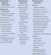
How can you tell the difference between inflammatory and degenerative/non-inflammatory joint disease?

What are some inflammatory joint diseases?
- RA
- SLE, Sjogren
- Seronegative arthritis e.g ankylosing spondylitis
- Gout
- Psoriatic
- IBD arthritis

How may rheumatoid arthritis present and what should you do if you suspect it?
- More common in women and presents around 30-50
- Symmetrical synovitis of the hand or feet (PIPJs) > 6weeks
- Pain worse at rest
- Swelling around the joint
- Morning stiffness over an hour
- Positive MTP, MCP squeeze or inability to make a fist
- Systemic features like malaise, weight loss, fever, sweats
Refer anyone within 3 working days if patient presents 3 months after symptoms arose, more than one joint affected or small hand/feet joints involved. Diagnosis within 3 weeks

What are some differential diagnoses for a presentation like RA?
- Fibromyalgia
- Connective tissue disorders
- Polyarticular gout
- Psoriatic arthritis
- Reactive arthritis
- Septic arthritis
- Sarcoidosis

How do we diagnose rheumatoid arthritis?
Do not wait for investigation results before treating!!
- Carry out blood test for rheumatoid factor and anti-CCP
- Carry out FBC and renal/liver function for treatment purposes
- Look at CRP and ESR as may be elevated
- Arrange xray of hands and feet
- Possible MRI/ultrasound

What drug treatment might be commenced by a specialist once a diagnosis of RA is confirmed?
- DMARD within 3 months of symptoms e.g methotrexate, leflunomide, sulfasalazine or hydroxychloroquine if palindromic
- Offer short term bridging glucocorticoid therapy until DMARD is working or switching DMARDs (2-3 months), or any flares
- Use step up therapy if needed and if not responding to conventional DMARDs consider biologics
- Regular monitoring due to DMARDs

What are the side effects of the following cDMARDs?
Methotrexate
Sulfasalazine
Leflunomide
Hydroxychloroquine
Methotrexate: pneumonitis, oral ulcers, hepatotoxicity
Sulfasalazine: rash, lowered sperm count, oral ulcers
Leflunomide: teratogenicity, oral ulcers, increased BP, hepatotoxicity
Hydroxychloroquine: irreversible retinopathy

What are some of the different biologics used for RA and what are the side effects of biologics?
Used if 2 DMARDs fail
1st line: TNFa inhibitiors: infliximab, etanercept, adalimumab, certolizumabpegol, golimumab
2nd line: B Cell depletion: rituximab
3rd line: IL1/IL6 inhibition: tocilizumab
4th line: Disruption of T Cell synthesis: abatacept
Side effects: reactivation of TB and hep B, worsening heart failure, hypersensisitivity

What is the role of primary care for a patient with diagnosed RA on specialist treatment?
- Drug monitoring: med reviews, blood tests, check concordance
- Ensure adult has access to specialist care when having a flare and has information about physios, OT, etc
- Check for development of comorbities e.g HTN, CVD
- Identify and manage flares
- Check anually if meeting treatment targets
- Offer more information on disease and how to manage it
- Assess need for surgery

How can a GP manage an RA flare?
- Exclude septic arthritis
- Look at inflammatory markers
- Offer short term corticosteroids either IM, oral or injection into local joint
- Offer NSAIDS
- Speak to specialists

What self-help and rehabilitative treatment can a GP offer to a patient with RA?
- Advise Med diet, stop smoking, limit alcohol
- Relative rest for acutely inflammed joint
- Stretching of other joints
- Aerobic conditioning
- Heat and Ice therapy

How would you diagnose a patient with OA if they have a history consistent with OA?
- X-ray imaging
- No imaging need if over 45, has activity related pain, no or <30minutes of morning stiffness

What are the features of osteoarthritis?
- Non-symmetrical
- Pain on movement, worse at end of the day
- Crepitus
- Stiffness after resting for 30 mins (joint gelling)
- Joint instability
- Loss of range of movement of joint

What are some risk factors of OA?
- Obesity
- Female
- Increasing age
- Trauma
- Joint malalignment
- Joint laxity
- Genetic

How can a GP initially manage a patient with OA non-pharmacologically?
- Provide information and resources e.g Arthritis Research UK
- Advise self-help e.g lose weight, local muscle strengthening, aerobic exercise exercises, prevent repetitive movements with hands, TENS, heat/ice packs, orthotics, advise no evidence for acupuncture/nutraceuticals
- Offer psychological support for any depression and work support e.g occupational health assessment

What pharmacological treatment can a GP offer to someone with OA?
- Paracetamol and topical NSAIDs regularly not as needed if not controlled
- If these don’t work stop topical NSAIDs and give oral NSAIDs
- If this doesn’t work consider weak opioid codeine or topical capsaicin
- Intrarticular steroid injections if all of this doesn’t work

When should you refer a patient with OA to an orthopaedic surgeon and what can they do for the patient?
- Symptoms of pain, stiffness and impairment not responding to core treatment
- Significant impact on the person’s quality of life.
- Uncertainty about the diagnosis or atypical symptoms.
- Sudden worsening of symptoms.
- Joint replacement or fusion e.g hip, knee, shoulder, elbow
- Arthroscopic Lavage and Debridement may be considered for people with knee OA with a clear history of mechanical locking
- Hand involvement may be treated by excision of the trapezium or joint fusion.

What are some complications with joint replacements?
- Aseptic loosening
- Pain
- Infection
- Fracture
- Dislocation

What is osteoporosis and the risk factors for it?
Progressive bone disease characterised by low bone mass measured by bone density, leading to an increased risk of fragility fractures, considered severe if 2 or more fractures
- Age (post menopausal)
- Smoking
- Low BMI
- Long term glucocorticosteroids
- Vit D and Ca deficiency
- Little exercise
- Previous fractures

How is osteoporosis investigated?
- DEXA scan
- X-ray following fracture with poor density can show osteopenia
- QFracture score

How is osteoporosis treated?
- Lifestyle: keep BMI 20-25, increase activity level, stop smoking, take vit D/Ca supplements or increase level in diet
- Post menopausal women/Men: oral bisphosphonates, alendronic acid and risedronate sodium. IV bisphosphonates like zoledronic acid if not tolerate orally.
Also HRT or Teriparatide if at severe risk of fracture.
- Give people undergoing glucocorticosteroid treatment or men undergoing prostectomy bone protection like above
- Review meds every 5 years

How do oral bisphosphonates work?
Inhibit osteoclast resorption by binding to hydroxyapatite crystals

What is the presentation of bursitis like and how would you tell if it was septic bursitis?
- Most common in shoulder, elbow, hip
- Swelling around a joint
- Often moveable swelling but tender/warm
- Develops over several hours or days
- History of repetitive trauma or movement
- No restriction of movement of the joint
Septic: fever, local cellulitis, increasing pain, limited mobility, tachycardia, abrasion over swelling.

How is non septic bursitis managed?
- Reassure patient it will go down conservatively
- Rest, ice, analgesia if any pain
- Avoid trauma, e.g wear elbow/knee pads
- Compression
- Consider aspiration if large or limiting function
- Corticosteroid injection or referral after 2 months if not responding

How is septic bursitis managed?
- Aspirate and give empirical antibiotic like flucloxacillin
- Consider repeated aspiration
- Review after 7 days
If signs of septic joint, e.g limited mobility, then refer to hospital immediately

What are some common sites of tendonitis and what are the symptoms?
- pain in a tendon that gets worse when you move
difficulty moving the tendon
- feeling a grating sensation when you move the tendon
- swelling, sometimes with heat or redness
- a lump along the tendon
TREAT WITH RICE, ANALGESICS, POSSIBLE CORTICOSTEROID INJECTION

What are the symptoms of psoriatic arthritis (type of inflammatory arthritis)?
- Mornings stiffness more than 30 minutes
- Sausage fingers
- History of psoriasis

How do we treat psoriatic arthritis?
- NSAIDs
- Steroids
- DMARDs (leflunomide first line then m/s)
- Injected biologics e.g adalimumab
- Drugs to lower CVD risk

What is pseudogout and how is it treated?
Deposition of calcium pyrophosphate dihydrate crystals, mainly in the cartilage of the knee causing chondrocalcinosis and acute synovitis
Treatment: rest, corticosteroid injections, joint aspiration, colchicine if can tolerate GI side effects, treat metabolic conditions like haemochromotosis
If chronic chondrocalcinosis then treat as OA

How do we treat septic arthritis?
- Surgical drainage and lavage
- High dose IV antibiotics

What are the differential diagnoses for knee pain depending on the patients age?

Why should you not order an X-ray for this lady?

- Would need an MRI to see soft tissue as Xray only views bones
- Refer to physio and last resort send to orthopaedic surgeon

