Arrhythmias Flashcards

1
Q

What are the 2 main sub-categories of arrhythmias?

A

Supravenricular and ventricular

2
Q

What are the 4 conducting pathways spreading from the SA node?

A

Anterior, middle, posterior intermodal tracts and then Bachmann’s bundle to the LA

3
Q

In which 2 ways can electrical dysfunction occur?

A

Defects in impulse formation eg. altered automaticity or defects in impulse conduction eg. re-entrant rhythms

4
Q

What are the 5 main mechanisms of arrhythmia?

A

Defects in impulse formation:

  • 1) Ectopic beats/Altered automaticity
  • 2) Triggered activity

Defect in impulse conduction:

  • 3) Re-entry
  • 4) Conduction block
  • 5) Accessory Tract Pathways
5
Q

Altered automaticity can be physiological or pathological, what are the physiological examples?

A

Modulation of SA node activity by the ANS (e.g. sinus tachycardia, sinus arrhythmia) or during respiration when HR increases on inspiration and decreases on expiration

6
Q

Altered automaticity can be physiological or pathological, what are the pathological examples?

A

Latent pacemaker subverts the SA node’s function as the normal pacemaker of the heart (overdrive suppression is lost)

7
Q

Arrhythmia

A

Abnormality of the heart rate or rhythm

8
Q

How can pathological altered automaticity occur?

A

Either SA node is firing pathologically low resulting in an escape beat/rhythm from the latent pacemaker. Or if the latent pacemaker fires at a rate faster than the SA node rate resulting in an ectopic rate/rhythm

9
Q

Ectopic beats

A

Beats that occur somewhere in the heart other than the SA node. The ectopic focus may cause single beats or take over and pace the heart, dictating its entire rhythm as a sustain arrhythmias.

10
Q

What is triggered activity as a cause of arrhythmias?

A

Afterdepolarisations triggered by a normal action potential (oscillations in the membrane potential which occur during depolarisation). Can be ether early afterdepoalrisation (EAD) or delayed after depolarisation (DAD). Occurs with digoxin toxicity, torsades de pointes or hypokalaemia

11
Q

What are early afterdepoalrisations (EAD)?

A

Occur during the exciting action potential (phase 2/3). Associated with prolonged QT interval, such as in QT syndrome

12
Q

What are delayed afterdepolarisations (DAD)?

A

Occurs after complete depolarisation and isa associated with raised intracellular Ca2+, such as during sympathetic stimulation

13
Q

What is re-entry as a cause of arrhythmias?

A

Self sustaining electrical circuit (anatomically may be two parallel conduction pathways), stimulates an area of myocardium repeatedly/rapidly

14
Q

**What causes re-entry?

A

Normally 2 signals go around either side of an area of non-excitable tissue, collide at the other side and go their separate ways. In re-entry, a unidirectional block on one side of the non-excitable tissue caused by trauma/ischaema etc means that there is no signal for the other signal on the other side to collide with, so it continues around as this area is no longer re-fractory like normal. so it creates a are re-entrant circuit

15
Q

What is conduction block as a cause of arrhythmias?

A

Any disease which disrupts electrical conduction may reduce conduction velocity or block conduction altogether causing bundle branch block, bradycardia or heart block

16
Q

What are the 3 types of conduction block?

A

1) Partial bock - Slowed conduction: tissue conducts all impulses, but more slowly than usual eg. First degree AV block
2) Intermittent - Tissue conducts some impulses, but not others eg. Second degree AV block
3) Complete - No impulses are conducted through the affected area e.g. Third degree AV block

17
Q

What are accessory tract pathways as a cause of arrhythmias?

A

Normally the only point of electrical continuity between the atria and the ventricles is the AV node – It is the only thing to pierce the fibrous cardiac skeleton. Some individuals possess electrical pathways that bypass the AV node eg. the Bundle of Kent. Ventricles receive impulses from both the normal and accessory pathways – can set up the condition for a re-entrant loop predisposing to tachyarrhythmias

18
Q

How to anti-arhythmic drugs generally work?

A

Generally inhibit specific ion channels with the intention of suppressing abnormal electrical activity e.g. the 3 main conductances

19
Q

How are anti-arrhythmic drugs classified pharmacologically?

A

Vaughn Williams classification. The classification defines four classes I, II, III and IV, with class I subdivided into subclasses Ia, Ib and Ic - characterised by their effects on the APs in Purkinje fibres

20
Q

**Which channels do Class I anti-arrhythmic drugs block and what effect do they therefore have? What differentiates the subtypes?

A

Block voltage active sodium channels. This slows the rate of rise of AP and prolongs the refractory period. The differences between the subtypes are the rate at which they unbind from the sodium channel

21
Q

What is a Class IA anti-arrhythmic drug?

A

Disopyramide

22
Q

What is a Class IB anti-arrhythmic drug?

A

Lignocaine

23
Q

What is a Class IC anti-arrhythmic drug?

A

Flecainide

24
Q

**Which channels do Class II anti-arrhythmic drugs block and what effect do they therefore have?

A

Act as beta –agonists by decreases the rate of depolarisation and rate of conductance through the AV node

25
Q

What is a Class II anti-arrhythmic drug?

A

Metoprolol, atenolol, propanolol, sotalol

26
Q

**Which channels do Class III anti-arrhythmic drugs block and what effect do they therefore have?

A

Block voltage activated potassium channel, so they prolong the AP and therefore the refractory period

27
Q

What is a Class III anti-arrhythmic drug?

A

Amiodarone, stool

28
Q

**Which channels do Class IV anti-arrhythmic drugs block and what effect do they therefore have?

A

Act on the calcium channels so affect the muscular AP plateau, and the upstroke in the nodal AP

29
Q

What is a Class IV anti-arrhythmic drug?

A

Verapamil

30
Q

Which state of the Na+ channel do Class I anti-arrhythmic drugs block?

A

The open state (as opposed to closed or inactive state). state. During high frequency firing (e.g. tachyarrhythmias) relatively more time is spent in the open and inactivated states than normal. This means there is more time for Class I agents to block the open state, and stabilise the inactive state. They spare the normal cardiac rhythm and target these areas of high-frequency firing

31
Q

Anti-Arrhythmic drugs can also be classed based on the site of the arrhythmia. Which class of drugs would you use for atrial arrhythmias?

A

Class IC and III - both as rate controllers of SVT

32
Q

Which drugs would you use for AV node arrhythmias?

A

Adenosine, digoxin and classes II and IV - rhythm control of SVT

33
Q

Which drugs would you use for ventricular arrhythmias?

A

Classes IA, IB and II

34
Q

What is the main aim of treatment of supra ventricular tachycardia?

A

To stop the spread of electrical activity from the atria to the ventricles. Can do this by blocking the AV node

35
Q

What drugs would you use to treat supra ventricular arrhythmias?

A

1) Adenosine (IV bolus) - Hyperpolarizes the AV node briefly, suppressing impulse conduction 2) Digoxin (IV infusion or oral) - Stimulates vagal activity and slows conduction and prolongs refractory period in AV node and bundle of His (esp. in AF) 3) Verapamil - class IV agent (oral) - Blocks L-type voltage-activated Ca2+ channel. Slows conduction and prolongs refractory period in AV node and bundle of His esp atrial flutter/fibrillation

36
Q

What drugs would you use to treat ventricular arrhythmias?

A

1) Lignocaine (Class IB) - Rapid block of voltage-activated Na+ channels. Used mainly (IV) in the treatment of ventricular arrhythmias following a myocardial infarction.

37
Q

What drugs would you use to treat atrial and ventricular arrhythmias?

A

Classes IA and IC, II and III:

1) Disopyramide and procainamide (Type Ia agents)
2) Flecainide (Type Ic agent) - mainly prophylaxis of paroxysmal AF
3) Propranolol and atenolol (Type II agents, β-blockers) - control SVT by suppressing AV conduction
4) Amiodarone and sotolol (Type Ill agents) - increase action potential duration and the effective refractory period - Supress re-entry

38
Q

What determines whether arrhymthias are symptomatic or not?

A

Whether they affect the cardiac output

39
Q

Wolf Parkinson White Syndrome

A

Presence of an abnormal accessory electrical conduction pathway between the atria and the ventricles: Bundle of Kent. Electrical signals traveling down this abnormal pathway may stimulate the ventricles to contract prematurely, resulting in a unique type of supraventricular tachycardia referred to as an atrioventricular re-entrant tachycardia.

40
Q

What does WPW Syndrome look like on ECG?

A

Presence of a delta wave: slurring slow rise of initial portion of the QRS

41
Q

Premature Ventricular complex

A

Relatively common event where the heartbeat is initiated by Purkinje fibers in the ventricles rather than by the sinoatrial node

42
Q

What are the main classes of causes of arrythmias?

A
  • Abnormal anatomy: left ventricular hypertrophy; accessory pathways,
  • Autonomic:
    • Excessive sympathetic stimulation:nervousness, exercise
    • Increased vagal tone
  • Metabolic:
    • Hypoxic myocardium
    • Ischaemic myocardium
    • electrolyte imbalances
  • Inflammation: viral myocarditis
  • Drugs -eg. those which block K channels and prolong QT segment
  • Genetic: mutations of cardiac ion channels e.g. long QT syndrome
43
Q

What are the symptoms of arrhtyhmias?

A
  • Asymptomatic
  • Palpitations
  • Shortness of breath
  • Dizziness
  • Syncope
  • Sudden Cardiac Death
  • Worsen pre-existing condition: angina, CHF
44
Q

What investigations can be done for arrythmias?

A
  • 12 lead ECG
    • This helps indicate the mechanism and assess rhythm
  • CXR
  • Echocardiogram
    • To assess for structural heart disease which may contribute
  • Stress/Exercise ECG
    • Look for myocardial ischaemia
  • 24 hour ECG Holter monitoring
    • Patients can press a button when they get their symptoms so they can see what it happening with the heart at the time
    • Used with paroxysmal arrhythmia
  • Event recorder
    • Implantable Loop Recorder (ILR) which get implanted under the skin
  • Electrophysiological (EP) study
    • Induce clinical arrhythmia to study mechanism and map pathway.
45
Q

Atrial ectopic beats

A

Atrial ectopic beats are caused by a premature depolarisation of the atria originating from a focus outside of the sinus node.

46
Q

What is the management of atrial ectopic beats?

A
  • Generally no treatment if they aren’t symptomatic
  • B-adrenergic blockers may help
  • Avoid stimulants (caffeine, cigarettes)
47
Q

Which HR define bradycardia?

A

<60bpm

48
Q

What are some of the non-physiological causes of bradycardia?

A
  • Drugs (B-Blocker)
  • Can also occur in acute ischaemia (common in inferior STEMIs)
49
Q

What is the managment for bradycardia?

A
  • Atropine (if acute, e.g. aMI)
  • Pacing if: Haemodynamic compromise- hypotension, CHF, angina, collapse
50
Q

What is the management for sinus tachycardia?

A
  • Treat underlying cause
  • B-adrenergic blockers
51
Q

What is supraventricular tachycardia and what are some of the causes?

A

Abnormally rapid heart rhythm arising from improper source of electrical activity of the heart, above the AV node. Covers several different conditions.

Can be caused by:

  • AV nodal re-entrant tachycardia
  • Accessory pathway tachycardia i.e. WPW
  • Ectopic atrial tachycardia
52
Q

What is the acute management for SVT?

A
  • Vagal manoeuvres:
    • carotid massage
    • Valsalva manouvre: Blowing hard whilst holding your nose and mouth closed - increases intrathoracic pressure which increases vagal tone to reduce HR
  • IV Adenosine
  • IV Verapamil (Calcium channel blocker which targets the SA and AV node)
53
Q

What is the chronic management for SVT?

A
  • Avoid stimulants
  • Radiofrequency ablation
  • Antiarrhythmic drugs (Class II or IV)
  • Ablation (Selective cautery of cardiac tissue to prevent tachycardia, targeting either an automatic focus or part of a re-entry circuit)
54
Q

1st Degree AV Block

A

P-R interval longer than normal (> 0.2 sec)

55
Q

What is the managment for 1st Degree AV block?

A

None - just monitor long term as more advanced block may develop

56
Q

2nd Degree AV Block

A

Intermittent block at the AVN (dropped beats) - Some of the P waves get conducted through, but some do not

57
Q

Second Degree Heart Block Mobitz Type I

A

Progressive lengthening of the PR interval, eventually resulting in a dropped beat.

58
Q

What is the management for 2nd degree AV block?

A

Permanent pacemaker indicated – ventricular pacing

59
Q

Second Degree Heart Block Mobitz Type II

A

PR interval is constant but every nth ventricular depolarization is missing (can be named in terms of ratio of missed – 2:1 block etc)

60
Q

3rd Degree AV Block

A

No action potentials from the SA node/atria get through the AV node. Have an escape rhythm, one which originates outwith the conducting system in the ventricles.

61
Q

What is the management for 3rd degree AV block?

A

Ventricular pacing

62
Q

What are the acute options for pacing?

A
  • Transcutaneous pacing - accomplished by delivering pulses of electric current through the patient’s chest
  • Transvenous pacing - achieved by threading a pacing electrode through a vein into the right atrium, right ventricle, or both.
63
Q

What are the options for implanted pacemakers?

A
  • Single chamber (paces the right atria or right ventricle only)
    • Atrial pacemakers: used in isolated Sino-atral node disease but normal AV node
    • Ventricular pacemakers
  • Dual chamber (paces the RA and RV)
    • Maintains A-V synchrony (preserves atrial kick)
    • Used for AVN disease
64
Q

If someone has broad complex tachycardia, what are you thinking until proven otherwise?

A

Ventricular tachycardia

65
Q

What are the ECG features of VT?

A
  • The QRS complexes are rapid, wide, and distorted.
  • The T waves are large with deflections opposite the QRS complexes.
  • The ventricular rhythm is usually regular.
  • P waves are usually not visible.
  • The PR interval is not measurable
66
Q

Ventricular fibrillation

A

Chaotic ventricular electrical activity which causes the heart to lose the ability to function as a pump. “bag of worms”

67
Q

What is the treatment for VF?

A

Defibrillation and Cardiopulmonary resuscitation (CPR)

68
Q

What is the acute treatment for VT?

A
  • DC cardioversion if unstable
  • If stable: consider pharmacologic cardioversion with anti-arrythmic drugs AAD
69
Q

What is the difference between defibrillationa and cardioversion?

A

Defibrillation - is the treatment for immediately life-threatening arrhythmias with which the patient does not have a pulse, ie ventricular fibrillation (VF) or pulseless ventricular tachycardia (VT).

Cardioversion - is any process that aims to convert an arrhythmia back to sinus rhythm.

70
Q

What is the chronic managment of VT/VF?

A
  • Look for causes
  • Correct ischaemia if possible (revascularisation)
  • Anti-arrhythmic drugs to date have been shown to be ineffective and are associated with worse outcome
  • Optimise CHF therapies (as many of these patients have CHF)
  • Implantable cardiovertor defbrillators (ICD) if life threatening
    • Delivers intracardiac current if fibrillation occurs
    • Acts as a defibrillator essentially
71
Q

Atrial fibrillation

A

A supraventricular tachyarrhythmia, specifically a chaotic and irregular atrial arrhythmia - results from multiple ectopic foci (as oppised to flutter which only 1 or 2)

72
Q

What are the 3 types of AF?

A
  1. Paroxysmal - lasting less than 48 hours and spontaneously come out of it on their own
  2. Persistant - lasting greater than 48 hours, which can still be cardioverted to NSR
  3. Permanent - cant get them back to sinus rhythm
73
Q

Lone (idiopathic) AF

A

AF with absence of any heart disease and no evidence of ventricular dysfunction. A diagnosis of exclusion.

74
Q

What are some of the associated diseases/causes of AF?

A
  • Valvular heart disease
  • Alcohol abuse
  • Congenital heart disease
  • Cardiac surgery
  • Pneumonia, Septicaemia, pericarditis, tumors
  • Vagal causes
  • Hypertension
  • Congestive heart failure
  • Coronary heart disease
  • Thyroid disease
  • Familial
75
Q

What is the mechanism of action of AF?

A

Multiple wavelets of reentry which do not allow the atria to organize.Ectopic focus around the pulmonary veins.

76
Q

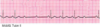
What characteristics are seen on ECG with AF?

A
  • Atrial Rate: > 300 bpm
  • Irregularly irregular rhythm
  • Absence of P waves
  • Presence of ‘f’ waves
77
Q

What are the clinical consequences of AF?

A

Lost ‘atrial kick’ and decreased filling times (reduced diastole) leading to reduced cardiac output

78
Q

What is the managment of AF?

A
  • Rhythm control first
    • Pharmacologic cardioversion (anti-arrhythmic drugs e.g. amiodarone)
    • Direct Current Cardioversion (DCCV)
    • Maintained by:
      • Anti-arrhythmic drugs
      • Catheter ablation of atrial focus/ pulmonary veins
      • Surgery (Maze procedure)
  • Rate control if cant restore NSR
  • Pharmacologic therapy to slow down AVN conduction eg. Digoxin, Betablockers, Verapamil, diltiazem
79
Q

Torsade de pointes

A

A rapid and distinct polymorphic VT with a twisting configuration of the QRS morphology, associated with prolonged repolarization.

80
Q

What is the CHADVASC score used for?

A

Determining risk of thromboembolism with AF

81
Q

Atrial flutter

A

Rapid and regular form of reentrant atrial tachycardia - resutls from 1 or 2 ectopic foci

82
Q

What does atrial flutter appear like on ECG?

A

Atrial bpm of over 300bpm with sawtooth ‘f’ waves

83
Q

What is the treatment for atrial flutter?

A
  • RF ablation (80-90% long term success)
  • Pharmacologic therapy
    • Slow the ventricular rate
    • Restore sinus rhythm
    • Maintain sinus rhythm once converted
  • Cardioversion
  • Warfarin for prevention of thromboembolism
84
Q

Channelopathies vs cardiomyopathies

A

Channelopathies are conditions which affect the channel conduction in the heart, while cardiomyopathies purely affect the heart muscle itself

85
Q

Abnormalities of which current cause most channelopathies?

A

Potassium current

86
Q

Long QT Syndrome

A

An inherited condition involving mutations that affect ion channels important in myocardial repolarisation, thus proloning the QT interval

87
Q

How many differnet mutations are associated with Long QT?

A

13

88
Q

Brugada Syndrome

A

Rare inherited heart rhythm disturbance that restricts the flow of sodium ions into the heart cells. As a result, the flow of electrical impulses through the heart is disrupted, which can lead to life-threatening heart rhythms eg. VT and VF

89
Q

Catecholaminergic Polymorphic Ventricular Tachycardia

A

Adrenergic induced bidirectional and polymorphic VT, SVTs, triggered by emotional stress, physical activity.

90
Q

Hypertrophic Cardiomyopathy (HCM)

A

Inherited disease of the myocardium in which a portion of the myocardium is hypertrophied without any obvious cause, creating functional impairment of the cardiac muscle.

91
Q

Which mutation is associated with HCM?

A

Myosin binding prptein C

92
Q

What are the clinical presentations for HCN?

A
  • Sudden death
  • Heart failure
  • Angina
  • Atrial fibrillation
  • Asymptomatic
93
Q

Dilated Cardiomyopathy

A

A condition in which the heart’s ability to pump blood is decreased because the heart’s main pumping chamber, the left ventricle, is enlarged and weakened

94
Q

Which mutations is associated with dilated cardiomyopathy?

A

Lamin A/C mutation

95
Q

Which intervention can be considered in channelopathies and cardiomyopathies?

A

Implanted cardioverter defibrillator ot avoid sudden cardiac death

96
Q

Arrhythmogenic right ventricular cardiomyopathy

A

Intercalated discs do not develop properly and so cannot keep the heart muscle cells together. The muscle cells become detached and fatty deposits build up in an attempt to repair the damage - Fibro-fatty replacement of cardiomyocytes.

97
Q

Which mutations is associated with arrhythmogenic right ventricualr cardiomyopathy?

A

Desmoglein

98
Q

What are the treatment options for Arrhythmogenic right ventricular cardiomyopathy?

A
  • ICDs for secondary prevention
  • Amiodarone can be used to improve symptoms in patients with PVC
  • Catheter ablation can also be used in this type, unlike others