Musculos Flashcards
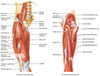
superficial muscles of the body (head) (6)

- Temporalis
- Masseter
- Epicranius, frontal belly
- Orbicularis oculi
- Zygomaticus
- Orbicularis oris

Superficial muscles of the body (neck) (3)

- Platysma
- Sternohyoid
- Sternocleidomastoid

Frontalis (frontal belly)
Origen, Insertion, Action

O = galea aponeurotica
I = skin of eyebrows and root of nose
A= raises the eyebrows. + wrinkles the forehead horizontally

Orbicularis oculi
(Origen, Insertion, Actions)
O = frontal and mazillary bones and ligaments around orbit
I = tissue of eyelid
A = bliking + squinting + draws eyebrows inferiorly

Orbicularis oris
(Origen, Insertion, Actions)
O = arises directly from maxilla & mandible
I = encircles mouth; inserts into muscle & skin @ angles of mouth
A = • closes lips • purses and protrues lips • kissing & whistling

Levator labii superior
(Origen, Insertion, Actions)
O = zygomatic bone & infraorbital margin of maxilla
I = skin & muscle of upper lip
A = raises & furrows the up- per lip

Risorius
(Origen, Insertion, Actions)
O = lateral facia assoc. with masseter muscle
I = skin @ angle of mouth
A = • draws corner of lip laterally + tense of lips • synergist of zygomaticus

Depressor anguli oris
(Origen, Insertion, Actions)
O = body of mandible below incisors
I = skin & muscle @ angle of mouth (below insertion of zygomaticus)
A = • draws corner of mouth laterally & downward • antagonist of zygomaticus

Platysma
(Origen, Insertion, Actions)
O = fascia of chest (over pectoral muscle & deltoid)
I = lower margin of mandible, and skin & muscle @ corner of mouth
A = • depresses mandible • pulls lower lip back & down

Temporalis
(Origen, Insertion, Actions)
O = temporal fossa
I = coronoid process of mandible
A = closes the Jaw + elevates & retracts mandible • synergist of pterygoids • maintains position of mandible at rest

Masseter
(Origen, Insertion, Actions)
O = zygomatic arch and maxilla
I = angle & ramus of mandible
A = • prime mover of jaw closure • elevates mandible

Zygomaticus
(Origen, Insertion, Actions)
O = zygomatic bone
I = skin & muscle @ corner of mouth
A = • raises lateral corners of mouth (smiling)

Sternocleidomastoid
(Origen, Insertion, Actions)
O = manubrium of sternum & medial portion of clavicle
I = mastoid process of temporal bone & superior nuchal line of occipital bone
A = • prime mover of active head flexion (when act together) • rotates head toward op- posite side (when act singularly

Pectoralis major
(Origen, Insertion, Actions)
O = sternal end of clavi- cle, sternum, cartilage of ribs 1-6, & aponeurosis of external oblique muscle
I = greater tubercle and by a short tendon into intertubercular groove of humerus
A = • agonist of arm flexion • rotates arm medially • adducts arm against resistance • pulls rib cage upward with scapula
fixed

Pectoralis minor
(Origen, Insertion, Actions)

O = anterior surface of ribs 3-5 (or 2-4)
I = coracoid process of scapula
A = • draws scapula forward & downward (ribs fixed) • draws rib cage superiorly (scaupla fixed)

Serratus anterior (boxer’s muscle)
(Origen, Insertion, Actions)
O = by series of muscle slips from ribs 1-9
I = anterior surface of vertebral border of scapula
A = • agonist to protract & hold scapula against rib cage • rotates scapula (infe- rior angle laterally & upward) • abduct & raise arm & horizontal arm move- ments

Diaphragm
(Origen, Insertion, Actions)
O = inferior, internal surface of rib cage & sternum, costal carti- lages of last six ribs & lumbar vertebrae
I = central tendon
A = • prime mover of inspira- tion, flattens on contrac- tion

External intercostals
(Origen, Insertion, Actions)

O = inferior border of rib above
I = superior border of rib below
A = • elevate rib cage, aids in inspiration
• synergist of diaphragm

Internal intercostals
(Origen, Insertion, Actions)

O = superior border of rib below
I = inferior border (costal groove) of rib above
A = • depress rib cage, aid in forced expiration • antagonist of external intercostals

External oblique
(Origen, Insertion, Actions)
O = outer surface of lower 8 ribs
I = Linea alba via aponeurosis
A = • when together, synergist to rectus abdominis, flex vertebral column & com- press abdominal wall • when alone, synergist to muscles of back, roate & lateral flexion of trunk

Internal oblique
(Origen, Insertion, Actions)
O = lumbar fascia, iliac crest, & inguinal liga- ment
I = linea alba, pubic crest, last 3 or 4 ribs, & costal margin
A = Same as External oblique (• when together, synergist to rectus abdominis, flex vertebral column & com- press abdominal wall • when alone, synergist to muscles of back, roate & lateral flexion of trunk)

Transversus abdominis
(Origen, Insertion, Actions)
O = inguinal ligament, lumbar fascia, carti- lages of last 6 ribs, iliac crest
I = linea alba, pubic crest
A = • compresses abdominal contents

Rectus abdominis
(Origen, Insertion, Actions)
O = pubic crest & sym- physis
I = xyphoid process & costal cartilages of ribs 5-7
A = • flex & rotate lumbar re- gion of vertebral column • fix & depress ribs • stabilize pelvis during walking • increase intra-abdominal pressure

Linea Alba
a median vertical tendinous line on the mammalian abdomen formed of fibers from the aponeuroses of the two rectus abdominis muscles and extending from the xiphoid process to the pubic symphysis

Tendinous intersections
The rectus abdominis muscle is crossed by three fibrous bands called the tendinous inscriptions (or tendinous intersections).
Usually three but occasionally four transverse fibrous bands or partial bands occurring at intervals as interruptions of the fleshy, contractile portions of the rectus abdominis muscle; they usually occur at and superior to the umbilicus.

Inguinal Ligament
the thickened lower border of the aponeurosis of the external oblique muscle of the abdomen that extends from the anterior superior iliac spine to the pubic tubercle, is continuous with the fascia lata near the thigh, and forms the external pillar of the superficial inguinal ring and a part of the anterior boundary of the femoral ring—

Deltoid
(Origen, Insertion, Actions)
O = lateral 3rd of clavicle, acromion & spine of scapula
I = deltoid tuberosity of humerus
A = • agonist of arm abduction with all fi- bers, antagonist of pectoralis major & latissimus dorsi • flexes & medially rotates humerus with anterior fibers, synergist of pectoralis major • extends & laterally rotates arms with posterior fibers

Biceps brachii
(Origen, Insertion, Actions)
O = long head (65-1): tuber- cle above glenoid cav- ity and lip of glenoid cavity of scapula. short head: (65-2): cora- coid process of scapula
I = by common tendon to radial tuberosity
A = • flexes elbor joint & supinates forearm (usually at the same time) • weak flexor of arm @ shoulder

Triceps brachii
(Origen, Insertion, Actions)
O = lateral head (68-1): pos- terior shaft of humerus long head (68-2) : infraglenoid tubercle of scapula medial head (68-3): posterior humeral shaft distal to radial groove.
I = by common tendon into olacrenon pro- cess of ulna
A = • agonist of forearm extension (medial head) • antagonist of forearm flexors • stablizes shoulder joint & assist in arm adduction (long head tendon)

Thenar group (ball of thumb)
(Origen, Insertion, Actions)
- Abductor pollicis brevis (Pollex = thumb) : abducts the thumb. This muscle is the most superficial of the thenar group.
- O = flexor retinaculum and nearby carpals. I = Lateral base of thumb’s proximal phalanx
- Flexor pollicis brevis, which lies next to the abductor, will flex the thumb, curling it up in the palm.
- O = flexor retinaculum and nearby carpals. I = Lateral base of proximal phalanx of thumb
- Opponens pollicis: lies deep to abductor pollicis brevis. As its name suggests it opposes the thumb, bringing it against the fingers.
- O = flexor retinaculum and trapezium I = whle anterior side of metacarpal I
- Adductor pollicis. It lies deeper and more distal to flexor pollicis brevis. Despite its name, its main action is mainly rotation and opposition
- O = capitate bone and bases of metacarpals II-IV; front of metacarpal III. I = medial side of base of proximal phalanx of thumb

Hypothenar group (ball of pinky)
(Origen, Insertion, Actions)

- Abductor digiti minimi
- O = pisiform bone. I = medial side of proximal phalanx of little finger.
- Flexor digiti minimi brevis
- O = Hamate bone and flexor retinaculum. I = same as abductor of digiti minimi.
- Opponens digiti minimi
- O = Same as flesor digiti mini brevis. I = most of length of medial side of metacarpal V

Quadriceps femoris
(Origen, Insertion, Actions)
Muscle group covering the front and sides of the thigh. It has four parts: rectus femoris, vastus lateralis, vastus medialis, and vastus intermedius.
O = They originate at the ilium (upper part of the pelvis, or hipbone) and femur (thighbone), come together in a tendon surrounding the patella (kneecap),
I = insert at (are attached to) the tibia (shinbone).
A = These muscles extend the legs at the knee and are important for standing, walking, and almost all activities involving the legs.

Rectus femoris
(Origen, Insertion, Actions)
O = anterior inferior iliac spine & superior margin of acetabulum
I = patella & tibial tuberosity via patella ligament
A = • extends knee • flexes thigh @ hip

Vastus Lateralis
(Origen, Insertion, Actions)
O = greater trochanter, inter- trochanteric line, linea aspera
I = patella & tibial tuberosity via patella ligament
A = • extends & stablizes knee

Vastus Medialis
(Origen, Insertion, Actions)
O = linea aspera, intertrochanteric line
I = patella & tibial tuberosity via patella ligament
A = • extends knee • stablizes patella (inferior fibers)

Vastus Intermedius
(Origen, Insertion, Actions)
O = anterior & lateral surfaces of proximal femur
I = Patella and tibial tuberosity via patella ligament
A = Extends the knee

Patellar ligament
(Origen, Insertion, Actions)
the continuation of the central portion of the tendon of the quadriceps femoris muscle distal to the patella, extending from the patella to the tuberosity of the tibia.

Sartorius
(Origen, Insertion, Actions)
the longest muscle in the human body
Sartorius comes from the Latin word sartor, meaning tailor,
O = anterior and superior iliac spine,
I = Proximal tibial, medial to tibial tuberosity .
A= flexes, abducts, and laterally rotates thigh hip

Biceps Femoris
(Origen, Insertion, Actions)
O = long head (a): ischial tuberosity. short head (b): linea aspera & distal femur
I = by common tendon into head of fibula & lateral condyle of tibia
A = • extends thigh & flexes knee • laterally rotates leg when knee is flexed

Semitendinosus
(Origen, Insertion, Actions)
O = iscial tuberosity
I = medial aspect of upper tibial shaft
A = • extends thigh & flexes knee • medially rotates leg with semi-membranosus

Semimembranosus
(Origen, Insertion, Actions)
O = ischial tuberosity
I = medial condyle of tibia
A = • extends thigh & flexes knee • medially rotates leg

Gluteus maximus
(Origen, Insertion, Actions)
O = dorsal ilium, sacrum & coccyx
I = gluteal tuberosity of femur, iliotibial tract
A = • major extensor of thigh • laterally rotates & abducts thigh • inactive during standing

Gluteus Medius
(Origen, Insertion, Actions)
O = lateral surface of ilium between anterior & posterior gluteal lines
I = via short tendon into lateral aspect of greater trochanter
A = abducts thigh • anterior part rotates hip medially • posterior part rotates hip lateraly

Iliotibial tract/band (fascia)
(Origen, Insertion, Actions)
a band of connective tissue that extends from the iliac crest to the knee and links the gluteus maximus to the tibia.

Tensor fasciae Latae
(Origen, Insertion, Actions)
O = anterior iliac crest & anterior superior iliac spine
I = iliotibial tract
A = • flexes & abducts thigh (synergist of iliopsoas & gluteus muscles) • rotates thigh medially • steadies the trunk by pulling ilio-tibial tract taut (locking the knee)

Occipitalis (occipital belly)
(Origen, Insertion, Actions)
O = arises from the highest nuchal lines
I = posterior edge of epicranial aponeurosis
A = pulls the aponeurosis posteriorly towards the occipital bone.

Rhomboid major
(Origen, Insertion, Actions)
O = spinous processes of T2-T5
I = medial border of scapula
A = • retract scapula (squar- ing shoulders), synergist with middle fibers of Trapezius • rotate glenoid cavity downward (lowering arm against resistence) • stablize scapula

Rhomboid minor
(Origen, Insertion, Actions)
O = spinous processes of C7 & T1
I = medial border of scapula
A = • retract scapula (squar- ing shoulders), synergist with middle fibers of Trapezius • rotate glenoid cavity downward (lowering arm against resistence) • stablize scapula

Latissimus dorsi
(Origen, Insertion, Actions)
O = via lumbodorsal fascia into spines of T7-L5, lower 4 ribs & iliac crest
I = floor of intertubercular groove of humerus
A = • agonist of arm extension • powerful arm adductor • medially rotates arm & shoulder • depresses scapula • pulls body upward & forward with arms fixed overhead

Supraspinatus
(Origen, Insertion, Actions)
O = supraspinous fossa of scapula
I = superior part of greater tubercle of humerus
A = • stabilizes shoulder joint • helps prevent downward dislocation of humerus

Infraspinatus
(Origen, Insertion, Actions)
O = infraspinous fossa of scapula
I = greater tubercle of humerus, posterior to supraspinatus
A = • helps to hold head of humerus in glenoid cavity • stabilizes the shoulder joint • rotates humerus laterally

Teres Major
(Origen, Insertion, Actions)
O = posterior surface of scapula @ inferior angle
I = intertubercular groove of humerus, tendon fused with tendon of latissimus dorsi
A = • posteromedially extends, medially rotates, & adducts arm • synergist of latissimus dorsi

Teres Minor
(Origen, Insertion, Actions)
O = lateral border of dorsal scapular surface
I = greater tubercle of humerus, inferior to infraspinatus
A = same as infraspinatus = • helps to hold head of humerus in glenoid cavity
• stabilizes the shoulder joint • rotates humerus laterally

Subscapularis
(Origen, Insertion, Actions)
O = subscapular fossa of scapula
I = lesser tubercle of humerus
A = • chief medial rotator of humerus, as- sisted by pectoralis major • helps to hold head of humerus in glenoid cavity, stablizes shoulder

Flexor carpi radialis
(Origen, Insertion, Actions)
O = medial epicondyle of humerus
I = base of 2nd & 3rd metacarpals (anterior)
A = • powerful flexor of wrist • abducts the hand • weak syngergist of elbow flexion

Palmaris longus
(Origen, Insertion, Actions)
O = medial epicondyle of humerus
I = palmar aponeurosis, skin & fascia of palm
A = • weak wrist flexor • weak synergist of elbow flexion • tenses skin of palm during hand

Flexor Carpi Ulnary
(Origen, Insertion, Actions)
O = medial epicondyle of humerus, olecranon pro- cess & posterior surface of ulna
I = pisiform & hamate bones & base of 5th metacarpal (anterior)
A = • powerful flexor of writs • adducts hand with extensor carpi ulnaris • stablized wrist during finger extension

Extensor carpi radialis longus
(Origen, Insertion, Actions)
O = lateral supracondylar ridge of humerus
I = base of 2nd metacar- pal (posterior)
A = • extends wrist with extensor carpi ulnaris • abducts write with flexor carpi radialis

Extensor carpi radialis longus
(Origen, Insertion, Actions)
O = lateral supracondylar ridge of humerus
I = base of 2nd metacar- pal (posterior)
A = • extends wrist with extensor carpi ulnaris • abducts write with flexor carpi radialis

Extensor carpi radialis brevis
(Origen, Insertion, Actions)
O = lateral epicondyle of humerus
I = base of 3th metacarpal (posterior)
A = extends and abducts wrist

Gracilis
(Origen, Insertion, Actions)
O = inferior ramus and body of pubis and ischial ramus
I = proximal medial surface of tibia
A = adducts, flexes and medially rotates leg
Adductor longus
(Origen, Insertion, Actions)

O = pubis near pubic symphysis
I = linea aspera of femur
A = adducts and flexes, and medially rotates thigh

Adductur Brevis
(Origen, Insertion, Actions)

O = Inferior ramus of pubis
I = linea aspera of femur above adductur longus
A = adducts and flexes, and medially rotates thigh

Adductor magnus
(Origen, Insertion, Actions)

O = Ischial and pubic rami, and ischial tuberosity
I = linea aspera and adductor tubercle of femur
A = adducts, flexes and medially rotates thigh

Calcaneal (Achilles) tendon
Largest tendon in the body. Is the thickest and strongest in the human body and is about 15 cm in length. This tendon connects the calf muscle to the heel bone.
