Paeds Non-Hip (Complete) Flashcards
What are risk factors for the development of clubfoot?
[CORR (2009) 467:1146–1153]
- Family history
- Boys
- Race (highest in Hawaiians and Maoris)
- Early amniocentesis (<13 weeks)
- Oligohydramnios
- Exposure to cigarette smoke inutero
What is the Pirani scoring system of clubfoot and what can it predict?
- Six signs are scored from 0 (no abnormality), 0.5 (moderate abnormality), 1 (severe abnormality)
- 3 signs related to the hindfoot
- Severity of the posterior crease
- Emptiness of the heel
- Rigidity of the equinus
- 3 signs related to the midfoot
- Curvature of the lateral border of the foot
- Severity of the medial crease
- Position of the lateral part of the head of the talus
- Predicts need for tenotomy
* 85% of feet with a score above 5 required tenotomy
What is the main radiographic feature in clubfoot?
[Orthobullets]
Hindfoot parallelism
- Talus and calcaneus are parallel/less divergent on AP and lateral
What are the 4 components of the clubfoot deformity?
[CORR (2009) 467:1146–1153]
CAVE
- Midfoot cavus
- Forefoot adductus
- Hindfoot varus
- Hindfoot equinus
Describe the pathoanatomy resulting in the deformity in clubfoot
[J Am Acad Orthop Surg 2003;11:392-402]
- Navicular displaces medially (articulates with the medial head of talus)
- Cuboid is adducted infront of the calcaneus
- Metatarsals are adducted on the midfoot
- Calcaneus is adducted and inverted around the talus medially
- Forefoot is pronated relative to the hindfoot (causing cavus)
- Tight muscles (gastroc/soleus, tib post, FHL, FDL)
- Tight posteromedial capsule and ligaments
Describe the Ponsetti method for clubfoot correction.
[J Am Acad Orthop Surg 2003;11:392-402] [J Am Acad Orthop Surg 2010;18:486-493]
- Serial foot manipulation followed by casting to maintain the correction with foot abduction orthosis as the final stage
- Manipulations are held for 1-3 minutes followed by above knee plaster cast with knee at 90° flexion
- Weekly cast change and manipulation
- ~6 cast changes required to correct most clubfeet
- Ponsetti method started ideally within the first month of life
- The order of foot deformity correction is cavus, adductus, varus, equinus (CAVE)
- Cavus correction
- Usually achieved with the first cast
- Technique – pressure under first metatarsal head to elevate it in line with other metatarsals
- Forefoot adduction and hindfoot varus corrected simultaneously
- With foot in slight supination and equinus the forefoot is abducted while stabilizing counterpressure is applied to the lateral head of the talus
- This will simultaneous correct adduction and hindfoot varus as the calcaneus abducts freely under the talus (important to avoid max dorsiflexion)
- Equinus correction
- Perform when hindfoot is neutral or slight valgus and forefoot is abducted 70° relative to the leg
- Technique – progressive dorsiflexion applied with broad pressure over sole of foot
- Heel cord tenotomy
- Required in ~75% of cases
- Performed after 4-6 weeks of casting
- Percutaneous tenotomy performed in clinic or OR followed by cast immobilization for 3-4 weeks
- Foot abduction orthoses (‘boots and bars’, Denis-Browne bar)
- Required to prevent relapse
- 15° dorsiflexion needed for proper fit
- Clubfoot placed in 70° abduction, unaffected foot in 40° abduction with feet shoulder width apart
- Worn full time (23 hours/day) for 3 months followed by bed and naptime use until age 4 (range 2-5)
- Patient should be followed every 3 months after bracing starts until 2 years of age
What is the most common factor related to clubfoot relapse?
[J Am Acad Orthop Surg 2010;18:486-493]
Failure to comply with foot abduction orthoses
By what age is clubfoot relapse most likely to occur?
[Am Acad Orthop Surg 2017;25:195-203]
Most frequently by age 5
- Rare after age 5 and extremely rare after 7
What are the signs of clubfoot relapse?
[Am Acad Orthop Surg 2017;25:195-203]
- Loss of dorsiflexion is the earliest sign
- Older infants – mild forefoot adductus, cavus, heel varus and limited abduction
- Walking child – increased lateral contact during stance phase, heel varus, inward deviation of toes, and dynamic supination during swing phase
How do you manage clubfoot relapse?
[Am Acad Orthop Surg 2017;25:195-203]
- Mild dorsiflexion loss
* If early, home exercise program and increased foot abduction orthosis use - If <10° dorsiflexion
- Repeat manipulation and casting as per Ponsetti (2-3 casts usually required changed weekly)
- Repeat tenotomy if 15° dorsiflexion not achieved
- Resume foot abduction orthosis
- If >2.5 years of age:
- Consider anterior tibial tendon transfer to 3rd cuneiform (now sufficiently ossified)
- First requires obtaining original correction with manipulation and casting (2-3 casts) and heel cord tenotomy if <10° dorsiflexion
- Anterior tendon should never be split (split weakens eversion power)
- Technique for anterior tibial tendon transfer:
- 3-4cm incision starting just distal to the navicular and extending proximal inline with tibialis anterior tendon is made
- The tibialis anterior tendon is released from the base of the 1st MT and a whip stitch is placed in the tendon
- A 2nd incision is made over the lateral cuneiform and localized with fluoro guidance, once confirmed a drill hole is made dorsal to plantar
- The tendon is tunneled subcutaneously from the medial to lateral incision
- Keith needles are threaded on to the sutures and passed through the drill hole and out through the plantar aspect of the foot
- The ankle is dorsiflexed and everted and the sutures are tied over a button
- The patient is casted for 6 weeks
- If age 4-9 with well-formed medial cuneiform ossific nucleus:
- Consider closing wedge osteotomy through the cuboid and medial opening wedge osteotomy through the cuneiforms (flip-flop technique)
5. Patients whose parents are unwilling to allow repeated cast and brace treatment and patients with feet that are otherwise refractory to the Ponseti method: - Consider posteromedial release (required in less than 5%)
- Highly associated with development of pain, stiffness and weakness in late adolescence and early adulthood
- Technique for posteromedial release [Lovell and Winter]:
- Cincinnati incision
- Extends medially from navicular, posteriorly just below medial malleolus and 1cm proximal to the posterior heel crease, laterally just below lateral malleolus ending at the sinus tarsi
- Releases include:
- Heel cord lengthening
- Posterior release of ankle and subtalar joint
- Plantar fascia
- Abductor hallucis
- +/- Tib post tendon lengthening
- +/- Talonavicular joint capsule release
- +/- FHL and FDL release
- Cincinnati incision
What are the associated conditions with congenital knee dislocation?
[JAAOS 2009;17:112-122]
- Ipsilateral DDH (70-100% of cases)
- Clubfoot
- Arthrogryposis
- Myelodysplasia
- Larsen syndrome
What is the classification of congenital knee dislocation?
[JAAOS 2009;17:112-122]
- Grade 1 = recurvatum
- Grade 2 = subluxation
- Grade 3 = complete dislocation
What is the clinical presentation of congenital knee dislocation?
[JAAOS 2009;17:112-122]
- Knee hyperextension
- Inability to flex knee in complete dislocation
What is the management of congenital knee dislocation?
[JAAOS 2009;17:112-122]
- Nonoperative
- Closed reduction and serial casting
- Closed reduction achieved by traction followed by knee flexion
- Serial casting in progressive knee flexion
- Operative
- Failure of nonoperative
- <30° of flexion after 3 months of casting [Orthobullets]
- Performed at ~6 months of age
- Involves open reduction and quadriceps lengthening
- Percutaneous quadriceps release
- Open V-Y quadriceps advancement
- Possible femoral shortening osteotomy (relative lengthening of extensor mechanism) [POSNA]
What is the management of ipsilateral congenital knee dislocation and DDH?
[JAAOS 2009;17:112-122]
- Treat congenital knee dislocation first
- Once adequate knee flexion achieved patient can be placed in Pavlik harness
* Pavlik harness helps to hold knee in flexion and maintain hip reduced
What is the definition of congenital dislocation of the patella?
Congenital, irreducible lateral patellar dislocation present at birth
What are the findings in patients with congenital dislocation of the patella?
- Anatomical
- Tight lateral structures (capsule, ITB, etc)
- Quadriceps contracture
- Lateralized patellar tendon insertion on tibia
- Hypoplastic patella
- Shallow trochlear groove
- Clinical
- Knee flexion contracture
- Genu valgum
- External tibial torsion
- Prominent femoral condyles
What is the management of congenital patellar dislocation of the patella?
Operative
- Principles:
- Extensive lateral release
- ITB, capsule, biceps femoris
- VY lengthening of quadriceps
- Medial capsule imbrication OR MPFL reconstruction
- Lateral patellar tendon insertion addressed via:
- Roux-Goldthwait procedure:
- Lateral half of patellar tendon detached from tibial tubercle, passed deep to remaining patellar tendon and attached medial to the medial half of the patellar tendon
- Patellar tendon periosteal sleeve medialization (complete medialization of patellar tendon)
- Roux-Goldthwait procedure:
- Extensive lateral release
What is developmental coxa vara?
[Lovell and Winter]
Decreased femoral neck shaft angle believed to be a result of a primary defect in endochondral ossification of the medial part of the femoral neck
What is the presentation of developmental coxa vara?
[Lovell and Winter]
Painless limp (unilateral) or waddling gait (bilateral)
- Due to abductor weakness and minor LLD in unilateral cases
What are the radiographic features of developmental coxa vara?
[Lovell and Winter]
- Decreased femoral neck-shaft angle (<120o)
- Vertical position of physeal plate
- Triangular metaphyseal fragment in inferior femoral neck with associated inverted Y appearance
- Shortened femoral neck
- Decrease in normal anteversion

How is the amount of varus deformity in developmental coxa vara quantified on plain films?
[Lovell and Winter]
Hilgenreiner epiphyseal angle (H-E)
- Angle between the physeal plate and Hilgenreiner line
- Normal = <25° (average 16°)
- Coxa vara = 40-70°

What is the management of developmental coxa vara?
[Lovell and Winter]
- Nonoperative
- H-E angle <45
- H-E angle 45-59 and asymptomatic
2.Operative
- H-E angle >60
- H-E angle 45-59 and symptomatic
- Symptomatic limp, Trendelenburg gait, or progressive deformity
- Neck shaft angle <100
- Technique:
- Valgus-producing proximal femoral osteotomy
What is the goal of correction in developmental coxa vara?
- <38° H-E angle [Orthobullets]
- 16° H-E angle [Lovell and Winter]
What are the associated conditions with PFFD?
[Lovell and Winter]
- PFFD is associated with fibular deficiency in 70% to 80% of cases
- Associated conditions are similar to that of fibular hemimelia
What is the most widely used classification for PFFD?
[Lovell and Winter]
- Aitken
- Class A
- Femoral head ossification delayed
- Acetabulum well formed
- Femur is short
- Proximal femur is at or above level of acetabulum
- Class B
- Femoral head ossification delayed
- Mild acetabular dysplasia
- Proximal femur is above level of acetabulum
- Subtrochanter region will not ossify and forms pseudoarthrosis
- Class C
- Femoral head does not form
- Severe acetabular dysplasia
- Femur is shorter than B
- Entire proximal femur does not form
- Class D
- Femoral head does not form
- Acetabulum does not form
- Distal femoral condyles are at level of acetabulum

What is the management of PFFD?
[Lovell and Winter]
- Nonoperative
- ‘bridge treatment’ from time patient begins to walk to surgical treatment (~3 years of age)
- Bilateral PFFD
- Operative
- 3 main procedures (stable hip)
- Knee fusion with foot ablation
- Indications:
- >20cm of LLD at maturity
- Foot above level of contralateral knee
- Ankle has <60° arc of motion
- Stable hip
- Foot ablation (amputation) through Boyd or Syme amputation
- Indications:
- Van Nes rotationplasty
- Indications:
- >20cm of LLD at maturity
- Foot at level of contralateral knee
- Ankle has >60° arc of motion
- Stable hip
- Technique:
- Leg is rotated 180° through the knee arthrodesis
- Indications:
- Limb lengthening
- Indications:
- <20cm of LLD at maturity
- Stable hip
- Good knee, ankle, foot function
- Indications:
- Knee fusion with foot ablation
- Unstable hip (Aitken C/D)
- Possible iliofemoral fusion (knee then functions as hip and ankle as knee)
What is the difference between true, apparent and functional LLD?
[Lovell and Winter]
- True (structural) LLD = anatomic difference in length of one of the segments of the lower extremity (femur, tibia, foot)
- Apparent (postural) LLD = discrepancies that are not true differences in anatomic segment lengths (eg. knee flexion contracture)
- Functional LLD = the sum of the true and apparent leg-length discrepancy (most important in treatment decisions)
What are the causes of LLD?
[Lovell and Winter]
- Congenital
* PFFD, fibular hemimelia, tibia hemimelia, posteromedial tibial bowing, clubfoot, hemihypertrophy - Acquired
* Trauma (growth arrest, overgrowth, malunion), radiation, tumor, infection
What are clinical methods to assess LLD?
[Lovell and Winter]
- True leg length
* Measure from ASIS to medial malleolus - Apparent leg length
* Measure from umbilicus to medial malleolus (affected by pelvic obliquity) - Galeazzi sign
* Difference in knee heights suggests difference in femoral lengths - Heel pad height
* With patient prone assess difference in heights of the heel pads, suggests difference in tibia/fibula length - Block method
* Place blocks under foot of short leg until pelvis level, height of block indicates LLD
What are the radiographic methods used to assess LLD?
[Lovell and Winter]
- Teleoroentgenogram (C)
- Standing alignment film (35cm × 90cm) taken with a single exposure at a distance of 2 m centered on the knee joint
- Advantages – angular deformity assessment
- Disadvantage – magnification error
- Orthoroentgenogram (A)
- Three separate exposures at the hip, knee, and ankle all placed on the same longstanding film with ruler centered over each joint
- Advantages – no magnification error
- Disadvantages – cannot assess angular deformity
- Scanogram (B)
- All three joints are placed on one smaller film; obtained having the film and x-ray source move
- Advantages – no magnification error
- Disadvantages – cannot assess angular deformity
- CT scanogram
- CT scan through hip, knee, and ankle to assess length
- Advantages – accurate length measurements in setting of joint contractures
- Disadvantages – cannot assess angular deformity

What is a method to assess skeletal age (rather than chronological age)?
[Lovell and Winter]
Greulich and Pyle
- Bone age x-ray (left hand)
- The clinician or radiologist can compare this child’s x-ray with those in the atlas and develop a bone age with a given standard deviation
What are methods used to predict the final LLD and timing of surgical intervention?
- Moseley Straight line graph [www.pedipod.com]
- Multiplier method
- Estimation method
What is the relative growth contribution of the growth plates of the lower extremity?
[Lovell and Winter]
- Proximal femur = 3mm/year
- Distal femur = 9mm/year
- Proximal tibia = 6mm/year
- Distal tibia = 5mm/year
What are the treatment options based on predicted LLD?
[Lovell and Winter]
- <2cm
- Observation
- Shoe lift
- 2-5cm
- Shoe lift
- Contralateral epiphysiodesis
- Contralateral skeletal shortening (usually for patients with inadequate growth remaining for epiphysiodesis)
3. >5-20cm - Limb lengthening +/- contralateral epiphysiodesis
4. >20cm - Amputation and prosthetic fitting
What are some principles for limb lengthening?
[Orthobullets]
- Initiation
- Ensure stable joint above and below prior to lengthening
- If joint subluxes during lengthening extend the frame across the joint
- Perform corticotomy and place fixator
- Metaphyseal corticotomy is preferred due to good blood supply
- Percutaneous corticotomy that minimizes trauma to the periosteum and preserves the blood supply of the marrow and periosteum
- Distraction
- Wait 5-7 days then begin distraction (allows for neovascularization of corticotomy site)
- Distract ~ 1 mm/day in four 0.25mm increments daily
- Do not distract more than 20% of the bones original length
- Following distraction keep fixator on for as many days as you lengthened
- Concurrent procedures
- May lengthen over a nail so ex-fix can be removed sooner
- Lengthening often combined with a shortening procedure (epiphysiodesis, ostectomy) on long side
- Note:
* Type of bone formation during distraction osteogenesis = intramembranous ossification
What are the causes of genu valgum?
[Orthobullets]
- Bilateral genu valgum
- Physiologic
- Renal osteodystrophy (renal rickets)
- Skeletal dysplasia (Morquio syndrome, Spondyloepiphyseal dysplasia, Chondroctodermal dysplasia)
- Unilateral genu valgum
- Physeal injury from trauma, infection, or vascular insult
- Proximal metaphyseal tibia fracture (Cozen fracture)
- Benign tumors
- Fibrous dysplasia
- Osteochondromas
- Ollier’s disease
During development when does maximum genu valgum occur?
[Lovell and Winter]
3-4 years of age (8-10 degrees)
- Corrects to stable adult valgus by age 6-7 (5-7 degrees)
What is the management of genu valgum?
[Lovell and Winter]
- Nonoperative
* Observation (bracing not indicated) - Operative
- Indications:
- Mechanical axis passes through zone 3 (beyond the lateral tibial plateau)
- .Mechanical axis passes through zone 2 (outer half of the lateral plateau) in presence of pain
- Timing :
- Usually deferred to 10-11 years of age
- Options
- Hemiepiphysiodesis
- Requires 1-2 years of growth remaining
- Eight-plate (guided growth plate) or staple placed extraperiosteally, parallel to physis, central on lateral view
- Placed medially in femur +/- tibia depending on location of deformity
- Normally the mLDFA and MPTA = 87°
- Monitor every 3 months with radiographs, remove once mechanical axis passes through central 1/3 knee joint
- Distal femur varus osteotomy
- Performed when inadequate growth remaining, severe deformity or immediate correction desired
- Hemiepiphysiodesis
What is the age of onset of infantile Blounts?
2-5
What percentage of patients with infantile Blount’s have bilateral involvement?
~50% (may not be symmetric)
What are the risk factors for infantile Blounts?
- Obesity
- Hispanic and black children
- ?early walkers
- ?Vit D deficiency
- ?zinc deficiency
What is the pathology and resulting tibial deformity in infantile Blount’s?
Spontaneous deceleration of growth occurs at the posteromedial proximal tibial physis resulting in:
- Varus/flexion/internal rotational deformity
- Medial and posterior “sloping” of the proximal tibial epiphysis
- In unilateral cases, variable relative tibial shortening
What is the histological change that occurs in the proximal tibial physis in infantile Blount’s?
[JAAOS 2013;21:408-418]
Disruption of the normal columnar architecture of the physis, replacement of physeal cartilage by fibrous tissue and, in the most severe form, osseous bridging (physeal arrest) between the epiphysis and metaphysis
What are the clinical features of infantile blounts?
[JAAOS 2013;21:408-418]
- Deformity:
- Proximal tibia varus
- Increased internal tibial torsion
- In unilateral cases, leg length inequality
- Palpable prominence or “beaking” of the proximal medial tibial epiphysis and metaphysis
- No tenderness, knee effusion, or restriction of joint motion
- Dynamic test:
- Lateral thrust may be noted in the child’s gait
- Single limb stance, the varus deformity acutely accentuates as if the knee were unstable
What are the classic radiographic features of infantile blount?
[JAAOS 2013;21:408-418] )
- Sharp varus angulation of the tibia metaphysis
- Widening and irregularity of the medial aspect of the growth plate
- Medial sloping and irregular ossification of the epiphysis
- Beaking of the medial part of the epiphysis
- The distal femur is usually normal (if abnormal it is a valgus deformity
What is the radiographic classification of infantile blounts?
[JAAOS 2013;21:408-418]
Langenskiold’s Classification
- Stage I: medio-distal beaking of the upper proximal tibial metaphysis.
- Stage II: wedging of the medial part of the upper tibial epiphyseal secondary ossification center plus a saucer shaped defect of the upper surface of the metaphyseal beak due to its dissolution, fragmentation & collapse.
- Stage III: stepping of the infero-medial border of the secondary ossification center but without extending distal to the physeal plate level plus deeping of the metaphyseal saucer into a step in the medial metaphysis.
- Stage IV: the epiphyseal secondary ossification center passes more distally and cross distal to the physeal level to fill the metaphyseal step.
- Stage V: separation of the most medial part of the ossification center from the bulk of the secondary ossification center and resides now in the depth of the metaphysiseal step below the physis.
- This is radiologically expressed as either a horizontal cleft (double epiphysis) or complete absence of the medial secondary ossification center as it will be overshadowed by the upper medial tibial metaphysis.
- Stage VI: medial epiphyseal plate closure with a bony bridge

What is the radiographic measurement of proximal tibia vara?
Metaphyseal-Diaphyseal Angle (Drennan)
- ≤9° suggest physiologic varus
- 95% chance of natural resolution of bowing
- ≥16° likely indicate infantile Blount disease
- 95% chance of progression
- >°9 and <16° are considered indeterminant and merit careful observation

What is the differential diagnosis of infantile blounts?
[JAAOS 2013;21:408-418]
- Persistent physiologic varus
- Vitamin D-deficiency rickets
- Renal osteodystrophy
- Vitamin D-resistant (hypophosphatemic) rickets
- Metaphyseal dysostosis
- SED or MED
- Thrombocytopenia-absent radius syndrome
- Focal fibrocartilaginous defect
- Proximal tibial physeal injury (eg. infection, fracture, irradiation)
What is the management of infantile blounts?
[JAAOS 2013;21:408-418]
Nonsurgical
- Anti-varus long leg bracing during ambulation
- Indications:
- Patients aged ≤3 years with unilateral involvement at stage 2, clear radiographic evidence of infantile Blount disease, or lateral thrust with ambulation
Surgical
- Indications:
- Patient is age ≥4 years
- Langenskiöld stage III or greater
- Demonstrates progressive radiographic deformity
- Options:
- High tibial and fibula osteotomy*
- Treatment of choice for progressive varus or brace failure
- Goal = Full to overcorrection of varus, flexion, and internal rotational deformities of the tibia
- Tibial osteotomy is performed below the tibial tubercle
- Closing wedge, opening wedge, dome, serrated, and inclined acceptable
- Stabilization with 1-2 pins plus long leg cast
- Lateral translation of the distal fragment to lateralize the mechanical axis of the limb is advisable
- Growth modulation
- Alternative to HTO
- Consider in young patients with less than Langenskiold stage IV
- Extraperiosteal tension band plate or staple across the lateral tibial physis
- Slight overcorrection such that the mechanical axis is lateral to the centre of the knee
- Note – does not correct the internal tibial torsion
- Physeal arrest resection
- Hemiplateau elevation
- Angular deformity correction and lengthening
- High tibial and fibula osteotomy*
What is the age of onset of adolescent blounts?
[JAAOS 2013;21:408-418]
>10
What percentage of adolescent blount patients have bilateral disease?
[JAAOS 2013;21:408-418]
Rare, usually unilateral
What are the clinical features of adolescent blounts?
[JAAOS 2013;21:408-418]
- Progressive varus deformity
- With or without knee pain
- Leg length discrepancy with unilateral or asymmetric bilateral
- Variable internal tibial torsion and proximal tibial flexion (procurvatum) deformities
- Limp or lateral thrust
What are the radiographic features of adolescent blounts?
[JAAOS 2013;21:408-418]
- Tibia findings
- Proximal varus deformity
- Widening or lucency of the medial tibial physis
- Proximal procurvatum
- Distal valgus
- Femur findings
- Distal femoral physeal growth disturbance
- Varus deformity
What is the management of adolescent blounts?
[JAAOS 2013;21:408-418]
- Nonsurgical management not indicated
- Surgical management
- Proximal tibial osteotomy with internal or external fixation
- Correction of the deformity, rather than overcorrection, is the goal of surgery because the proximal tibial physis typically grows symmetrically postoperatively
- Distal femoral varus deformity and distal tibia valgus deformity may need to be addressed
Compare and contrast infantile and adolescent blounts

What is the classification of tibia hemimelia?
[Lovell and Winter][Orthobullets]
Jones Classification
- Type 1a
- No proximal tibia visible on radiograph
- Extensor mechanism absent
- Hypoplastic distal femoral epiphysis
- Type 1b
- Proximal tibia eventually ossifies and extensor mechanism will often function
- Distal femoral epiphysis appears normal
- Type 2
- Proximal tibia present at birth but short tibia
- Distal tibia fails to ossify
- Type 3
- Diaphyseal and distal tibia present but proximal tibia absent
- Type 4
- Short tibia
- Fibula migrated proximal
- Diastasis of distal tib-fib joint
What are the clinical features of tibia hemimelia?
[Lovell and Winter]
- Shortened tibia
- Rigid equinovarus-supinated foot pointing toward the perineum
- Prominent fibular head
What is the treatment of tibia hemimelia?
[Lovell and Winter]
Nonoperative
- Indications:
- Bilateral tibial deficiency with active knee extension and acceptable foot position
Operative
- Type 1A and other types with no extensor mechanism
- Knee disarticulation with prosthetic fitting
- Type 1B and 2 with intact extensor mechanism
- Tibiofibular synostosis with modified Syme amputation
- Type 3
- Rare (limited data)
- Ankle disarticulation and prosthetic fitting
- Type 4
- Projected LLD <5cm = soft tissue correction of foot deformity + later contralateral epiphysiodesis
- Projected LLD >5cm = Syme amputation and prosthetic fitting*
What is the most common congenital long bone deficiency?
[JAAOS 2014;22:246-255]
Fibular hemimelia
What are the associated anomalies of fibular hemimelia?
[JAAOS 2014;22:246-255]
- Foot and ankle
- Absent lateral rays
- Tarsal coalition
- Ball and socket ankle
- Ankle instability
- Equinovalgus (equinovarus less frequently)
- Lower extremity
- Fibular hemimelia/amelia
- Anteromedial tibial bowing
- Hypoplastic lateral femoral condyle
- Genu valgum
- ACL deficiency
- PCL deficiency
- Patella alta
- Hypoplastic patella
- PFFD
- Varus and valgus femoral neck
- Femoral retroversion
- Acetabular dysplasia
3. Upper extremity - Ulnar hemimelia/amelia
- Syndactyly
- Other
- Renal anomalies
- Cardiac anomalies
What are the classification systems for fibular hemimelia?
[JAAOS 2014;22:246-255]
- Achterman and Kalamchi
- Type IA
- Fibula is present
- Proximal fibular epiphysis is distal to the level of the tibial growth plate
- The distal fibular growth plate is proximal to the dome of the talus
- Type IB
- Partial absence of the fibula
- The fibula is absent for 30% to 50% of its length proximally
- Distally, the fibula is present but does not support the ankle.
- Type II
- Complete absence of the fibula
- Birch classification
- Complete absence of the fibula
- Type 1 = foot preservable (3 or more rays present)
- Subdivided based on overall percentage limb-length inequality compared with the contralateral side
- Type 1A = <6% (correlates to a projected expected inequality at maturity of ≤5 cm)
- Type 1B = 6-10%
- Type 1C = 11 to <30%
- Type 1D = ≥30%
- Type 2 = foot is not preservable
- Subdivided based on presence or absence of upper extremity deficiency requiring the use of the foot to substitute for upper extremity prehension
- Type 2A = functional upper extremity
- Type 2B = nonfunctional upper extremity

Based on the Birch Classification what is the recommended treatment of fibular hemimelia?
[JAAOS 2014;22:246-255][JBJS 2011;15;93(12):1144-51]
Type 1A
- No treatment OR orthosis OR epiphysiodesis
Type 1B
- Epiphysiodesis ± lengthening
Type 1C
- 1 or 2 lengthenings ± epiphysiodesis or extension orthosis
Type 1D
- >2 lengthenings OR amputation OR extension orthosis
Type 2A
- Amputation
Type 2B
- Consider salvage

What are the general treatment options and indications for fibular hemimelia?
[JAAOS 2014;22:246-255]
- Primary problems are LLI, foot deformity, and ankle instability
* Goal of achieving normal WB, normal gait and equal limb length - Orthoses and/or epiphysiodesis candidates:
* Mild LLI (<6%) and functional plantigrade foot - Limb lengthening +/- epiphysiodesis candidates:
- Less severe foot deformities (eg. 3 or more present rays)
- Predicted LLI <30%
- Amputation candidates:
- Severe foot deformity (eg. 3 or more absent rays)
- Predicted LLI ≥30% at the age of skeletal maturity
- >5cm discrepancy at birth
What type of amputation is performed if indicated in fibular hemimelia?
[JAAOS 2014;22:246-255]
Syme or boyd
- Performed at the time the child attempts to walk
What conditions are associated with anterolateral tibial bowing?
[JAAOS 2010;18:346-357]
- Neurofibromatosis Type I
- Of patients with anterolateral tibial bowing – 50% have NF
- Of patient with NF – 5-10% have anterolateral tibial bowing
- Fibrous dysplasia (15% of cases)
What is the diagnostic criteria of NF-1?
[JAAOS 2010;18:346-357]
The diagnostic criteria for NF-1 are met when two or more of the following are found:
- ≥6 café-au-lait macules >5 mm in greatest diameter in prepubertal persons and >15 mm in greatest diameter in postpubertal persons
- ≥2 neurofibromas of any type or one plexiform neurofibroma
- Freckling in the axillary or inguinal region
- Optic glioma
- ≥2 Lisch nodules (dome-shaped gelatinous masses developing on the surface of the iris)
- A distinctive osseous lesion, such as sphenoid dysplasia or thinning of long bone cortex, with or without pseudarthrosis
- A first-degree relative (parent, sibling, or offspring) with NF-1 as diagnosed using the listed criteria
CAFÉ SPOT (mnemonic for diagnostic criteria)
- Café au lait spots (coast of california, smooth)
- Axillary and inguinal freckling
- neuroFibromas
- Eye (lisch nodules)
- Skeletal abnormality (bowing/thinning of long bone, pseudoarthrosis of tibia)
- Positive Family History
- Optic Tumor (optic glioma)
What is the natural history of anterolateral tibial bowing?
[JAAOS 2010;18:346-357]
- Anterolateral bowing of the tibia may be apparent at birth or may progress with weight bearing
- Spontaneous resolution is uncommon
- Fracture with resultant pseudarthrosis typically occurs in the first 4 to 5 years of life
- Fracture risk decreases at skeletal maturity
- Once established, the natural history of a pseudarthrosis is that of persistent instability and progressive deformity
What is the management of anterolateral tibial bowing?
[JAAOS 2010;18:346-357]
Nonoperative
- Indicated for anterolateral bowing in absence of fracture
- Involves bracing when weightbearing until skeletal maturity
- Goal is to prevent progressive deformity and fracture/pseudoarthrosis
Operative
- Osteotomies to correct bowing is contraindicated
- Surgery is indicated once pseudoarthrosis develops
- No surgical technique has proven superior
- Principles include:
- Resection of the pseudoarthrosis
- Stable fixation
- Correction of angular deformity
- Surgical options include:
- IM rod and bone grafting
- Circular fixator with bone transport
- Vascularized fibular graft
- Adjunctive use of bone morphogenetic protein
- Amputation
- Consider after persistent pseudoarthrosis after 2-3 failed surgeries
- Syme amputation preferred
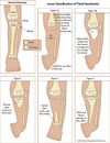
Compare and contrast anteromedial, anterolateral and posteromedial tibial bowing

What are the radiographic findings in cavovarus foot?
[Lovell and Winter][Orthobullets][SPORC][JAAOS 2014;22:512-520]
- Calcaneal pitch >30
- Meary angle >4 (apex dorsal usually centered at the medial cuneiform)
- Fibula overlies the posterior 1/3 of the tibia on lateral view
- Vertically oriented midfoot with a “stacked” conformation of the talonavicular joint and calcaneocuboid joint (see picture with sinus tarsi see-through sign)

What are the radiographic features of pes planovalgus?
[Instr Course Lect 2015;64:429]
- Lateral
- Meary angle – apex plantar
- Calcaneal pitch (normal = 10-30°; <10° = pes planus)
- Talocalcaneal angle (normal = 25-55°; >55° = hindfoot valgus)
- AP
- Talonavicular uncoverage
- Talocalcaneal (Kite) angle (normal = 15-30°; >30° = hindfoot valgus)
- Oblique
* Assess for calcaneonavicular coalition

What angles should be evaluated on AP and lateral in CVT?
- AP talocalcaneal angle
- AP TAMBA (Talar axis-first metatarsal base angle)
- Lateral TAMBA
- Lateral talocalcaneal
- Lateral tibiocalcaneal

What should be evaluated on each radiographic view to diagnose CVT?
- Neutral lateral view
- Talus vertically oriented
- Calcaneus in equinus (high tibiocalcaneal angle)
- TAMBA >35° diagnostic for CVT
- Plantarflexed lateral view
* Persistent vertical talus - Dorsiflexed lateral view
* Persistent hindfoot equinus - AP view
* Talocalcaneal angle increased (no angle pathognomonic)

What are the 3 types of accessory naviculars?
[Orthobullets]
Geist classification
- Type 1 = “sesamoid”
- Sesamoid in the tibialis posterior tendon
- Type 2 = “synchondrosis”
- Accessory bone attached to native navicular by synchondrosis
- Type 3 = “synostosis”
- Complete bony enlargement

What are common associated findings with a congenital hallux varus?
[Lovell and Winter]
- Short, thick first metatarsal
- Fibrous medial band
- Longitudinal epiphyseal bracket
- The condition is characterized by a shortened and angulated first metatarsal. The medial diaphysis and metaphyses of the bone are bracketed by a continuous epiphysis
- Suggested by the D-shape of the metatarsal with no cortical differentiation along the convex medial border of the diaphysis
- Accessory metatarsals/phalanges, duplication of the hallux

What is the treatment of congenital hallux varus?
[Lovell and Winter]
Operative
- Surgery is mandatory
- Technique depends on the associated findings:
- Medial soft tissue release – fibrous medial band
- With or without syndactylization (creates syndactyly between the 2nd toe and hallux)
- Farmer technique (see photo)
- With or without syndactylization (creates syndactyly between the 2nd toe and hallux)
- Resection (of central portion) and interposition grafting of a longitudinal epiphyseal bracket
- Medial soft tissue release – fibrous medial band

What is Vicker’s ligament?
[JAAOS 2013;21:372-382]
Abnormal volar ligament that tethers the lunate to the volar distal radius

What is the resulting bony deformity in Madelung’s deformity?
[JAAOS 2013;21:372-382]
- Ulnar and volar curvature (increased radial inclination and volar tilt)
- Positive ulnar variance
- Proximal subsidence of the lunate

Clinically, on observation how does the hand and wrist appear in Madelung’s Deformity?
[JAAOS 2013;21:372-382]
The hand appears to be translated volarly and ulnarly relative to the wrist, and dorsal prominence of the ulnar head is a distinguishing feature

List the criteria to differentiate between septic arthritis and transient synovitis
[Pediatr Clin N Am 61 (2014) 1109–1118]

What are the radiographic features of Rickets?
[Lovell and Winter]
- Wide and indistinct growth plates (HALLMARK)
- Lateral expansion of growth plates
- Cupped and splayed metaphysis
- Short long bones for age
- Angular deformity (coxa vara, genu varum/valgum)
- Looser zones
- Transverse bands of unmineralised osteoid (‘Pseudofracture’)
- Typically appear in the medial aspect of the proximal femur and at the posterior aspect of the ribs
- Acetabular protrusio
- Pathological fracture

What are the types of Rickets?
[Lovell and Winter]
- Nutritional
- Causes:
- Vit D deficiency (most common)
- Profound calcium deficiency (rare)
- Combined Vit D and calcium deficiency
- Presents at 6m - 3y
- Pathophysiology [Orthobullets]
- Low Vitamin D levels lead to decreased intestinal absorption of calcium
- Low calcium levels leads to a compensatory increase in PTH and bone resorption
- Bone resorption leads to increased alkaline phosphatase levels
- Treatment = Vit D and calcium
2. X-linked hypophosphatemic rickets - AKA Familial hypophosphatemic rickets (x-linked dominant)
- Cause = renal phosphate wasting AND low or normal kidney production of 1,25-dihydroxyvitamin D3
- Inability of renal tubules to absorb phosphate
- Presents at 1 - 2y
- Treatment = phosphate and Vit D (calcitriol)
- Renal osteodystrophy
- Causes = renal failure
- Insufficient 1,25-dihydroxyvitamin D3 activation
- Reduced phosphate excretion
- Hyperphosphatemia causes hypocalcemia (reduced renal uptake) which causes secondary hyperparathyroidism
- Treatment = dietary phosphate restriction, phosphate binding agent, Vit D3
- Hypophosphatasia
- Causes = ALP deficiency (autosomal recessive)
- Treatment = no medical treatment
- 1-Alpha-Hydroxylase deficiency (‘Vitamin D dependent’-Type 1)
- Causes = unable to convert 25-hydroxyvitamin D3 to its biologically active form of 1,25-dihydroxyvitamin D3
- Treatment = 1,25 Vit D3
- End organ insensitivity (‘Vitamin D dependent’-Type 2)
- Causes = lack receptor for 1,25 Vit D3
- Treatment = high dose 1,25 Vit D3 and Calcium

What is the classification system for OI?
Silence Classification
- TYPE I (non-deforming)
- Prevalance = ∼50% (most common)
- Severity = most mild (minimally deforming)
- Inheritance = AD
- Features:
- Blue sclera – YES
- DI – variable
- Ambulatory, normal/slightly short stature, minimal kyphoscoliosis, variable hearing loss
- Hallmark is multiple childhood fractures
- Likely underdiagnosed
- TYPE II (lethal)
- Prevalance = N/A (rare)
- Severity = lethal
- Inheritance = AR
- Features:
- Fatal in the perinatal period secondary to thoracic bony insufficiency and respiratory complications
- Type III (severe deforming)
- Prevalance = ∼20%
- Severity = most severe form compatible with life
- Inheritance = AR
- Features:
- Blue sclera = NO
- DI = YES
- Wheelchair-bound or assistive devices, short stature, severe kyphoscoliosis, frequent hearing loss, shortened and bowed limbs, triangular facies, chronic pain
- Type IV (intermediate)
- Prevalance = ∼20%
- Severity = moderate
- Inheritance = AD
- Features:
- Blue sclera = NO
- DI = variable
- Moderately short stature, moderate kyphoscoliosis, variable hearing loss
Modification to Sillence classification
- Type V
- Prevalance = ∼5-10%
- Severity = moderate
- Inheritance = AD
- Features:
- Blue sclera = NO
- DI = NO
- Normal hearing, mild to moderate short stature, variable kyphoscoliosis
- Congenital bilateral anterolateral radial head dislocation with synostosis, hyperplastic callus formation in long bones following fracture
- Type VI-XIII
- Variable
