Parkinson's disease Flashcards

1
Q

What is Parkinson’s disease?

A

A progressive neurodegenerative brain disorder that causes a range of neurological symptoms associated with movement

2
Q

How is PD subdivided?

A

Sub-divided into young onset (<40) and late onset disease (>40)

3
Q

What proportion of PD cases are sporadic vs inherited?

A
  • 95% are sporadic i.e. no genetic linkage
  • The other 5% are inherited
4
Q

List some of the symptoms assocaited with PD

A
  • Muscle rigidity, stiffness
  • Resting rhythmic tremor
  • Bradykinesia (slowing of physical movement)
  • Postural instability
  • Depression, dementia, speech and swallowing difficulties, impotence, urinary frequency and constipation
5
Q

What are basal ganglia?

A

A collection of subcortical nuclei situated within each cerebral hemisphere and upper brain stem

6
Q

Name the basal ganglia (there are 5)

A

Caudate nucleus, putamen, globus pallidus, subthalamic nucleus, substantia nigra

7
Q

What is the key function of the basal ganglia?

A

Initiation and direction of voluntary movement

8
Q

How do motor signals travel within the CNS, starting at the cerebral cortex?

A

Cerebral cortex -> basal ganglia and cerebellum -> thalamus -> cerebral cortex -> brain stem -> spinal cord

9
Q

How do basal ganglia initiate and direct movement?

A

They receive input from all cortical areas (not just motor) and project to the thalamus, then to cortical regions involved in motor planning

10
Q

Describe some of the basal ganglia connections

A
  • The major input to the striatum (caudate nucleus + putamen) comes from the cerebral cortex
  • Cortical info is processed in the striatum and passed to BG output nuclei (internal segment of GP and SNpr)
  • BG influence motor behaviour by projections from these output nuclei to the thalamus and then back to the cortex
11
Q

What are some of the neuropathological hallmarks of Parkinson’s disease?

A
  • Loss of nigrostriatal dopamine neurons
  • Presence of Lewy bodies (intraneuronal proteinacious cytoplasmic inclusions)
12
Q

Where are the cell bodies of nigrostriatal dopamine neurons found and where do they project to?

A

Found in the substantia nigra pars compacta and project to the putamen

13
Q

What makes the substantia nigra pars compacta a dark colour and what causes the depigmentation seen in PD?

A
  • Neuromelanin is contained within the nerve cells in the SNpc
  • Loss of dopamine neurons results in classical neuropathological trait of SNpc depigmentation
14
Q

What happens as a result of loss of projection to the putamen in PD and when do PD symptoms typically begin?

A
  • Loss of projection to putamen results in dopamine depletion in the putamen.
  • Onset of symptoms typically ocurs when around 80% of putamental dopamine is depleted or 60% of SNpc dopamine neurons have been lost
15
Q

The substantia nigra isn’t the only area of the brain affected by PD. Name some other areas of the brain affected by PD

A

Neurodegeneration and Lewy body formation occurs in:

  • Noradrenergic neurons in the locus coeruleus (nucleus in the pons)
  • Serotonergic neurons in the Raphe nucleus (in the brainstem)
  • Cholinergic neurons in dorsal motor nucleus of vagus
  • Cerebral cortex, olfactory bulb, autonomic nervous system and hippocampus
16
Q

Describe the appearance of Lewy bodies histologically

A

Usually circular, with a dense protein core surrounded by a peripheral halo

17
Q

Lewy bodies are composed of filaments, name some of the key filaments

A
  • Ubiquitin, neurofilament proteins
  • Alpha-synuclein
18
Q

Describe some of the properties of alpha-synuclein

A
  • It is a natively unfolded protein with significant structural plasticity
  • Can aggregate to form insoluble filaments
  • Fibrillar forms of a-synuclein are a major component of Lewy bodies in PD
19
Q

Familial Parkinson’s disease is much rarer than familial Alzheimer’s disease however there is still a strong genetic component to PD. What proportion of PD patients have autosomal dominant trait PD and when does this usually present?

A
  • 1% of PD patients have pure autosomal dominant trait familial PD
  • This usually has an early onset (<40)
20
Q

How many genetic loci have been associated with familial PD?

A

10 distinct genetic loci and mutations in more than 4 genes

21
Q

Variations in alpha-synuclein levels can predispose individuals to disease. What mutations have been identified in alpha-synuclein to cause familial PD?

A
  • An A53T missense mutation on chromosome 4q21-q23 has been identified in gene coding for alpha-synuclein
  • A 2nd mutation in alpha-synuclein gene (A30P) and E46K mutation has also been found
22
Q

What is the physiological function of alpha-synuclein?

A

May play an important role in regulating synaptic vesicle size and recycling with particular relevance to storage of neurotransmitter dopamine

23
Q

What does mutant alpha-synuclein do that wild type alpha-synuclein doesn’t?

A

The mutant type self-aggregates more readily than wild type, suggesting that aggregation of alpha-synuclein is a key event in pathogenesis of PD

24
Q

It has been proven that aggregation of a-synuclein is a key event in PD but what else can mutant alpha-synuclein cause that contributes to PD?

A
  • Mice overexpressing A53T mutation develop intraneuronal inclusions, mitochondrial DNA damage and apoptosis of neocortical, brainstem and motor neurons
  • Over expression of wild type or mutant alpha-synuclein in vitro increaes ROS production and results in enhanced cell death
25
Q

How are Lewy bodies formed?

A
  1. Unfolded or disordered a-synuclein monomers form beta-sheet rich oligomers
  2. The protofibrils give rise to more stable amyloid like fibrils
  3. Alpha-synuclein fibrils aggregate and form Lewy bodies in vivo
26
Q

How common is a mutation in the gene encoding Parkin in familial PD?

A

Relatively common with 50% of early onset cases having a Parkin mutation

27
Q

Parkin is an E3 ubiquitin ligase. What do E3 ubiquitin ligases do?

A

It is an important enzyme that tags target proteins with ubiquitin for proteosomal degradation

28
Q

What do Parkin mutations do to Parkin’s E3 ligase activity?

A
  • They usually cause loss of function of Parkin which impairs Parkin’s E3 ligase activity.
  • This results in improper targeting of substrates for proteosomal degradation leading to their potentially toxic accumulation in neurons
29
Q

UCH-L1 catalyzes hydrolysis of C-terminal ubiquityl esters and is involved in recycling ubiquitin ligated to misfolded proteins. In what ways are mutations in UCH-L1 associated with PD?

A

I93M and S18Y polymorphisms alter the activity of UCH-L1 which suggests that impaired activity of the ubiquitin-protease system is key for PD pathogenesis

30
Q

Mutations in DJ-1 gene has also been linked to PD. What does DJ-1 do in the brain?

A
  • Localised to mitochondria and may be important in mitochondrial function.
  • DJ-1 is also thought to be a cellular monitor of oxidative stress
31
Q

Describe how Parkin and UCH-L1 are related to each other in the ubiquitin-proteasome system

A
  • Parkin acts as an enzyme that tags polyubiquitin protein conjugate to be broken down into ubiquitin monomers.
  • If there is a mutation in UCH-L1 then ubiquitin will not be broken down
32
Q

LRRK2 gene encodes for a protein called dardarin. How does this relate to PD?

A
  • Dardarin comes from the basque word for tremor.
  • Mutations have been found throughout the LRRK2 gene in patients with PD and they are thought to interfere with cytoskeletal motility and vesicular trafficking events which may play a role in PD
33
Q

What are the 2 main hypotheses of the mechanisms of neurodegeneration in PD?

A
  • Misfolding and aggregation of proteins are instrumental in death of SNpc DA neurons
  • Mitochondrial dysfunction and consequent oxidative stress, lead to cell death
34
Q

What is the evidence supporting the theory that misfolding and aggregation of proteins leads to neurodegeneration?

A

Abnormal deposits of protein in brain tissue is a feature of several neurodegenerative diseases inc. PD, indicating that protein deposition is toxic to neurons

35
Q

What is the evidence of abnormal protein conformation causing neurodegeneration in inherited PD?

A
  • Pathogenic mutations may directly induce abnormal protein folding (a-synuclein)
  • Indirect interference with processing of misfolded proteins e.g. parkin, UCHL-1
36
Q

What is the evidence of abnormal protein conformation causing neurodegeneration in sporadic PD?

A
  • Direct protein damaging modifications and indirect changes in processing of misfolded proteins have also been detected
  • Reactive oxygen species are thought to trigger dysfunctional protein metabolism
37
Q

What is the evidence supporting the theory that mitochondrial dysfunction and oxidative stress lead to neurodegeneration?

A
  • Defects in oxidative phosphorylation in PD suggested as MPTP blocks mitochondrial electron transport chain by inhibiting complex I (NADH dehydrogenase)
  • Abnormalities in mitochondrial complex I have also been identified in PD
38
Q

NADH binds to complex I which triggers a sequence of events leading to reduction of Fe3+ to Fe2+. How does this happen?

A
  • NADH binds to complex I and passes 2 electrons to FMN group.
  • FMN is reduced to FMNH2.
  • Electrons are passed to iron sulphur proteins (FeS).
  • Electron is accepted by Fe3+ which is reduced to Fe2+.
39
Q

What is the effect of inhibiting complex I?

A

Increases reactive oxygen species (ROS) superoxide which forms toxic hydroxyl radicals or reacts with NO to form peroxynitrite. –> cellular damage by reacting with proteins, lipids and nucleic acids

40
Q

What evidence is there of ROS in PD?

A
  • Biological markers of oxidative damage are elevated in PD.
  • Content of anti-oxidant glutathione is reduced in PD brains
41
Q

What 2 genes are involved in removal of ubiquitin proteins?

A

UCHL-1 and Parkin

42
Q

What evidence is there for programmed cell death (PCD) in PD?

A
  • Increase in number of bax positive SNpc DA neurons in PD.
  • Increased neuronal expression of Bax in PD, suggesting that cells are undergoing PCD.
  • Other molecular markers of PCD are also altered in PD incl. caspase-8, caspase-9 and Bcl-xl
43
Q

How does the neurotoxin 6-OHDA cause dopaminergic neuronal death in PD?

A

6-OHDA accumulates in cytosol, generates ROS and inactivates various molecules by generating quinones that attack nucleophillic groups

44
Q

How does the neurotoxin MPTP cause dopaminergic neuronal death in PD?

A
  • Dopaminergic cell death occurs in SNpc and neuromelanin containing cells are more susceptible to damage.
  • In humans and monkeys, MPTP produces irreversible and severe PD syndrome, characterised by tremor, rigidity, bradykinesia and postural instability
45
Q

What difference from PD in humans do both 6-OHDA and MPTP induced PD share?

A

Lack of Lewy bodies

46
Q

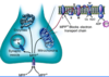
Describe the mechanism by which MPTP causes dopaminergic neuronal death

A
  • MPTP crosses the BBB where it is then converted to MPDP+ by MAO-B in non-DA/glial cells, and then into MPP+.
  • MPP+ is released into extracellular space and concentrated into DA neurons via dopamine transporter where it causes damage to DA neurons
47
Q

What happens to MPP+ inside dopamine neurons?

A
  1. Concentrate in mitochondria where it blocks complex I. This enhances ROS production and reduced ATP synthesis (toxic)
  2. Interact with cytosolic enzymes (toxic)
  3. Sequester into synaptic vesicles via vesicular monoamine transporter
48
Q

What makes paraquat toxic to DA neurons?

A

It causes formation of superoxide radicals via inhibition of mitochondira complex I and causes formation of Lewy bodies

49
Q

What makes rotenone toxic to DA neurons?

A
  • Binds to and inhibits mitochondria complex I.
  • Causes selective degeneration of DA neurons and a-synuclein inclusions –> abnormal postures and bradykinesia