0818 - anatomy of the respiratory system - AHF Flashcards
(32 cards)
What constitutes the upper respiratory tract?
nose
nasal cavity
oral cavity
pharynx
What constitutes the lower respiratory tract?
larynx
trachea
bronchus
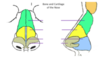
Label the external features of the nose.


Label the bones and cartilages of the external nose.

Name the recesses between the conchae, and what paranasal sinus drains where.


What are the 3 major functions of the nose?
- warming, moistening and filtering air (inferior and middle turbinate covered with respiratory epithelia)
- olfactory sensation (superior turbinate covered with olfactory epithelia)
- speech modification (sinuses provide resonance)
What is the pharynx?
- muscular tube (“funnel”) extending from base of skull to oesophagus
- nasopharynx, oropharynx, laryngopharynx
What, and where is the larynx?
- “voice box”
connects laryngopharynx with trachea
- between C4 - C6
- comprises 9 cartilages
- singles
- thyroid
- epiglottis
- cricoid
- pairs
- arytenoid
- corniculate
- cuneiform
- singles

What are the vestibular and vocal folds?
Vestibular fold: superior pair; “false vocal chord”; mucosa of larynx that folds around the vestibular ligament (thickened free lower margin of quadrangular membrane)
Vocal fold: inferior pair; “true vocal chord”; mucosa of larynx that folds around the vocal ligament (thickened free upper margin of cricothyroid membrane)
Label this diagram:
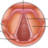

What is the position of the vocal folds during forced inspiration? During phonation?
Inspiration: vocal folds abducted, rima glottides open, vestibule open
Phonation: vocal folds adducted, vestibule open
What is the shape of the vocal folds when making a low-pitched sound? High-pitched sound?
Low: short, wide
High: long, narrow
At what level does the trachea bifurcate?
T4, sternal angle
How many U-shaped tracheal cartilages are there? What is between the rings?
14 - 17
Anular ligaments
Which bronchus is essentially a continuation of the trachea?
Right; it is more vertical, shorter and wider than the left, so more likely to get foreign objects lodged there.
What is the lung root?
All the structures (primary bronchus, pulmonary artery, pulmonary vein, bronchial artery, bronchial vein, lymphatic vessels, lymphatic node, nerve) that pass through the hilum and the pleural sleeve they are wrapped in.
Name the fissures and lobes of the left and right lungs.
Fissures:
L - oblique fissure
R - oblique fissure, horizontal fissure
Lobes:
L - superior lobe, inferior lobe
R - superior lobe, middle lobe, inferior lobe
Outline the structures within each lung lobule.
- single terminal bronchiole and its branches (i.e. respiratory bronchioles, alveolar ducts, alveolar sacs, alveolar
- associated blood vessels (i.e. arteriole, capillaries, venules) and lymphatic vessels
How many segments does each lung have? Describe them.
10 segments (10 segments of bronchi; each 1 supplies a different lung segment)
Segments are
- wedge-shaped
- segmental bronchi and pulmonary arteries are centralised
- intersegmental veins between segments
- separated by connective tissues
- functionally independent, so can remove a segment and still have a functional lung
What is the difference between type I and type II alveolar cells?
Type I:
- thin, squamous epithelial cells
- constitute 97% of alveolar surfaces
- provide air-blood barrier for gas exchange
Type II:
- cuboidal epithelial cells
- constitute 3% of alveolar surfaces
- secrete surfactant (thin layer of salt) to lower surface tension and hence ease lung expansion
- also repair and replace type I cells after injury
Outline the structures that gas must diffuse through between an alveolus and a capillary.
- epithelial alveolar cells
- basal lamina (basement membrane)
- endothelial cells of capillary
Where do the bronchial arteries branch from?
Thoracic aorta
Where do the bronchial veins drain to?
Azygos vein (R), hemiazygos vein (L), but they communicate with the pulmonary veins via a shunt.
Trace the lymph drainage out of the lung.

