1. Steatohepatopathy, Ionophore toxicosis, Chronic demarcation, Tuberculosis Flashcards
(30 cards)
What is the basic structural unit of the liver?
Liver Lobule
Center: Central vein (vena centralis) = liver damage or hepatocyte degneration begins here due to blood supply coming from portal vein and hepatic artery (already filtered of nutrients) i.e. in worse metabolic state.
Edges: Portal Triad = portal vein, hepatic artery, bile duct.

Fatty liver infiltration (steatohepatopathy) of a cat.
Yellow and shiny due to presence of fat.

Microvesicular Fatty Infiltration:
- Less severe (smaller holes)
- The holes used to be filled with fat but are now empty as the slide was dehydrated via formaldehyde and xylene which dissolved the fat.
- We can tell fat used to be present due to the size, shape, and location of the vesicles.

Microvesicular Fatty Infiltration:
- Less severe (smaller holes)
- The holes used to be filled with fat but are now empty as the slide was dehydrated via formaldehyde and xylene which dissolved the fat.
- We can tell fat used to be present due to the size, shape, and location of the vesicles.

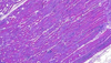
Macrovesicular Fatty Infiltration
- More severe. Larger vesicles, more damage.
- Most fat accumulation (larger vesicles) around the central vein.
- This can be so severe that the vesicles can be larger than the hepatocytes (liver cells) and fuse with them, leading to their degeneration and dysfunction.

Macrovesicular Fatty Infiltration
We can see lighter and darker areas i.e. some areas are effected more than others.
What causes Steatohepatopathy?
- Bovine: Negative energy balance post-partum causes fat to be mobilised for energy. This fat is depositied in the liver (ketosis)
- Canine: Diabetes mellitus
- Geese and Ducks: Foie gras production (force feeding)
Explain how you would prepare a frozen slide and why you would use that technique of preparation?
It is prepared in a cryostat machine and stained with Sudan staining or Oil-Red-O staining to see the fat content.
We use this method if we want to see the actual fat instead of where it used to be and also because it is quick.
It does not show the structure of the tissue.
Also used intraoperatively to check if a tissue is tumorous.
What are Ionophore antibiotics for and what happens when they are overdosed?
They are anticoccidiostatic antibiotics used in poultry and rabbits.
Their overdose causes the dysfunction of calcium channels in striated muscles, leading to muscle degeneration and dystrophy.
In the histological slide, we see decrustation, coagulative necrosis, and an increased number of macrophages.

Ionophore antibiotic toxicosis
Ionophore antibiotic toxicosis

Ionophore antibiotic toxicosis
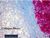
Explain Chronic Demarcation
The separation between angiofibroblastic and necrotised tissue.
The necrotised area is too big to be replaced by connective tissue so the body separates it from the live tissue via collagenous connective tissue (scar tissue).
What is angiofibroblastic tissue?
‘Angio’ represents the new blood vessels. These blood vessels leak blood into the tissue. If this tissue is on the skin surface i.e. a wound, then we must be careful of physical damage to avoid bleeding.
‘Fibroblast’ represents the collagen produced.

On the left, we see necrotised tissue
On the right, we see angiofibroblastic tissue with blood leaked into the tissue itself.
What is the difference between a fibroblast and a fibrocyte?
Fibroblast
Cigar-shaped with a light, achromatic, very active nucleus. There is constant DNA transcription to allow for dividing and collagen production.
Fibrocyte
Darker, flatter nucleus. Fibroblast becomes fibrocyte when more matured.
More mature = more collagen produced, more blood
What are collagen fibres stained with to show chronic demarcation?
Azan staining stains them blue
What causes tuberculosis?
Mycobacterium infection
What is seen in tuberculosis?
Microbronchi (no cartilage in their walls), and a granuloma known as a tuberculum

What is the tuberculum?
An epithelioid-type granuloma
Describe the tuberculum
- Concentric-circle structure with the centre being the necrotic zone with neutrophil granulocytes that disappear once the granuloma has matured. Moving outward, there are modified macrophages and epithelioid cells.
- It also contains langhans-type giant cells which are multinucleated and formed through the fusion of epithelioid cells and macrophages.
- Calcification can occur in the centre in mammals: common in cattle, does not occur in poultry.
In which mammal does calcification NOT occur in the tuberculum?
Poultry
(common in bovine)
What stain is used for TB?
Ziehl-Neelsen staining
(stains it red)
How does Ziehl-Neelsen staining work?
It uses properties of mycobacteria e.g. acid-fast and alcohol-fast. It is based on mycobacteria’s resistant cell wall.
We pour the red stain onto the slide, boil it to allow the stain enter the bacteria and then add sulfuric acid and concentrated alcohol to remove the stain from everywhere except the bacteria i.e. bacteria remains red.
