30. Oesophagus Flashcards

1
Q

List 10 diseases that static positive contrast oesophagrography is helpful for

A

Stricture

Mass

Vascular ring

Perforation

Diverticulum

Tracheo / Bronchooesophageal fistula

Hiatal hernia

Oesophagitis

FB (non-radio)

Dysphagia

2
Q

List 1 indication for pneumooesophagography

A

Oesophageal ST mass

3
Q

List 3 uses for oesophageal scintigraphy

A

Oesophageal transit time

Motility

Reflux

4
Q

List 3 indications for use of endoscopic oesophageal US

A

Infiltrative disease

Perioesophageal masses

fistula / diverticula

5
Q

What is another name for the cranial oesophageal sphincter?

A

Cricopharyngeal sphincter!

6
Q

Which 2 muscles form the cricopharyngeal sphincter?

A

Criocpharyngeus

Thyropharyngeus

7
Q

What 2 muscles run dorsal to the cranial thoracic oesophagus?

A

Longus coli

Longus capitis

8
Q

Where are the vagal nerves relative to the oesophagus?

A

Run bilaterally on the sides of the oesophagus -> Travel dorsocaudally, joining to form VAGAL TRUNK, which passes through oesophageal hiatus

9
Q

What are the four layers of the oesophagus?

A

Fibrous, muscularis, submucosa, mucosa

10
Q

How does feline and canine oesophaeal anatomy differ ?

A

Dogs: ALL STRIATED muscle -> Lonitudinal folds

Cats: Cranial 2/3 striated, caudal 1/3 SMOOTH -> Oblique striated pattern (HERRINGBONE)

11
Q

What 3 structures comprise the caudal oesophageal sphincter?

A

Thickening of oesophageal muscularis layer

Gastric folds

Muscular sling (formed by diaphragmatic crus and lesser curvature)

12
Q

Describe the arterial supply to the oesophagus (4 different bits!)

A

Cervical: Thyroid arteries

Thoracic cranial 2/3rds: Main supply bronchooesophageal artery

Thoracic caudal 1/3rd: Oesophageal branches of aorta / intercostal arteries

Terminal: Left gastric artery

13
Q

Describe the venous drainage of the oesophagus

A

Only mentions thoracic portion….

Left gastric vein and azygous vein

14
Q

Describe innervation of the oesophagus / swallowing

A

Complex!! 25 paired ganglia from C2 to L5

5 x CN: V, VII, IX, X, XII

15
Q

A small amount of oesophageal fluid can be seen normally in which lateral?

A

LEFT!!!! Makes sense…

16
Q

BOX: Survey features of oesophageal disease

A
17
Q

How do pneumomediatinum and oesophageal gas differ radiographically?

A

Pneumo: Highlights ADVENTITIAL surface of oesophagus and vessels

Oeso: Gas more contiguus, tracheal stripe, visualisation of longus coli

18
Q

What creates the dorsal indentation pictured?
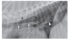
A

Azygous vein

19
Q

How can the significance of oesophageal redundancy be established?

A

Dynamic oesophagography -> Motility can be ++ reduced

20
Q

What is the tracheal stripe sign a reliable indicator of?

A

Oesophageal GAS (not megaO)

21
Q

List pertinent complications of oesophageal contrast agents

BOX ATTACHED

A

Barium paste: Contraindicated if aspiration risk -> respiratory obstruction

All barium agents: Pneumonia and granuloma RARE complication of aspiration, CONTRAINDICATED if perf suspected

-> If alveolar, will stay permanently

IONIC: oedema if aspirated, GI influx

22
Q

BOX on Oesophgram technique

A
23
Q

BOX phases of swallowing including control mechanisms and rx features

A
24
Q

What frame rate is required for videofluroscopic evaluation of swallowing?

A

30-60 frames per sec

25
Q

BOX Types of dysphagia

A
26
Q

What variables have been shown to be affected by variation in bolus form and size (in normal dogs in lateral)?

A

Degree of pharyngeal contraction

Opening of cranial sphincter

Thoracic oesophageal transit time

Peristalsis

27
Q

What clinical features are consistent with oral phase dysphagia?

A

Difficulty with prehension

Failure of bolus formation

Failure of transport to pharynx

=> CS: Dropping food, drooling

28
Q

Features of pharyngeal dysphagia

A

Challenging! Nonspecific, e.g. gagging, retching, multiple swallow attempts

Dx = When oral bolus is propelled inadequately across pharynx to cricopharyngeal sphincter

RARE as a SOLE ABNORMALITY

29
Q

What measure has been established for assessing pharyngeal contraction? How is it interpreted?

A

Pharyngeal constriction ratio

=> Pharyngeal area in contraction : area at rest

Dogs with pharyngeal dysphagia have markedly HIGHER ratio than healthy dogs

NB: CAN BE INCREASED WITH CRICOPHARYNGEAL DYSPHAGIA

30
Q

What differs between pharyngeal and cricopharyngeal dysphagia if similar constriction ratio present?

A

Time to criopharngeal sphincter opening -> delayed in CP dysphagia (sig shorter in pharyngeal)

31
Q

Features of cricopharyngeal dyphagia

A
  • Failure of CP sphincter to open OR dyssynchrony
  • CS: Similar to pharyngeal
  • TOY BREEDS, shortly after weaning (?congenital)
  • Rx: pharyngeal stasis / barium retention; hypertrophy cricopharyngeaus mm, oesophageal contrast retention, airway contrast! abnormal swallows interspersed with normal ones

CRITICAL: DELAY OR FAILURE OF SPHINCTER OPENING

LACK OF COORDINATION BETWEEN PHAR CONTRACTION AND OPENING

32
Q

What is the difference in timings of swallow -> Sphincter opening in achalasia vs normal?

A

TIME DELAY from closure of epiglottis to opening of sphincter

Kibble: 0.31 vs 0.09

Wet: 0.37 vs 0.1

**ALSO difference in sphincter closure times**

33
Q

DIfference between cricopharyngeal achalasia and chalasia?

A

Achalasia: Delayed / dysynchronous opening

Chalasia: Lack of positive pressure (remains open) -> Can happen with myasthenia gravis. INCREASED ASPIRATION RISK, do not use barium. May see gas at rest in sphincter.

34
Q

What is the most common type of dysphagia in cats (RARER THAN IN DOGS)? 3 most common causes?

A

OESOPHAGEAL

  • Hiatal hernia, dysmotility, stricture
35
Q

How do quatitative measures of swallowing (phar constric etc) compare between cats with oesophageal dysphagia and NORMAL dogs?

A

Similar! Data lacking on normal cats

36
Q

What fluroscopic features of oesphageal studies differ in dogs depending on recumbency?

A

Sternal:

  • SHORTER oesophageal transit time
  • GREATER % primary waves (kibble and liquid)

Lateral

  • GREATER % swallows with no peristaltic waves
37
Q

Oesophageal dysfunction has an association with which disease in dogs?

A

Layrngeal paralysis

38
Q

List 4 measures of oesophageal dysmotility (In the absence of megaO)?

A
  • Abnormal 1ry waves -> bolus <5cm
  • Abnormal 2ry waves -> retenion in oesophagus after 2 swallows
  • Retrograde bolus motion >10cm
  • Prolonged transit time >5secs
39
Q

List Ddx for cause of segmental (++) vs generalised O dilation

A

Generalised

  • FUNCTIONAL DISEASE

Segmental

  • Hiatal
  • infiltrative
  • FB
  • Redundant
  • stricture
  • vascular ring
  • Segmentlal motor
40
Q

What does mega O describe?

A

DILATED ANDHYPOMOTILE!

-> resulting from neuromuscular dysfunction, often idiopathic

41
Q

Megaoesophagus in the cat has been described in association with what?

A

Pylorospasm

-> MegaO rare entity in the cat

42
Q

What is the most common cause of regurg in the dog?

A

MegaO

=> ALSO most common O motility disorder

43
Q

Which components of the neuro system can be affected to produce MegaO?

A
  • Muscle (myopathy)
  • Neuromuscular (MG)
  • Peripheral (Polyneuropathy)
  • CNS (inflamm, tox, neoplasia)
44
Q

Which breed has congenital oesophageal hernia?

A

Shar pei

45
Q

List the 3 described types of hiatal hernia

A

Sliding: Movement of caudal sphincter into thorax

Paraoesophageal: Movement of fundus into thorax (sphincter in abdomen)

GO intussuception: Stomach (or other organ) telescopes into oesophagus

46
Q

What risk factors have been associated with GO intussception?

A
  • Oesophageal dilation
  • Caudal oesophageal sphincter surgery -> resultant chalasia
47
Q

What breeds are predisposed to O FBs?

A

Terriers

Dogs>Cats

48
Q

What are the 3 most common sites of O FB?

A

Thoracic inlet

Heart base

Cranial to diaphragm

49
Q

What are contraindications for barium in O FB?

A

INdicators of perf!

  • Pneumomediastinum
  • Pneumothorax
  • Pleural effusion
50
Q

Which structure forms the aortic arch in the NORMAL dog?

A

Left fourth aortic arch

51
Q

What is the classification system for vascular rings?! How many types are there?

A

SEVEN!

  • 1-3 = PRAA variants
  • 4 = DOUBLE aortic arch
  • 5-7 = LEFT aorti arch with combos of persistent right lig art and R subclavian
52
Q

In approx terms, how do PRAA and normal anatomy differ?

A

Normal: Aorta, MPA and lig art ALL LEFT SIDED

PRAA: Aorta RIGHT SIDED (r side of trachea), other left sided -> Compression of oesophagus against trachea by lig art

PRAA MOST COMMON VARIANT

53
Q

What argument is there for performing videofluoro in PRAA cases?

A

Check caudal oesophageal motility -> Affects px

54
Q

Which CONCURRENT vascular malformation is commonly seen with PRAA, and can effect management?

A

PERSISTENT LEFT CRANIAL VENA CAVA!

-> Prevent left thoracotomy for correction

55
Q

Where does an aberrant right subclavian artery originate? And normally?

A

Normally: Brachiocephalic trunk

Aberrant: Direct from AA (three vessels instead), distal to LSA

56
Q

Where do oesophageal strictures tend to occur when they result from GO reflux under GA?

A

Caudal to level of heart base

57
Q

Oesophageal neoplasia is rare! List some (9)!

A

OSA, FSA (in S.lupi areas)

SCC

Adenocarcinoma

Branchioma

Branchial cleft cysts

Papilloma

Angioleiomyosarcoma

Leiomyosarcoma

58
Q

Features of Spirocerca lupi

A
  • Tropical and subtropical locations
  • Mainly affects oesophagus, aorta -> GI, resp and circulatory signs
  • Caudal mediastinal (oesophageal) mass => GRANULOMA
  • ALSO:

ventral changes thoracic vertebra dorsal to mass

Enlargement of descending Ao (aneurysm)

=> NEOPLASTIC TRANSFORMATION: OSA / FSA

59
Q

Which radiographs are advised for S.lupi?

A

R lateral

DV

=> avoid interpretaton of normal o fluid in LL as mass, better Ao visualisation

60
Q

What CT features have been demonstrated in S.lupi?

A

Variable vascularity in non-neoplastic vs neoplastic lesions

OESOPHAGEAL SARCOMAS SIG LESS ENHANCING THAN GRANULOMAS

61
Q

How are oesophageal diverticulae categorised?

A

1) Congenital vs acquired
2) Acquired - Pulsion vs traction

PULSION

  • e.g. obstruction
  • Most commonly between heart and diaphragm

TRACTION -> e.g. mediastinal adhesions

  • Most commonly cranial and midthoracic oesophagus
62
Q

What are the reported risk factors for complications associated with Oesophageal FB? What is the reported complication rate for O FBs?

A

Bony FBs

Bodyweight <10kg

>3 days duration

12.7%!

=> perf, stricture, diverticula, abscess, pneumo, pleural effusion, resp arrest

CERVICAL PERF BETTER PX THAN THORACIC

63
Q

Oesophageal varices may result from which pathological states?

A

Portal hypertension -> specifically, flow via L gastric into venous plexus of oesophagus

Obstruction of cranial cava -> oesophageal / paraoesophageal varices

64
Q

What condition should be included as a DDx for oesophageal varices?

A

BRONCHOESOPHAGEAL ARTERY HYPERTROPHY

  • occurs secondary to chronic pulmonary or TE disease
  • Can occur concomittantly with varices