Structure of the lungs Flashcards
What structures are involved in the conducting zone?
What structures are involved in the respiratory zone?
Trachea position?
- Palpable anteriorly, above suprasternal notch
- C-shaped rings of hyaline cartilage supporting a fibro-elastic and muscular air-transport tube
- Starts at C6, ends at T 4/5 (sternal angle) at carina
- Trachealis muscle (posteriorly positioned) alters tracheal diameter

Tracheobronchial tree
Do the left lung and right lung have same amount of lobes?
Is foreign bodies more likely to enter one side than the other?
Why is this the reason?
Trachea and Bronchi: The left lung has 2 lobes while the right lung has 3 lobes, however both have only 1 main bronchus. The right main bronchus is slightly more vertical, shorter and wider than the left, hence f.b. entry.

Lobar bronchi
Where does the bronchi get supplied with oxygenated blood?
Lobar Bronchi: Since the left lung has 2 lobes it has 2 (secondary) lobar bronchi while the right lung with its 3 lobes therefore has 3 (secondary) lobar bronchi. Each lobar bronchus then divides into segmental bronchi.
The bronchi are supplied with oxygenated blood via the bronchial arteries.

Bronchioles
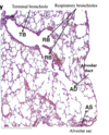
Bronchi continue dividing into smaller and smaller branches, becoming conducting bronchioles, then terminal bronchioles, and then respiratory bronchioles decreasing in diameter along the way before finally becoming alveoli.

Histology of the resipiratory tract in terms of the trachea?
The histology of the trachea is characterised by pseudostratified, ciliated columnar epithelium, with goblet cells for mucous secretion (‘respiratory epithelium’).Volume of mucus produced per day?

Histology of the respiratory tract in terms of the bronchi?
Similar to the trachea, the histology of the bronchi still has pseudostratified, ciliated columnar epithelium but the height is decreased (flattened) compared to the trachea. As the bronchi branch into the lungs the C-shaped cartilage rings are replaced by cartilage plates.

Histology of the respiratory tract in terms of bronchioles?
As the bronchial tree divides and branches, it eventually forms bronchioles with very thin lumen of <1mm in diameter. The epithelium changes to become ciliated columnar (thinner/flatter) and there is a surrounding band of smooth muscle. The cartilage and glands disappear and the bronchiole is held open by the surrounding lung tissue.
In asthma, the smooth muscle in the wall may excessively narrow the lumen.

Histology of the respiratory tract in terms of term and respiratory bronchioles?
n the Terminal and Respiratory Bronchioles the epithelium becomes non-ciliated cuboidal (thinner and flatter) and goblet cells disappear.
Gas exchange begins to occur in the respiratory alveoli that bud from the respiratory bronchioles.
Histology of the respiratory tract in terms of alveoli?
Alveoli are the basic structural and functional unit of the lungwhere gaseous exchange takes place.
Alveoli are found as outpocketings of respiratory bronchioles, alveolar ducts and alveolar sacs.
Alveoli are separated from one another by septae (alveolar wall) which is a thin membrane containing capillaries. The septae is the air-blood barrier for gas exchange.

Lungs pleura and pleural cavities
Pleura are the membranes which cover/line the organs (lungs) within the cavity as well as the cavity walls.
The pleural cavity is the space between the layers of pleura. It contains fluid to lubricate the pleural surfaces allowing for smooth, gliding movements between surfaces.
Visceral/pulmonary pleura refers to the pleura covering the lungs.

Lungs, pleura and pleural cavities - parietal pleura
Parietal pleura are the membranes which cover/line the cavity walls. Depending on where the pleura is located, a different name is used (cervical, costal, mediastinal, diaphragmatic)

What do we mean by parietal and visceral layers?

Lungs: Left
The lungs are separated from each other by the mediastinum.
The left lung is slightly longer and narrower than the right lung, predominantly to make room for the heart and pericardium.
From this viewpoint, we see the costal surface.

Lungs left - mediastinal surface
The diagram on the right shows the hilum of the left lung. The hilum is the ‘root of the lung,’ whereby structures (main bronchus, pulmonary artery, and pulmonary veins) pass into and out of the lung. De-oxygenated blood enters the lungs from the right ventricle via the pulmonary arteries. Oxygenated blood leaves the lungs towards the left atrium via the pulmonary veins.
Notice something odd about above two sentences??

Lungs : right
The right lung is slightly wider but shorter than the left lung. This is mainly due to the right dome of the diaphragm being higher than on the left side.
From this viewpoint, we see the costal surface.

Lungs right - mediastinal surface
The diagram on the right shows the hilum of the right lung. As before, the hilum is the ‘root of the lung,’ whereby structures (main bronchus, pulmonary artery, and pulmonary veins) pass into and out of the lung.
De-oxygenated blood enters the lungs from the right ventricle via the pulmonary arteries. Oxygenated blood leaves the lungs towards the left atrium via the pulmonary veins.

Lung relations : Mediastinum from left

Lung relations : Mediastinum from right

Lungs : surface anatomy
Superiorly, notice that the apex of the lung and pleura are above the clavicle.
Inferiorly, the pleura extend down to the costal margin, but the lungs end 2 rib spaces higher.
During normal/quiet respiration the lungs do not extend to the lower parts of the pleural cavity.

Respiration
Intercostal muscles between the ribs assist in respiration by expanding the size of the thoracic cavity during inspiration. They raise the ribs (and therefore push the sternum upwards as well), but may also act in forced expiration to help lower the ribs.

Respiration in relation to the diaphragm
The diaphragm is muscular at its periphery, but is tendinous centrally and it has left and right domes as seen in these superior and coronal views. Motor and sensory supply is by the PHRENIC NERVE (C3, 4, 5). (“C3, 4, and 5 keep the diaphragm alive.”)
During inspiration the domes descend, causing negative intrathoracic pressure, but raising intra-abdominal pressure.

Chest radiography from normal to abnormal
