Introduction to Body Cavities Flashcards
what plane is this?
sagittal
what plane is this?

transverse
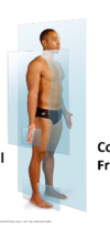
what is the name of this plane?

coronal plane
how do u look at an axial CT or MRI?
is if standing by bedside at feet

SO: Left hand site of CT = right hand side of patient
what is the cranial cavity continious with?
cranial cavity continues into vertebral canal

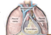
which subcavities are found in the thoracic region?
thoracic cavity:
- pleural cavity (lungs)
- mediastinum (heart, esophagus, trachea, thoracic nerves and systemic blood vessels)
- pericardial cavity (inside the mediastinum) (heart)

what surrounds and protects the brain (inside the skull) - where do they go?
3 membranous layers: meninges - continue down vertebral column:
- dura mater (outer layer)
- arachnoid mater (middle layer - spider leg layer)
- pia meter (closest to brain - follows the contours of the brain and spinal cord

what is found betwen the arachnoid and pia meter layers
role of ^?
sub arachnoid space: filled with cerbrospinal fluid
- role: buffers and protects the brain and spinal scord
label this correctly


what is meningitis?
how test?
inflammation of the meninges
test: test CSF via lumbar puncture
what are serous membranes?
serous membranes: sealed, two layered internal cavities of the body. filled with serous fluid:
2 layers are continous:
a) partietal: line body cavity and share nerve supply to body wall: somatic
b) visceral: cover the organ and share same NS to organ: autonomic
which are the main three serous membranes?
- pleural cavity: serous membranes of lung
- pericardium: serous membrane o fheart
- peritoneum: serous membrane of (continuous membrane which lines the abdominal cavity and covers the abdominal organs)
how many pleural cavities are there?
whats in the mediastinum?
whats the pericardium
- 2 - 2 lungs lol
- mediastinum: where following are collected: heart, great vessels, trachea, oesophagus etc
- peridcardium: a thin sac that surrounds your heart.
what is inbetween the viesceral and parietal pleura?
function of ^?
- pleural fluid
-
function:
- reduces friction of expansion / depression
- help stick viseral pleura to parietal pleura

describe the structure of the pericardium
- viseral pericardium (surface of heart)
- parietal pericardium (ourtside of visc)
- parietal cavity: space between ^. filled with fluid
- fibrous pericardium: fibrous sack

what are these?

A - left lung
B - pleural cavity
C - oesophagus
D - thoracic aorta
E - peridcardium

how do you split up the regions of the abdomen?

label these

phyloric sphincter controls movement of food into duodenum

which organ does duodenum curl around?
head of the pancreas - pancreatic and bile ducts drain into duodenum

which organ is connected to the liver?
connected to gall bladder: liver produces bile, stored in gall bladder (which then goes to duodenum)

what are the names for the different parts of the small intestine?
duodenum -> jejunum -> ileum
jejunum: upper left
ileum: lower right quadrant

what are the different parts of the colon?

what does spleen do ?
what type of organ?
location: upper left hand side of abdomen. behind stomach. quite posteriorly
organ type: lympathic organ

which kidney sits higher than the other? why?
which gland is located superiorly to the kidney?
what is pathway from kidney to urethra?
right kidney is lower than left: liver pushes it down
above kidneys: adrenal glands
kidneys -> ureter -> bladder -> urethra

how do you describe the different organs that located with the peritoneum?
- organs stuck to the posterior abdomen wall: peritoneum covers the anterior surface of the organ (these organs = retroperiotneal organs)
- organs that are full covered: intraperitoneal organs

GO: greater omentum fold
LO: lesser omentum fold
M: mesentery folds
MC: mesocolon fold

label this correctly pls xox


what colours on the diagram are the lesser sac and the greater sac?

greater sac: green and peach
lesser sac: blue

identify A-E :)

A: right kidney
B: liver
C: gall bladder
D: pancreas
E: lumbar vert

what seperates the thoracic and abdomenal organs?
what seperates the abdomen and pelvic organs?
- diaphragm
- nothing: abdominal peritoneum drapes over the pelvic organs
