S2) Cardiac Cycle & Valvular Problems Flashcards
Distinguish between systemic and pulmonary circulation
- Pulmonary circulation where the right side of the heart pumps blood through the lungs where it is oxygenated
- Systemic circulation where the left side of the heart pumps blood through the rest of the body to provide oxygenated blood
Which type cells are found in the myocardium?
The myocardium consists of individual specialised muscle cells joined by low electrical resistance connections
Which cellular event causes the cardiac myocytes to contract?
The contraction of each cell is produced by a rise in [Ca2+]i triggered by an the action potential in the cell membrane
How long is an action potential in the heart?
A single action potential will produce a sustained contraction of the cell lasting about 200 - 300 ms
In 5 steps, outline the conduction system
⇒ Pacemaker cells in the SAN generate an action potential
⇒ Activity spreads over atria producing atrial systole
⇒ Action potential reaches AVN and is delayed for ~ 120 ms to prevent simultaneous atrial and ventricular contraction
⇒ Excitation spreads down IV septum & through ventricular myocardium from endocardial to epicardial surface
⇒ Ventricle contracts from the apex up
Define the terms systole and diastole
- Systole is the period when the myocardium is contracting
- Diastole is the period of relaxation between contractions

What is the cardiac cycle?
The cardiac cycle is the sequence of pressure flow changes and valve operations that occur with each heartbeat
In four steps, outline the stages of the cardiac cycle from early diastole until reduced filling
⇒ Ventricular muscle relaxes & intraventricular pressure falls
⇒ Atrioventricular valves open as atrial pressure > ventricular
⇒ Blood is forced rapidly from the atria into the ventricles due to atrial distension by venous return
⇒ Filling of the ventricles continues and steadily decreases as IVP rises
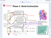
In four steps, outline the stages of the cardiac cycle from atrial systole until reduced ejection
⇒ Atrial systole forces a small extra amount of blood into the ventricles
⇒ Ventricles contract ‘isovolumetrically’ (the volume if blood in ventricles stays the same, the valves to the arteries are still closed. contraction increases pressure so valves open) and IVP rises rapidly until outflow valves open
⇒ Rapid ejection of blood out of ventricles
⇒ Towards the end of systole, IVP falls & the outflow valves close
What are heart sounds?
- Heart sounds are produced by sudden acceleration and deceleration of structures or by turbulent flow relating to the preceding events in the cardiac cycle
- It can be used to assess the state of the heart
When are the first and second heart sounds heard?
- First heart sound: closure of atrioventricular valves (‘lub’ sound)
- Second heart sound: closure of semi-lunar outflow valves (pulmonary and aortic valve) (‘dub’ sound)
When are the third and fourth heart sounds heard?
- A 3rd sound may be heard early in diastole
- A 4th sound is sometimes associated with atrial contraction
What are murmurs?
Murmurs are heart sounds associated with disturbed flow through a narrowed valve or back flow through an incompetent valve
When are heart murmurs normally expected?
In exercise, turbulent flow generates ‘murmurs’ in normal individuals
Stenosis is abnormal valve function.
When does it occur?
Stenosis occurs when the valve doesn’t open enough and there is a resultant obstruction to blood flow when then valves normally open

Regurgitation is abnormal valve function.
When does it occur?
Regurgitation occurs when the valve doesn’t close all the way and there is a resultant back leakage when the valve should be closed

What is aortic valve stenosis?
Aortic valve stenosis is the narrowing of the aortic valve, obstructing blood flow into the aorta

What sound can be heard in an aortic valve stenosis?
Crescendo-decrescendo murmur

Identify three causes of aortic valve stenosis
- Degenerative (senile calcification/fibrosis)
- Congenital (bicuspid aortic valve)
- Chronic rheumatic fever (inflammation - commissural fusion)

Identify two consequences of aortic valve stenosis
- Increased left ventricle pressure ⇒ LV hypertrophy
- Left sided heart failure ⇒ syncope, angina

What is aortic valve regurgitation?
Aortic valve regurgitation is the prolapse of the aortic valve, resulting in the backflow of blood from the aorta

What sound can be heard in aortic valve regurgitation?
Early decrescendo diastolic murmur

Identify two causes aortic valve regurgitation
- Aortic root dilation (leaflets pulled apart)
- Valvular damage (endocarditis, rheumatic fever)

A primary consequence of aortic valve regurgitation is LV hypertrophy.
Describe 3 ways in which this manifests
- Stroke volume increases - blood trickling in
- Systolic pressure increases
- Diastolic pressure decreases (bounding pulse)
- aortic pressure drops (because there is less blood so lower pressure)

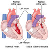
What is mitral valve stenosis?
Mitral valve stenosis is the narrowing of the mitral valve, obstructing blood flow into the left ventricle
What sound can be heard in mitral valve stenosis?
Diastolic rumble (snap as valve opens)

What is the primary cause of mitral valve stenosis?
Rheumatic fever (fusion of valve leaflets)

A primary consequence of mitral valve stenosis is increased LA pressure.
Describe 3 ways in which this manifests
- Pulmonary oedema, dyspnea (shortness of breath), pulmonary hypertension → RV hypertrophy
- LA dilation → atrial fibrillation → thrombus formation
- LA dilation → oesophagus compression → dysphagia (problem swallowing)

What is mitral valve regurgitation?
Mitral valve regurgitation is the prolapse of the mitral valve, resuting in the backflow of blood from the left ventricle

What sound can be heard in mitral valve regurgitation?
Holosystolic murmur

Identify four causes of mitral valve regurgitation
- Myxomatous degeneration of chordae tendineae & papillary muscle
- Damage to papillary muscle after AMI (acute myocardial infarction)
- LV dilation after left-sided heart failure
- Rheumatic fever disrupts seal formation

What is the consequence of mitral valve regurgitation?
Increased preload causes LV hypertrophy

label the heart


label the coronal section of the heart


wiggers diagram

wiggers diagram
phase 1 atrial contraction
wiggers diagram
phase 2 isovolumetric contraction

wiggers diagram
phase 3 rapid ejection

wiggers diagram
phase 4 reduced ejection
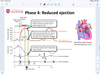
wiggers diagram
phase 5 isovolumetric relaxation

wiggers diagram
phase 6 rapid filling

wiggers diagram
phase 7 reduced filling
