11 Congenital pathology Flashcards
(34 cards)
When during embryogenesis do most heart anomalies arise? What are the usual causes?
3-8 weeks
**90%= idiopathic (can be genetic causes like trisomies/turner syndrome, or environmental causes as well)
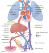
Describe the fetal circulation
- placenta -> umbilical vein -> liver/ductus venosus -> IVC
- IVC -> RA -> RV -> pulmonary trunk -> ductus arteriosus -> aorta
- IVC -> RA -> LA -> LV -> aorta
- aorta -> organs (gut/kidneys/legs/etc) -> umbilical arteries
Describe a structural versus functional shunt
Structural= actual physical connection
Functional= abnormal pressures alter flow
What are 4 examples of L->R shunt CHD?
- ASD
- VSD
- AVSD
- PDA
**notice late cyanosis
What are 3 examples of obstructive CHD?
- pulmonary stenosis
- aortic stenosis
- coarctation
**NO cyanosis
What are 5 examples of R->L shunt CHD?
- tetralogy of fallot
- transposition of the great arteries
- truncus arteriosus
- TV atresia
- TAPVR
**notice cyanosis (right away)
What is an example of valvular regurg CHD?
Ebstein’s anomaly
Describe the etiology of late cyanosis in L->R shunts
- initially oxygenated blood flows to the right sided circulation (no cyanosis)
- however this increases pulmonary flow beyond its capacity
- results in pulmonary HTN and RV hypertrophy
- increased right sided pressure reverses the blood flow
- becomes a R->L shunt
- causes late cyanosis
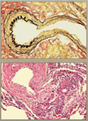
Describe plexogenic pulmonary HTN
- medial hypertrophy
- intimal proliferation
- plexiform lesions (irreversible damage)
- VSD>PDA>>ASD
- increased flow (ASD) better tolerated than increased pressure (VSD)
Describe an ASD
- may be asymptomatic until adulthood
- allows paradoxical embolism
- <10% lead to pulmonary HTN
- location
- 90% secundum (at fossa ovalis)
- 5% primum (adjacent to AV valves)
- 5% sinus venosum (near SVC entrance)

Contrast an ASD and PFO

What are the long term effects of an ASD?
Right ventricular hypertrophy and dilation
AND
Dilated RA and LA
Describe a VSD
- most common congenital heart anomaly
- 90% at septum (membranous VSD)
- usually associated with other anomalies
Contrast membranous and muscular VSDs
- membranous
- usually large
- <10% spontaneously close by septal TV leaflet
- requires surgery
- muscular
- usually small
- spontaneous closure by fibrosis >60% by 1 yo (most don’t require surgery)
- less common than membranous
Describe a PDA
- normal closure:
- functional ~12hr
- structural ~3mo
- delayed by prostaglandin E
- causes harsh, continuous “machinery like” murmur
- usually in isolation (90%)
Describe an AVSD
- deficient AV septum, associated with MV and TV anomalies
- two types
- partial= primum ASD + cleft MV (w/ mitral regurg)
- complete= AVSD + common AV valve
- down syndrome 40% with complete AVSD
- needs early surgical correction

What are some clinical symptoms of R->L shunting?
- early cyanosis
- digital clubbing
- polycythemia
- paradoxical emboli
- decompression sickness (gas not filtered by lungs)
Describe tetralogy of fallot
- most common form of cyanotic CHD
- anteriosuperior displacement of the infundibular septum leads to…
- VSD
- subpulmonary stenosis (protects lungs, but results in small pulmonary outlet)
- overriding aorta
- RVH (heart may appear “boot shaped” on xray)

What determines the clinical outcome of tetralogy of fallot?
**based on severity of subpulmonary stenosis:
- pink tetralogy (less cyanotic)
- less severe stenosis
- BAD; doesn’t protect the lungs from high pressures
- classic tetralogy
- more cyanotic
- subpulmonary stenosis protects lungs

Describe TGA
- transposition of the great arteries
- aorta from the RV
- pulmonary artery from the LV
- RVH and pulmonary HTN develop
- two types
- intact ventricular septum (65%); unstable, need prompt surgery
- with VSD (35%); stable

Describe truncus arteriosus
- origin of aorta and pulmonary artery from common truncal artery
- most have large VSD
- mixing of blood= cyanosis
- increased pulmonary flow -> pulmonary HTN

Describe tricuspid atresia
- complete occlusion of the tricuspid orifice (from unequal division of the AV canal; mitral valve is enlarged)
- needs ASD/PFO and VSD for survival
- causes right ventricular hypoplasia

Describe TAPVR
- total anomalous pulmonary venous return (pulmonary veins don’t directly drain into LA -> LA hypoplasia)
- pulmonary veins connect with left innominate/brachiocephalic or coronary sinus
- ASD/PFO allows oxygenated blood to enter systemic circulation

Describe aortic coarctation
- constriction/narrowing of aorta
- 2 types
- preductal/infantile (tubular hypoplasia with PDA)
- postductal/adult (ridgelike infolding at ligament without PDA)


