1.1 General organisation Flashcards

(51 cards)

1
Q

Which cranial nerve innervates the muscles of facial expression?

A

Facial nerve CN VII

2
Q

What are the 5 extra cranial branches of the facial nerve? What do they innervate?

A

(Superior to inferior) Temporal Zygomatic Buccal Marginal Mandibular Cervical branch Innervate the muscles of facial expression

3
Q

Which cranial nerve innervates the muscles of mastication?

A

Trigeminal nerve, CN V

4
Q

The trigeminal nerve has 2 functions in the face. What are they?

A

Innervate the muscles of mastication Main sensory nerve of face and scalp (transmits sensation to brain)

5
Q

What are the 3 key branches of the trigeminal nerve? Describe the regions they innervate?

A

Ophthalmic Va Maxillary Vb Mandibular Vc Ophthalmic is the temporal lobe and superior nose Maxillary is the cheeks Mandibular is the jaw and lateral face

6
Q

Which main artery supplies the head and neck by its terminal branches? Which terminal branch supplies the face?

A

Common carotid artery Terminal branches are internal and external branches (Facial artery branch of the) external carotid artery

7
Q

Which 2 veins drain the head and neck region? Which is the main one?

A

External and internal jugular veins Internal jugular vein

8
Q

What drains into the IJV?

A

Facial vein

9
Q

Which of the jugular veins in the neck is more superficial?

A

External jugular vein

10
Q

Which 2 muscles does the accessory nerve innervate? What number cranial nerve is the accessory nerve?

A

Trapezius Sternocleidomastoid CN IX (eleven)

11
Q

Which nerve innervates the platysma muscle?

A

Cervical branch of the facial nerve/ CN VII

12
Q

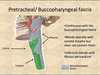
PIC Which hyaline cartilage structure sits in front of the larynx and above the thyroid gland? Which other cartilage structure sits more inferiorly in the neck?

A

Thyroid cartilage Cricoid cartilage

13
Q

Where is the cricoid cartilage? PIC

A

Surrounds the trachea (ring shaped cartilage) Above the thyroid gland and below the thyroid cartilage

14
Q

What’s the proper name for the adam’s apple?

A

Laryngeal prominence of the thyroid cartilage

15
Q

The supra-hyoid muscles are innervated by which group of nerves? How does the differ for the infra-hyoid muscles?

A

Supra-hyoid = cranial nerves I

nfra- hyoid (strap muscles) = cervical nerves

16
Q

What are the 3 borders of the anterior triangle?

A
  • Inferior margin of the mandible - Midline of neck - Anterior margin of sternocleidomastoid
17
Q

What are the 3 borders of the posterior triangle?

A
  • anterior margin of trapezius - posterior margin of sternocleidomastoid - middle 3rd of the clavicle
18
Q

What are the borders of the carotid triangle?

A
  • posterior belly of the digastric muscle - medial border of sternocleidomastoid - superior belly of the omohyoid
19
Q

The anterior triangle can be subdivided into how many triangles?

A

4 the carotid triangle is an example

20
Q

What forms the floor of the posterior triangle?

A

The scalene muscles (3)

21
Q

What’s the proper name for the ‘strap’ muscles? Where are they found?

A

The infra-hyoid muscles Beneath the hyoid bone, overlying the larynx and trachea

22
Q

Where do the supra-hyoid muscles attach to? What effect do they have on the mandible and hyoid bone?

A

Superiorly; the mandible or base of skull Inferiorly; the hyoid bone Depress the mandible (open mouth) and elevate the hyoid

23
Q

Which muscles’ actions depress the hyoid?

A

The infra-hyoid / strap muscles

24
Q

In which triangle of the neck is the carotid sheath and its contents accessible? Why?

A

Carotid triangle Because it’s not covered by sternocleidomastoid in the area

25
What are the 4 main structures inside the carotid sheath?
- Common carotid artery - Internal jugular vein - Vagus nerve, CN X - Cervical lymph nodes
26
In what fascial compartment does the common carotid artery bifurcate and into which vessels?
Carotid sheath Onto the internal and external carotid arteries
27
Why is an infection or abscess in the retropharyngeal space so dangerous? What is there a risk of developing?
The potential space opens into the mediastinum so infection can track downwards into the chest Mediastinitus
28
Which fascial layer sits anteriorly to the retro-pharyngeal space?
Buccopharyngeal fascia (invests the constrictor muscles of the pharynx)
29
What is the mediastinum? What does is it contain? Give some examples
The central compartment of the thoracic cavity / Space between the 2 pleural sacs Great vessels (Aorta + Superior vena cava) Vagus nerve, phrenic, carotid nerves Sympathetic trunk Trachea and oesophagus Thymus gland
30
Which immune cells develop in the thymus?
T-lymphocytes
31
How many cervical fascial layers are there?
4 One superficial layer and 3 deep cervical fascial layers
32
Which is the most superficial of the deep cervical fascial layers? Describe the region of the neck that it covers.
Investing layer Wraps around the whole neck like a collar. The superior margin is attached on either side to the lower border of the mandible. Inferior margin exends to the clavicle and acromion anteriorly. Posteriorly attaches to the spinous process and ligamentum nuchae.
33
Which is the deepest layer of the 3 deep cervical fasica? Which layer is neither the most superficial nor deepest of the deep cervical fascia?
Pre vertebral layer Pre-tracheal layer is between the investing and pre-vertebral layers
34
Which structure in the neck can be described as a 4th deep cervical fascia? What is it formed from?
Carotid sheath Parts of all three layers of deep cervical fascia
35
Which deep cervial fasica invests the muscles of the pharynx and oesophagus as it continues posteriorly? What is it now referred to as?
Pre-trachial layer Buccopharyngeal fasica (fascial associated with oesophagus and pharynx)
36
What are the borders of the retropharngeal space?
Superior; base of skull Anterior; buccopharngeal fascia Posterior; alar fascia Inferior; extends into mediastinum and space ends at T1-T2
37
Which structures are enclosed by the the muscular and visceral layers of the pre-tracheal deep cervical fascia?
muscular layer encloses the strap/ infrahyoid muscles visceral layer encloses the thyroid, trachea and oesophagus
38
Why is the retropharngeal space good?
Allows free movemnt of the pharynx on the vertebral column and expansion during swallowing
39
Where is the pharynx?
Immediately posterior to the mouth and nose, it connects them to the oesophagus. It is posterior to the larynx and infron of the vertebral column
40
In what type of patient is a retropharyngeal abscess most commonly seen? What usually precedes the abscess?
Children under 5 Upper respiratory tract infection \> retropharngeal space infection \> abscess
41
What does it mean if a neck lump moves upwards with swallowing? Explain.
The lump is associated with the thyroid gland. The thyroid gland is enclosed by the pre-tracheal deep cervical fascial layer which is attached to the hyoid bone superiorly. The hyoid bone and larynx move upwards with swallowing so the thyroid does too
42
Why does the larynx move upwards whilst swallowing? CHECK
Connected to the hyoid bone which is pulled anteriorly and superiorly by the suprahyoid muscles contracting
43
How do the buccinator muscles help during chewing?
Compress the cheeks against the teeth/ keep cheeks taut so that food remains within the oral cavity
44
Injury to the parotid gland risks damage to which cranial nerve? Explain
the facial nerve CN VII The facial nerve exits the base of the skull to become extracranial. It then runs through the parotid gland before branching into its 5 extracranial branches
45
How can a goitre cause stridor (wheezing sound) or facial oedema?
A goitre is an enlarged thyroid gland. The thyroid gland is enclosed in the pre tracheal deep cervical fascia which extends into the mediastinum. The goitre can extend retrosternally (behind the sternum) through the root of the neck and compress the trachea (causing stridor/ breathlessness) or compress/ impede venous drainage from the head and neck causing facial oedema
46
What is Bell's Palsy? How does it usually present?
Inflammation of the facial nerve Sudden weakness in one side of the face, all movements using muscles of facial expression are affected
47
Why would would an infected parotid gland not cause ipsilateral facial muscle weakness?
Note; The facial nerve runs through the parotid gland It is (usually) only malignancies/ parotid cancers which damage the facial nerve. Inflammation, infection or benign conditions of the parotid gland wil not cause damage.
48
How many muscles of mastication are there? Name them.
four lateral pterygoid, medial pterygoid, masseter, temporalis
49
Which of the two pterygoid muscles opens the mouth and how can you remember this?
The lateral pterygoid Saying 'laaaa'teral requires you to open your mouth
50
Which of the muscles of mastication acts in opposition to the majority?
They all move the mandible at the temperomandibular joint (TMJ) but the LATERAL PTERYGOID depresses the mandible (so your mouth opens) whilst the medial pterygoid, masseter and temporalis all elevate the mandible (close the mouth) which is important whilst chewing
51
The scalene muscles form the floor of which cervical triangle? Which structures pass between the medial and anterior scalene muscles? Describe the position of the phrenic nerve in relation to the scalene muscles.
Posterior triangle The subclavian artery and brachial plexus The phenic nerve runs along the surface of the anterior scalene before entering the thorax