11. Head and Neck - Development of the Midline Structures (Pituitary G, Tongue, Thyroid G) Flashcards
(22 cards)
What is the pituitary gland and where is it located?
- A ‘two in one’ gland
- Tissues in both lobes are completely different to each other
- Posterior lobe = neuroendocrine secretions
- Anterior lobe = endocrine secretions
- Size of a pea, sits in the sphenoid bone surrounded by the sella tursica

How does the pituitary gland develop?
- Ecotderm differentiates into neuroectoderm (which forms the neural tube –> CNS tissue)
- Neuroectoderm gives rise to infundibulum (downward out-growth of the forebrain which grows toward the roof of the pharynx), which gives rise to the posterior pituitary
- Outpocketing of ectoderm (or invagination of roof of oropharynx) grows up and thickens as it differentiates
- Gives rise to ‘Rathke’s pouch’, which gives rise to anterior pituitary
- i.e. infundibulum growing down to oropharynx, rathke’s pouch up from oropharynx
- Grow and form around each other forming two discrete lobes
- Rathke’s pouch pinches off and sphenoid bone continues to ossify beneath it, walling it off from cavity of the oropharynx

Summarise what parts of the pituitary glands are derived from what parts of the embryology
- Infundibulum = pos pit and stalk
- Rathke’s pouch = ant pit
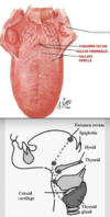
Describe the anatomy of the tongue
- Lies partly in oral cavity and partly in the pharynx
- Needs to be highly mobile for its function of mastication and swallowing
- Tethered to floor of oral cavity by short cord called the lingual frenulum
- Boundary of anterior 2/3 and posterior 1/3 separated by sulcus terminalis (V shaped) – foramen cecum means a blind ending pouch – when we look at thyroid, we see significance of this anatomical landmark
- Comprised of intrinsic muscles (work and contract to change shape of tongue) and extrinsic muscles (move tongue around mouth)

How does the tongue develop?
- Gets contribution from all pharyngeal arches
- Pharyngeal arches:
2 lateral lingual swellings - derived from 1st arch
Tuberculum impar - derived from 1st arch
Cupola - derived from 2nd, 3rd, little bit of 4th arch
Epiglottal swelling - derived from 4th arch
- Lateral lingual swelling overgrow tuberculum impar (lose blue shading)
- 3rd arch over grows 2nd arch component
- Apoptosis - frees up tongue from floor of cavity - what remains = lingual frenulum allowing tongue to stay attached to the oral cavity

What happens if the degeneration that frees up the tongue from the oral cavity (normally leaving the lingual frenulum) is not complete?
- lingual frenulum = tight
- ‘tongue tied’ so movement of tongue not optimal
- easy procedure to loosen
4.
What is the sensory innervation of the tongue?
- Mucosa of anterior 2/3 derived from first and third pharyngeal arches (as we lose the contribution from the 2nd). The nerves that innervate these arches = 5th trigeminal and 9th glossopharyngeal CN
- Posterior 1/3 derived from third and 4th therefore = 9th glossopharyngeal and 10th vagus CN
- Taste - develop in discrete structures called papillae - derived from second arch therefore = facial CN = branch is chorda tympani (pass from 2nd arch into first arch structure which derives middle ear and therefore chorda tympani passes through middle ear)
What is the motor innervation of the tongue?
Both intrinsic and extrinsic muscles of the tongue develop from myogenic precursors (small occipital somites) that migrate into the developing tongue - innervated by 12th hypoglossal CN
What and where is the thyroid gland?
- The thyroid gland is an endocrine gland
- In the neck
- Consists of two lobes connected by an isthmus
- TSH (from ant. pit) stimulates TRH (from hypo) stimulates T3 and T4 from it –> inc metabolic rate, inc rate and strength of heart beat, inc growth rate in young people
How does the thyroid gland develop?
- Begins development in the floor of the oropharynx between the tuberculum impar and cupola
- Descends to its final position in the anterior neck
What is the path of descent of the thyroid gland?
- Foramen caecum marks point of origin for descent (boundary of pos1/3 and ant2/3 of tongue)
- bifurcates and descends as a bilobed diverticulum connected by the isthmus
- during descent, developing thyroid gland remains connected to the tongue by thyroglossal duct (atrophys in adult life)
- migration is ant to pharyngeal gut, hyoid bone and laryngeal cartilages
What is a thyroglossal cyst?
- a fibrous cyst that forms from a persistent thyroglossal duct
- anywhere along path from foramen caecum to position of thyroid gland in neck
- maintained part of duct - can collect fluid

What is ectopic thyroid tissue?
- anywhere along path from foramen caecum to position of thyroid gland in neck
- presence of thyroid tissue in locations other than the normal anterior neck region between the second and fourth tracheal cartilages
- can lead to congenital hypothyroidism - is a condition of thyroid hormone deficiency present at birth.
What is cleft lip and palate?
- Palate formed by palatal shelves (which grow medially into oral cavity from maxillary prominence)
- Once mandible sufficiently enlarged, tongue drop on palatal shelves, meets in midline and fuses
- Abnormality = failure of FNP to fuse with maxillary prominence and failure of palatal shelves to fuse

What is first arch syndrome?
- Problems related to development of eyes, ears, mandible and palate
- Example: Treacher- Collins syndrome – mandible is hypoplastic, hypoplasia of facial bones, low set ears, abnormal appearance of external ear, haploinsufficiency of treacle protein (doesn’t work) = neural crest cells cannot adequately arrive in first arch and begin normal program of development. Doesn’t stop completely but ability to function properly is. Impacts on neural crest migration.

What is di-george’s syndrome?
- Deletion of chromosome 22 = Disruption of development of 3rd and 4th pharyngeal pouches = abnormal development of neural crest = congenital thymic aplasia, absence of parathyroid glands = hypoparathyroidism
What is charge syndrome?
- CHD7 protein heterozygous mutation
- Like treacle, impacts ability of neural crest cells to behave normally and differentiate and multipotency (treacle affects migration)
- = wide spectrum of defects related to function of neural crest cells
What embryonic structures contribute to the development of the face?
Frontonasal nasal prominence and the 1st and 2nd pharyngeal arches
What structures in the developing face must fuse to form the upper lip and jaw
Frontonasal prominence and medial nasal prominence
To what structures in the head and neck do neural crest cells contribute?
Cartilage bars in each of the pharyngeal arches leading to development of the skeleton of the face
What structures develop from the 3rd and 4th pouches?
Parathyroid glands and thymus
Which cranial nerves are associated with the pharyngeal arches?
CN V, VII, IX and X
