11. The Thigh :) Flashcards
(36 cards)
What forms the linea aspera?
Linea aspera: gluteal tuberosity+pectineal line
Identify the following bones (colored) of the lower body.

blue: femur
green: patella
purple: tibia (more medial)
orange: fibula (more lateral)
What is the patella?
sesamoid bone: loose sitting in tendon
Which bone does all the weight bearing: tibia or fibula?
tibia
Identify the structures on the ANTERIOR aspect of the femur.

a) medial epicondyle
b) lateral epicondyle
RED DOT near medial epicondyle: adductor tubercle
Identify the following parts of the POSTERIOR aspect of the femur.

a) medial condyle
b) lateral condyle
c) linea aspera
d) lateral supracondylar ridge
e) medial supracondylar ridge

What is found between the tibia and fibula?
interosseus membrane: distribute force and weight to fibula
Idenitfy the following parts of the ANTERIOR aspect of the tibia.

top: medial condyle
middle: lateral condyle
bottom: tuberosity
Identify the various structures of the ANTERIOR aspect of the fibula and tibia.


What is found on the superior view of the tibia?
medial and lateral plateau that articulates with femur
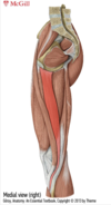
Name the different posterior thigh muscles. Whats another name for this group.
Posterior thigh muscle=hamstring muscles
- bicep femoris (most lateral)
- semitendinous
- semimembranosus (most medial)
(all inn by sciatic n)
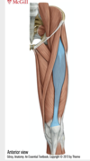
Identify the muscle in blue.

Hamstring: BICEP FEMORIS
Long Head
ori: ischial tuberosity
ins: head of fibula
inn: tibial division of sciatic n
act: extension of hip
Short head: (deep)
ori: linea aspera & lateral supracondylar ridge
ins: head of fibula
inn: common fibular division n
act: flexion @ knee & lateral rotation @ knee
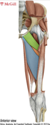
Identify the muscle in green.

Hamstring:
SEMITENDINOUS
(1/2 tendon)
ori: ischial tuberosity
ins: surface of anterior tibia (medial to tibial tuberosity)=Pes anserinus (duck foot)
inn: tibial division sciatic n
act: flexion @ knee, medial rotation @ knee, extension hip
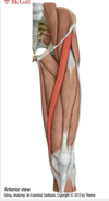
Identify the muscle in orange

Hamstring:
Semimembranosus
ori: ischial tuberosity
ins: medial condyle of tibia
inn: tibial divion of sciatic n
act: flexion @ knee, medially rotate knee, extend hip
Name the 6 different muscles of our anterior thigh.
anterior thigh (all inn by femoral n)
Quadratus Femoris group: rectus femoris, vastus lateralis, vastus intermedialis, vastus medialis
sartorius and iliopsoas
Where do the quadriceps femoris group all insert on?
Quadriceps femoris ( rectus femoris, vastus lateralis, vastus intermedius, vastus medialis)
all insert on quadriceps tendon=patellar ligament =knee cap (sesamoid bone)
(all inn by femoral n)
Identify the following muscle.
(part of quadriceps femoris-ant thigh compartment)
RECTUS FEMORIS
ori: AIIS (anterior inferior iliac spine)
ins: patella & tibial tuberosity
inn: femoral n
act: extension @ knee (kicking)
Identify the muscle in green.

(ant thigh: quadriceps femoris)
VASTUS LATERALIS
ori: greater trochanter & lateral lip of linea aspera
ins: patella & tibial tuberosity
inn: femoral n
act: extension at knee
Identify the muscle in orange.

(ant thigh-quadriceps femoris)
VASTUS MEDIALIS
ori: intertrochanteric line & medial lip of linea aspera
ins: patella & tibial tuberosity
inn: femoral knee
act: extension @ knee
Identify the muscle in purple.

(ant thigh- quadriceps femoris)
VASTUS INTERMEDIUS (deep to rectus femoris)
ori: shaft of femur
ins: patella & tibial tuberosity
inn: femoral n
act: extension at knee
What are the two diseases due to abnormal q angles?
q angle: angle between femur and knee
- GENU VARUM: ‘bow legged’
more p on medial
- GENU VALGUM: ‘knock kneed’
more pressure on lateral
Identify this muscle.
(ant thigh)
SARTORIUS
ori: ASIS (anterior superior iliac spine)
ins: Pes anserinus (medial to anterior tibial tuberosity)
inn: femoral n
act: flexion @ hip, lateral rotation of hip, flexion @ knee
Name the 5 muscles part of the medial thigh.
medial thigh=adductor muscle group
- pectineus (inn by femoral n)
- adductor longus
- adductor brevis
- adductor magnus
- gracilis
- Obturator externus
(all inn by obturator n)
What is the border between the medial and anterior thigh compartment?
border: between ilipsoas and sartorius