11.03 Bones and Joints of the Pelvis Flashcards
• Bones forming the pelvis • Pelvic types and their clinical significance • Apertures of pelvis & structures transmitted • Mechanism of the pelvis • Structure & function of sacroiliac joint • Structure & function of pubic symphysis • Applied anatomy (51 cards)
What bones form the pelvis?
What does pelvis roughly translate into?
The hip bone and the sacrum
Pelvis roughly translates into basin
What are the two divisions of the pelvis?
What delinetaes them?
The upper part of the basin is called the false (greater pelvis) which holds some of the abdominal viscera.
The area below that is the true (lesser) pelvis which has walls anteriorly, laterally and posteriorly.
The Iliopectineal line (pelvic brim) is the line between them

What makes up the walls that surround the lesser (false) pelvis
Anterolateral walls are formed by the hip bone and posterior wall by the sacrum.

What are important structures arising from the walls of the lesser (false) pelvis?
Muscles arise from the sacrum and get out of the pelvis and muscles from the lateral wall of the pelvis which have an important role in relation to the pelvic floor.
What is the ileopectal line?
(also called the pecten pubus)
A line/ridge from the ileum posteriorly to the pubis anteriorly. It demarcates false and true pelvis
The pectineal line forms part of the pelvic brim. Lying across it are fibers of the pectineal ligament and the proximal origin of the pectineus muscle.

What is the general difference between the male and female pelvis?
Female pelvis is broad, wide and is shallower from above downwards (sacrum is broad and width equivalent to the length).
Male pelvis is narrower but is longer.

The two hips joints home together at the midline in a typical midline joint arrangement. Describe this
Anteriorly the two pubic bones come together in a midline joint
(All midline joints in the body are symphiseal joints with a fibrocartilaginous discs).

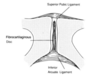
Describe the surfaces that make up the sacroileac joint
The second joint of the pelvis.
The ileum has an ear shaped surface covered by haline cartilage called the auricular surface.
The sacrum also has an auricular surface on it lined by hyaline

Describe the sacrum
- It has a coccyx (variable structure)
- Anterior foramine which has nerves (ventral rami and sacral plexus) going through them.

Structures other than the nerves pass through the ventral foraminae of the sacrum. What are these structures and what is the significance of this?
These foraminae are also roots for veins draining pelvic viscera (plexuses of veins).
Normally this drainage is back into the inferior vena cava; but if there is interference of flow then there is passage through anterior sacral veins into spinal canal itself (also common route of cancer spread)

Describe the bones of the pelvis in terms of epiphyseal sites.
What is the importance of this?
There are many epiphyseal sites in the pelvis
Children: multiple sites of empiphysise: acetabulum, ishium, crest of the ileum

What are the two parts of the main pelvic hole?
Pelvic inlet (doing down from above)
Pelvic outlet (coming out from above)

What are the four main types of pelvic inlet shapes? What proportion of women have that particular shape?
- Gynaecoid - 50%
- Android - 30%
- Anthropoid - 18%
- Platepelloid - 2%

Describe the gynaecoid pelvic inlet shape
The pelvic inlet is broad to accommodate the head of the baby as it passes through birth canal. The true pelvis the dimension of width is retained down the pelvis and the pelvic outlet is also broad (subpubic angle)

Describe the android pelvic inlet shape \
Narrow inlet and narrow true pelvis and narrow outlet (not designed for transmission of the foetus).
The male pelvic inlet is heart shaped (is narrow).
The outlet is diamond shaped in both males and females but is more broader in females.

Describe the anthropoid and platapelloid pelvis shapes
Anthropoid (narrow from side to side but enlongated in the AP direction).
The remainder are playpelloid which is wide from side to side and narrow in the AP direction.
Describe the line of gravity through the pelvis
What is the net effect of this?
Lumbar lordosis provides flexibility in stance and locomotion.
The line of gravity passes through the vertebrae and passes anteriorly to the sacrum.
The net effect of gravity is to force the sacrum to tilt forwards and inferior in the inverse direction of the lumbar spine.

Describe the alignment of the major anterior structures of the hip bone (ASIS and pubic tubercle) in normal stance
In normal orientation the ASIS is in line with the pubic tubercle (vertical alignment).

Describe the horizontal alignment betwen the sacrum and the hip bone (pelvis)
The top of the pubic symphisis is in the same horizontal plane as the tip of the coccyx

Compare the forward/inferior tilt in females with that in males
The tilt is eccentuated in females due to increased lumbar lordosis
What does the pelvic outlet transmit?
Pelvic outlet is diamond shaped and it transmits tubular viscera to the exterior (as well as important muscles and ligaments)
How is the pelvic outlet divided?
Divided into 2 triangles
- Urogenital triangle (uretha and vagina in females and beginning of membranous urethra in males).
- Boundaries are boney structures
- Anal triangle which contains the opening for the anal canal.
- Boundaries are mainly ligamentous: sacrotuberous ligament .

Describe the anterior and posterior definitions of:
- Pelvic Inlet
- Pelvic Outlet
Pelvic Inlet:
- Top of pubic symphisis to the first sacral vertebrae.
Pelvic outlet:
- From the inferior pubic symphisis to tip of the cocyx

Where and what is the narrow pelvic plane?
The narrow pelvic plane runs from inferior part of pubic symphis through the ischeal spine to S4.
It is the narrowest part of the pelvis that has to be negotiated by foetal head in childbirth.