Anaemia Flashcards

1
Q

What is the definition of anaemia in men and women?

A

Men: <135 g/L

Women: <115 g/L

2
Q

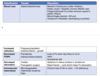
3 broad causes of anaemia based on mechanism and size

A
  1. decreased hB production
  2. Increased Hb consumption
  3. Dilutional anaemia
    size: microcytic, macrocytic, normocytic
3
Q

Causes of microcytic anaemia (FAST)

A
  1. F- Fe- iron deficiency anaemia
  2. A: anaemia of chornic disease
  3. S-siderbalstic anaemia
  4. T- thalassamiea (may not have anaemia if it is mild- eg thalassameia triat)
4
Q

Causes of normocytic anaemia

A

All Hoes Fuck Prostitutes

A: anaemia of chronic disease

H: hypothyoridism

F: failure of bone marrow

P: pregnancy causing dilutional anaemia due to increase in plasma volume

5
Q

Causes of macrocytic anaemia

A

FATRBC

Fetus (pregnancy)

Antifolates (eg phenytoin)

Thyroid (hypothyroidism)- thyroid hormone is required to produce EPO

Reticulocytosis -

B12 or folate deficiency

Cirrhosis (alcohol excess OR liver disease)

Other haem causes: Myelodysplastic syndromes, myeloproliferative disorders, multiple myeloma

6
Q

Iron deficiency anaemia: iron studies, FBC, blood film, management

A
  • microcytic anaemia

Iron studies

  • low Fe
  • low ferritin
  • high transferrin
  • high TIBC

Blood film

  • pencil cells (aka cigar cells)

FBC

reactive thrombocytosis

Management

Ferrous sulphate and investigate underlying cause

7
Q

Thalassaemia: key features

A

Blood film

basophillic stippling - aggregation of ribosomes

target cells

micocytic anaemia

iron studies

NORMAL

management

transfusions

iron chelation

8
Q

Sideroblastic anaemia: key features

A

causes: congenital or acquired

**you have enough iron but body cannot incorporate it into haemoglobin so it builds up in diff places**

  • iron accumulation within the bone marrow leading to ineffective erythropoiesis. you also get iron deposition in other parts leading to endocrine, liver and cardiac damage.

causes- myelodysplastic disorders, following chemotherapy, irradiation, alcohol excess, lead poisoning, anti-TB drugs or myeloprolfierative disease

blood film

basophilic stippling

bone marrow

ring sideroblasts

iron studies

  • high iron
  • high ferritin
  • low transferrin
  • low TIBC

management

treat underlying cause

PYRIDOXINE (vitamin B6) - aids with erythrpoiesis

9
Q

anaemia of chronic disease

A

causes- infection, inflammation, malignancy

iron studies

  • low iron
  • raised ferrtin
  • low transferrin- low TIBC

(same as sideroblastic anaemia)

+ RAISED CRP AND ESR

10
Q

causes of megaloblastic anaemia and how to differentiate between them

A
11
Q

causes of non-megaloblastic anaemia and how to differentiate between them

A
12
Q

how to classify normocytic anaemia??

A
  1. Haemolytic
    a) inherited - membrane, haemoglobin, metabolism
    b) acquired
    - autoimmune - warm vs cold
    - alloimmune - rehsus or abo incomaptibility
  2. Non-haemolytic
    - anaemia of chronic disease
    - failure of erythropoiesis
13
Q

How to split up haemolytic anaemias?

A

**don’t forget that metallic heart valves can cause haemolytic anaemia**

**cancers can cause MAHA**

14
Q

how to classify haemolysis (DIFF FROM CLASSIFICATION OF HAEMOLYTIC ANAEMIAS!!)

A

*with intravascular haemolysis the rbc get excreted through kidneys

15
Q

classification of inherited haemolytic anaemias

A
16
Q

types of red blood cell membrane disorders

A
  1. vertical interaction: hereditary spherocytosis
  2. horizontal interaction: hereditary elliptocytosis

**alphabetical order- he, vs**

17
Q

guidelines for when to suspect rbc membrane disorders?

A

basc when you’ve ruled out everything else!!

18
Q

when do you see retiuclocytes on blood film?

A
19
Q

features of hereditary spherocytosis

A

inheritance- autosomal dominant

**in qs: often father has splenectomy**

blood film- spherocytes, polychromasia

test- positive osmotic fragility, positive eosin-5-maelimide test (MOST SENSITIVE)

treatment - folate supplementation, splenectomy (as you get extravascular haemolysis)

20
Q

G6PD: inheritance

A

x linked recessive

**usually affects males**

21
Q

g6pd: pathophysiology

A

G6PD generates NADPH via pentose phosphate pathway

Episodes of acute haemolysis following exposure to oxidative stress (e.g. fava beans, mothballs, drugs)

**NB: it’s generaly ACUTE haemolysis, very rarely chronic

Clinical consequences: common cause of neonatal jaundice

22
Q

g6pd: blood film

A

bite cells (because macrophages take a BITE), heinz bodies, hemighost cells

*heinz bodies only seen when you stain with methyl-violet.*

(this is during an episode of acute haemolysis)

23
Q

g6pd: treatment

A

avoidance of triggers

24
Q

g6pd: type of haemolysis

A

intravascular haemolysis

low haptoglobins,increased unconjugated bilirubin, haemoglobinuria, high LDH

25
Q

Which drugs can cause haemolysis in G6PD deficiency?

A

basically all sulfa drugs can cause haemolysis

sulph- drugs: sulphonamides, sulphasalazine and sulfonylureas can trigger haemolysis

26
Q

what antibodies present in warm aha

A

IgG

27
Q

antibodies in cold aha

A

IgM

28
Q

causes of cold vs warm aha

A

warm: associated with CLL, SLE, Methyldopa (basc non infectious)
cold: associated with mycoplasma, EBV, Hep C​ (basc infections)

_____________________

passmed:

Warm AIHA

Warm is the most common type of AIHA. In warm AIHA the antibody (usually IgG) causes haemolysis best at body temperature and haemolysis tends to occur in extravascular sites, for example the spleen.

Causes of warm AIHA

idiopathic

autoimmune disease: e.g. systemic lupus erythematosus*

neoplasia

lymphoma

chronic lymphocytic leukaemia

drugs: e.g. methyldopa

Management

treatment of any underlying disorder

steroids (+/- rituximab) are generally used first-line

Cold AIHA

The antibody in cold AIHA is usually IgM and causes haemolysis best at 4 deg C. Haemolysis is mediated by complement and is more commonly intravascular. Features may include symptoms of Raynaud’s and acrocynaosis. Patients respond less well to steroids

Causes of cold AIHA

neoplasia: e.g. lymphoma
infections: e.g. mycoplasma, EBV

*systemic lupus erythematosus can rarely be associated with a mixed-type autoimmune haemolytic anaemia

29
Q

what type of haemolysis does cold vs warm aha cause

A

warm- extravascular

cold- intravascular

30
Q

how to treat AHA?

A

same for cold and warm

treat underlying cause

steroids

rituximab (anti CD20)

31
Q

pathophysiology of MAHA

A

non-immune mediated small vessel disease

endothelial damage–>platelet aggretation, fibrin aggregation. RBC get stuck within the small blood vessels and haemolysed.

32
Q

blood film in MAHA

A

schistocytes, thrombocytopaenia

33
Q

which disorders do you see maha in?

A

hus, ttp, dic

34
Q

how to distinguish causes of maha using clotting tests?

A

TTP+HUS: normal PT, APTT and fibrinogen

DIC: prolonged PT and APTT, low fibrinogen

35
Q

what causes HUS?

A

Escherichia coli O157:H7 – Shiga-like toxin

36
Q

epideiology of HUS

A

More frequent but less severe in children

37
Q

triad of symptoms in HUS + management

A

MAHA, thrombocytopaenia, acute renal failure. self limiting in children

often after an episode of diarrheoa

managemebt- supportive

38
Q

TTP pentad

A

MAHA, thrombocytopaenia, acute renal failure, fever, neuroligical symptoms

39
Q

what defect causes TTP

A

deficiency of ADAMSTS enzyme.

this causes overactiivty of vWF–> too much clotting

**this can be inherited or acquired*

40
Q

why is it important to catch TTP?

A

because it has high mortality rate.

need to catch early to treat quickly with supportive care and plasma exchange (to remove the antibodies that are attacking ADAMSTs13 enzyme)

**the reason it affects the brain selectively to cause neuro symptoms is because the brian is particularly susceptible to ischameia*

41
Q

summarise the physiology of haemoglobin

A
  • 4 globin chains surround haem group
  • globin chains coded for by alpha and beta genes

- we have 4 alpha genes and 2 beta genes

  • types of haemoglobin:
    a) HbA: 2 alpha, 2 beta (>95% of adult Hb)
    b) HbA2: 2 alpha, 2 gamma (<3% of adult Hb)
    c) HbF: 2 alpha, 2 delta (<1% if adult Hb, more common in babies)
42
Q

Which part of Hb does alpha thalassaemia, beta thalassaemia, SCD and HbH disease affect?

A

Alpha thalassaemia and HbH disease affect the alpha globin gene.

Beta thalssaemia and SCD affect the beta globin gene

43
Q

What is the embryonic form of haemoglobin?

A

Hb gower/ portland.

ζ2ε2

(2 zeta, 2 epsilon)

44
Q

Levels of different haemoglobin genes over time

A

Summary:

  • alpha stays high throughout
  • beta was ow in foetal stage, increases ina dulthood
  • gamma was high in foetal stage and then decreases in adulthood
  • delta was nonexistent in foetal stage, increases in adulthoot but levels are always low
  • zeta and episilon only present in foetal stage
45
Q

Describe the pathophysology of sickle cell disease

A
  • substitution mutation in codon 6 of the beta globin gene
  • GAG–>GTG (glutamine–>valine)
  • gives rise to HbS instead of HbA

–> in conditions of low oxygen tension, you get polymerisation of HbS and resultant sickling.

46
Q

What are the 4 forms of sickle cell disease?

A
  1. SCA: Hb SS
  2. SCT: Hb AS
  3. Sickle beta thalassaemia: HbS/B. Inherit HbS from one parent and B thalassaemia trait from the other parent. severity similar to Sickle cell anaemia
  4. Sickle haemoglobin C disease: HbSC: one HbS from one parnet and HbC from the other parent
47
Q

What age does sickle cell anaemia manifest?

A

3-6 months as before this HbF is still present

48
Q

What are the two main features of sickle cell disease?

A

1) haemolysis:
2) vasoocclusion + (SICKLED)

49
Q

What is the age of onset of the different types of consequences of sickle cell disease?

A

Childhood- stroke, splenomegaly + splenic crises, dactylitis

Teenage- impaired growth, priapism, gallstones, psych

Adults - hyposplenism, CKD, retinopathy, pulmonary hypertension

50
Q

Diagnosis of sickle cell disease

A

Blood film: sickle cells, target cells

Sickle solubility test

Hb electrophoresis

Guthrie test at birth to administer pneumococcal prophylaxis

51
Q

How to treat sickle cell disease?

A

Acute: opioid analgesia and exchange transfusions for severe crises (NOT TOP UP TRANSFUSIONS!)

Chronic:

  • pneumovax, penicillin V, HIB vaccine
  • folic acid and hydroxycarbamide (increases HbF %)
  • regular exchange transfusions
  • carotid doppler screening in childhood followed by prophylactic exchange transfusion
  • new drugs - crizanulizimab
  • allogeneic stem cell transplant - not funded in UK currently

-

52
Q

Pathophysiology of thalassaemia

A

Unbalanced globin chain production

Leads to precipitation of the globin chains

Leads to haemolysis and ineffective erythropoiesis

53
Q

Pathophysiology of beta thalassaemia

A

point mutations in beta globin gene

Leads to decreased beta chain production

So you get excess alpha chains

Increase in HbA2 and HbF (as a compensatory mechanism due to decrease in HbA)

54
Q

What are the different phenotypes of beta thalssaemia?

A

Beta thalssaemia major: B0B0- absent beta globin chain production

  • severe anaemia at 3-6 months
  • failure to thrive
  • hepatosplenomegaly (extramedullary haematopoiesis)
  • bony defmority
  • severe anaemia and heart failure

Beta thalassaemia intermedia: BB0 or BB+

  • moderate anaemia
  • splenomegaly
  • bony deformity
  • gallstones (due to bilirubin release from haemolysis)

Beta thalassaemia minor: B+B0 or B+B+

  • asymoptomatic carrier
  • mild anaemia

____________________

  • bony deformity = frontal bossing, maxillary hypertrophy, hair on skull
  • hepatosplenomegaly
55
Q

Treatment of beta thalssaemia

A

Blood transfusions with iron chelation

depends on severity

56
Q

Pathophsyiology of alpha thalassaemia

Treatment?

A

Deletions - reduced α-chain synthesis, excess β-chains • 4 α genes, severity depends on number deleted
o α- thalassaemia trait (1/2 deleted) → Asymptomatic, mild anaemia o HbH (Bart’s) disease (3 deleted) → Moderate anaemia, splenomegaly o Hydrops Foetalis (4 deleted) → Incompatible with life

Treatment: same as beta thalssaemia

57
Q

List some causes of intravascular haemolysis

A
  • G6PD deficiency
  • malaria
  • paroxysmal nocturnal haemoglobinuria
  • cold autoimmune haemolytic anaemia
  • MAHA- eg HUS and TTP
  • drugs
  • ABO incompatibility
58
Q

List some causes of extravascular haemolysis

A
  1. autoimmune
  2. alloimmune
  3. hereditary spherocytosis
59
Q

Clinical consequences of chronic haemolytic anaemia

A
  • anaemia (may or may not have depending on whether the bone marrow is able to replace the cells that are being destroyed)
  • folate deficiency - due to increased turnover
  • parvovirus b19 infection (reduced red cell lifespan)
  • gallstones (because RBC broken down–>bilirubin released)
  • iron overload (because of RBC breakdown–>compensatory increase in iron absorption. or because of iron overload from transfusions)
  • osteoporosis

**eg parvovirus B19 infection can cause haemolytic crisis even in disorders like hereditary spherocytosis

60
Q

Clinical features of haemolytic anaemia

A
  • Pallor- if patient’s anaemic
  • Jaundice- increased bilirubin
  • Splenomegaly
    • particularly prominent in extra-vascular haemolysis or extra-medullary haemopoiesis
  • Pigmenturia, dark urine
    • useful for diagnosis, especially when there’s haemoglobinuria
  • Family Hx- evidence of inherited phenotype
61
Q

Clinical features that help to diagnose haemolytic anaemia

A
  • Age of onset (e.g. neonatal is more likely to be inherited)
  • Pattern of haemolysis - episode or chronic:
  • Episodic haemolysis characterises G6PD deficiency and some drug-induced haemolysis
  • Mode of inheritance
  • Other somatic effects:
    • e.g. some glycolytic disorders are associated with neuromuscular disease (such as triosephosphate isomerase and phosphofructokinase deficiencies
62
Q

Hereditary spherocytosis vs elliptocytosis

A

Spherocytosis: defects in vertical interaction (Band 3 and ankyrin)

Elliptocytosis: defects in horizontal interaction ( alpha and beta spectrin)

63
Q

What type of haemolytic anaemia is caused by g6pd deficiency vs pyruvate kinase deficiency?

A

g6pd deficiency: acute episodic

pk: chronic haemolysis

64
Q

Features of intravascular haemolysis

A
  • increased LDH
  • low haptoglobins
65
Q

First line investiigations for haemolysis

A
  • FBC:
    • Anaemia (not always present)
    • Increased reticulocytes (except in Parvovirus)
  • Bilirubin:
    • Hyperbilirubinaemia
  • Blood film:Polychromasia :
    • an appearance where cells take up both the eosinophilic and basophilic dye
    • have a bluish appearance
    • Due to the presence of reticulocytes
  • Identify intravascular haemolysis:
    • Increased LDH (enzyme in RBCs)
    • Reduced or absent haptoglobins (protein binds to Hb)
    • Haemoglobinuria can be assessed by visual inspection
    • Urinary haemosiderin/ haemoglobin:
      • requires a special stain for iron
      • e.g. Perl’s stain or Prussian blue
      • detects iron in urine excreted from tubular cells
      • implies intravascular haemolysis
  • Direct Antiglobulin Test:
    • Detects presence of immunoglobulin on RBCs
    • Check for autoimmune haemolysis
  • Osmotic fragility:
    • Suggests hereditary spherocytosis
    • The dye binding test is being used more frequently now
  • G6PD +/- PK activity:
    • G6PD tends to be characterised by episodic haemolysis following exposure to oxidants
    • PK deficiency causes chronic haemolytic anaemia
  • Haemoglobin separation A and F%:
    • to exclude Hb disorders- Hb separation via electrophoresis or more commonly HPLC, along with blood count
  • Heinz body stain:
    • Suggests oxidative haemolysis- G6PD deficiency
  • Ham’s test/ Flow cytometry of GPI-linked proteins:
    • Ham’s test looks at the sensitivity of red cells to lysis by acidified serum
    • These are tests for paroxysmal nocturnal haemoglobinuria
  • Thick and thin blood film: malaria
66
Q

WHen is splenectomy indicated for haemolytic anaemia?

A
67
Q

Causes of increased and decreased reticulocyte count

A
68
Q
A
69
Q

DDx for basophilic stippling

A
  • sideroblastic anaemia
  • lead poisoning
  • thalassamiea
70
Q

Thalassamiea minor vs major

A

major

  • severe anaemia
  • marked blood film reticulocytosis and anisopoikilocytosis.
  • Paradoxically, HbA2 is often only mildly elevated or normal.

minor

  • mild hypochromic, microcytic anaemia - microcytosis is characteristically disproportionate to the anaemia
  • HbA2 raised (> 3.5%)
71
Q
A

Folate deficiency

(B12 usually takes a longer time to develop)

72
Q
A
73
Q
A

eosin 5 maelimide test

74
Q
A

G6PD deficiency

75
Q
A

Raised conjugated bilirubin

76
Q
A

TTP

77
Q
A
78
Q

In which condition do you see reactive thrombocytsois?

A

IRON DEFICIENCY ANAEMIA

mechanism is unclear

79
Q

What are some clinical signs of iron deficiency anaemia?

A

Koilonychia, atrophic glossitis, angular cheilosis, post-cricoid webs (Plummer-Vinson syndrome), brittle hair and nails.

80
Q

What are the causes of iron deficiency anaemia?

A

**bleeding until proven otherwise**

81
Q

In which scenario can blood transfusions be better than oral iron supplements to treat irion defieincy anaemiaS?

A

severe infection or sepsis

82
Q

What is the mechanism of anaemia of chronic disease?

A

Cytokine driven inhibition of rbc production

  • Inflammatory markers like IFNs, TNF and IL1 reduce EPO receptor production (and thus EPO synthesis) by kidneys.
  • Iron metabolism is dysregulated. IL6 and LPS stimulate the liver to make hepcidin, which decreases iron absorption from gut (by inhibiting transferrin) and also causes iron accumulation in macrophages.
83
Q

What are some causes of ACD?

A

Chronic infection (e.g. TB, osteomyelitis)
• Vasculitis
• Rheumatoid arthritis
• Malignancy etc.

84
Q

In whcih cells does iron accumulate in in ACD?

A

in macrophages - almost like a protectvie mechanism to stop bacteria from having iron

85
Q

Whaty are the two broad mechanisms of macrocytic anaemia?

A

Megaloblastic: folate or vitamin B12 deficiency

*To differentiate between them- folate deficiency onset much quicker; neurological sx with vitamin B12 deficiency (subacute combined degeneration of spinal cord), ELEVATED SERUM METHYLMALONIC ACID in Vit B12 deficiency, Schilling test positive in pernicious anaemia,

Non-megaloblastic: all the other causes

*differentiate between the causes via history and examination findings*

86
Q

Summary of plasma iron studies

A
87
Q

Clinical sx of vitamin B12 and folate deficiency

A
88
Q

WHich protein is defective in hereditary spherocytsosis?

A

spectrin

89
Q

Hereditary elliptocystosis

A
90
Q
A
91
Q

Where is G6PD deficiency prevalent?

A

Prevalent in areas with high malaria endemecity eg africa and medetirranean

92
Q

How do you diagnose G6PD deficiency?

A

Need to do an enzyme 2-3 months after

*during an acute peisode the levels may be normal to reflect increased RBC production*

93
Q

WHat is the mode of inheritance of PK deficiency?

What type of haemolysis do you get?

A

Autosomal recessive

results in a chornic haemolytic anaemia (as oppoed to G6PD where you get an acute haemolytic anaemia)

94
Q

What tests are done in people with warm AIHA?

A

Tend to do CT-CAP because it tends to be in cancers like CLL

95
Q

What is paroxysmal cold haemoglobinuria?

A

Heamoglobin in the urine usually caused by a viral infection eg: measles, syphilis, VZV
Donath-Landsteiner antibodies → stick to RBCs in cold → complement-mediated
haemolysis on rewarming (self-limiting as IgG so dissociate at higher temp than IgM).

**this is also a form of autoimmune haemolytic anaemia so would. be DAT positive**

96
Q

What is a key infection that causes acquired haemolytic anaemia?

A

Malaria

*non-immune

97
Q

WHat is paroxysmal NOCTURNAL haemoglobuuria?

A

(Dat negative)

Acquired loss of protective surface GPI markers on RBCs (platelets + neutrophils) →
complement-mediated lysis → chronic intravascular haemolysis especially at night.
• Morning haemoglobinuria, thrombosis (+Budd- Chiari syndrome – hepatic v thromb). • Diagnosis: immunophenotype shows altered GPI or Ham’s test (in vitro acid-induced
lysis).
• Treatment: iron/folate supplements, prophylactic vaccines/antibiotics. Expensive
monoclonal antibodies (eculizumab) that prevents complement from binding RBCs

98
Q

What happens in DIC?

A

**rmb amniotic fluid embolism can cause this**

99
Q

Ham’s test positive

A

paroxysmal nocturnla haemoglubinuria

**ppl go HAM at night**

100
Q

Which proteins are deficient in PNH?

A

GPI proteins

these are ofund on the cell membrane

when lacking this makes them suspcetible to breakdown

101
Q

Which of these is true about alpha thalssaemia?

A

4

102
Q

hair on end appearance?

A

beta thalssaemia

103
Q

what test is used to diagnose beta thalassaemia?

A

high performance liquid chromatography

104
Q

how many genees

A
105
Q

What infections are sickle cell disease patients susceptible to?

A

1) parvovirus B19–> can cause aplastic crisis
2) pneumoccocus- pneumonia and SEPSIS
3) salmonella- osteomyelitis

106
Q

What coagulation abnormality do you see in anti phospholipid syndrome?

A

Prolonged APTT

107
Q
A
108
Q

What underlying disorders could MAHA be due to?

A

underlying cancers

109
Q
A
110
Q

if somoene has vitamin B12 and folate deficiency what do you treat first?

A

must treat vitamin B12 deficiency first as if you treat folic acid deficiency first this will precipitate subacute combined degeneration of the spinal cord

111
Q

A 52-year-old woman attends clinic for investigation of abdominal pain and constipation. On examination you note blue lines on the gum margin. She mentions that her legs have become weak in the past few days. What is the most likely diagnosis?

A

LEAD POISONING

112
Q

2 main ddx for abdo pain and neuro symptoms

A
  1. acute intermittent porphyria
  2. lead poisoning
113
Q

What abnormlaity of the spleen is associated with coeliac disease?

A

hyposplenism–>can lead to howell jolly bodies in the bloodfilm