Embryology 1 Flashcards

1
Q

What are the stages of heart development?

A

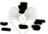
Bilateral heart primordia

Primitive heart tube

Heart loobing

Atrial and ventricular septation

Outflow tract and septation

2
Q

What happens in the 3rd week in cardiovascular development?

A

Lateral plate splanchnic mesoderm forms circulatory system (and other viscera)

Angiogenic cell islands collect in the lateral plate splanchnic mesoderm, move towards the midline and coalesce to form the two primitive heart tubes

3
Q

What is the first major system to function in the embryo?

A

Cardiovascular system

4
Q

When does the heart start to function?

A

Beginning of week 4

5
Q

Why does the embryo need a functioning cardiovascular system at an early stage?

A

Rapidly growing embryo –

Nutrition by diffusion is not

Enough to satisfy the growing embryo

6
Q

Label this diagram

A
7
Q

Where do blood vessels first appear?

A

Wall of yolk sac, allantois, connecting stalk and chorion

8
Q

Where and when do angioblastic cords form?

A

In the cardiogenic mesoderm in the 3rd week

9
Q

What do angioblastic cords canalize to form?

A

Heart tubes

10
Q

What happens does tubular heart join to?

A

Blood vessels in other areas to form primordial cardiovascular system

11
Q

Label this diagram

A
12
Q

What does cranial folding of the embryo do?

A

Reorientates the heart tube dorsal to pericardial cavity

13
Q

What is the pericardium dervied from?

A

Intra-embryonic coelom

14
Q

What is dervied from the somatic mesoderm in the cardiovascular system

A

The parietal layer of serous pericardium and fibrous pericardium

15
Q

What happens to the perocardial cavity?

A

Dorsal to ventral

16
Q

What happens to the cardiac tube?

A

Goes from ventral to dorsal

17
Q

What is derived from splachnic mesoderm?

A

Visceral layer of serous pericardium

18
Q

Label this diagram

A
19
Q

Label this

A
20
Q

Label this

A
21
Q

Label this

A
22
Q

How many horns are there?

A

2- left and right

23
Q

Where does each horn get venous blood from?

A

Yolk sac

Placenta

Body of the embryo

24
Q

Where does the yolk sac get blood from?

A

Vitelline vein

25
Q

Where does the placenta get blood from?

A

Umbilical vein

26
Q

Where does the body of the embryo get blood from?

A

The common cardinal vein

27
Q

Label this

A
28
Q

Where does the aortic arches arise from?

A

Aortic sac

29
Q

Where do the aortic arches terminate?

A

The dorsal aorta

30
Q

Label this

A
31
Q

What grows the fastest in the growing heart?

A

Bulbis cordis and ventricle grow faster than other regions forming a U-shaped bulboventricular loop

32
Q

When does the cardiac crescent occur?

A

Day 15

33
Q

When does the linear heart tube form?

A

Day 21

34
Q

When does the looped heart tube form?

A

Day 28

35
Q

When does the mature heart form?

A

Day 50

36
Q

What happens in dextrocardia?

A

Heart tube loops to the left side (instead of right), thus coming to lie facing the right (= dextro)

Most frequent positional abnormality of the heart

Dextrocardia can be associated with situs inversus (transposition of viscera)

37
Q

When does the partitioning of the primordial heart form?

A

Seen around 27th and 37th days of embryonic development.

Involves one or two actively growing masses of tissues.

38
Q

What happens during endocardial cushion formation?

A

Separates right atrium + ventricle from left atrium +ventricle to form L&R AV canals

39
Q

What happens during septum formation?

A

Separates right atrium from left atrium + right ventricle from left ventricle

40
Q

What are many cardiac malformations associated with?

A

Defective formation of endocardial cushion and septum formation, e.g. atrial septal defect (ASD) & ventricular septal defect (VSD)

41
Q

Label this

A
42
Q

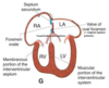
What are the stages in the partitioning of primitive atrium into left and right atrium

A
43
Q

How does the septum primum form?

A

End of 4th week

Sickle shaped crest grows from roof of atrium

44
Q

What is the ostium primum?

A

Opening between septum primum and enocardial cushion

45
Q

When is foramen secundum formed?

A

Formed at the upper end of septum primum

Forms on the right side of the septum primum

Septum secundum grows down and overlaps the foramen secundum.

46
Q

What is the septum secundum perforated by?

A

Oval foramen

47
Q

What is the role of foramen ovale before birth?

A

Allows most of the blood to pass from the right atrium to the left atrium (non functioning lung)

Prevents the passage of blood in the opposite direction

48
Q

What is the role of foramen after birth?

A

Normally closes (increased pressure in the left atrium due to increased pulmonary circulation)

Septum primum (= valve of oval foramen) fuses with the septum secundum

Oval fossa (fossa ovalis) of adult heart is a remnant of foetal oval foramen

49
Q

What happens if foramen ovale does not close?

A

Patent Foramen Ovale = Atrial Septal Defect (ASD) – “Hole in the heart”

50
Q

What is an atrial septal defect?

A

Common congenital heart anomaly

More common in females than in males

Common form – patent foramen ovale

51
Q

What are the 4 clinically significant types of ASD?

A

Foramen secundum defect

Endocardial cushion defect with foramen primum defect

Sinus venosus defect

Common atrium

The first two types are more common

52
Q

What are the steps in partitioning of the primitive ventricle?

A

Muscular ventricular septum forms. Opening is called interventricular foramen.

Bottom of spiral aorticopulmonary septum fuses with muscular ventricular septum to form membranous interventricular septum, closing interventricular foramen. (Aorticopulmonary septum divides bulbis cordis and truncus arteriosus into aorta and pulmonary trunk).

Growth of endocardial cushions also contributes to membranous portion of the interventricular septum.

53
Q

Label this

A
54
Q

Label this

A
55
Q

When does the aorticopulmonary septum develop?

A

5th week

56
Q

What does the aorticopulmonary septum divide the bulbus cordis and truncus arterios into?

A

Aorta and pulmonary trunk

57
Q

Label this

A
58
Q

What is a ventricular septal defect?

A

Most common type of CHD (congenital heart disease) – 25% of defects

Common in males

Can appear in any part of septum

Small VSDs close spontaneously (30 -50%)

Membranous type of VSD is most common

59
Q

How does the conducting system of the heart work?

A

Early pacemakers - primitive atrium and then sinus venosus

SA node (pacemaker) develops during 5th week

Adult location of SA node – High in the right atrium near the entrance of the SVC

AV node and bundle (bundle of His) develops from cells of AV canal and sinus venosus

60
Q

What is the clinical syndrome of the conducting system of the heart?

A

Cot death or sudden infant distress syndrome

61
Q

What are the causes of the conducting system abnormality

A

Abnormalities of conducting tissue

62
Q

What does the truncus arteriosus form?

A

Aorta

Pulmonary trunk

63
Q

What is the bulbis cordis?

A

Smooth part of the right ventricle (conus arteriosus)

Smooth part of left ventricle (aortic vestibule)

64
Q

What is the primitive ventricle?

A

Trabeculated part of right ventricle

Trabeculated part of left ventricle

65
Q

What does the primitive atrium form

A

Trabeculated part of right atrium

Trabeculated part of left atrium

66
Q

What does the sinus venosus form?

A

Smooth part of right atrium

Coronary sinus

Olique vein of left atrium

67
Q

What is aetiological factors in congenital heart disease?

A

Rubella infection in pregnancy (PDA)

Maternal alcohol abuse (septal defects)

Maternal drug treatment and radiation

Genetic - 8%

Chromosomal – 2% (Down’s and Turner’s syndrome)

68
Q

What percentage of congenital heart disease are accounted for by VSDs and ASDs

A

30% of congenital heart disease: VSD for 20% and ASD for 10%.

Girls have ASDs twice as often as boys

VSDs are also slightly more commonin female patients than in male patients

69
Q

What is transposition of great vessels?

A

Common cause of cyanotic disease in newborn infants

Associated with ASD and VSD

Permit exchange of systemic and pulmonary circulation

70
Q

What are the causes of transposition of great vessels?

A

Failure of aorticopulmonary septum to take a spiral course

Defective migration of neural crest cells

71
Q

What are the 4 cardiac defects of the teratology of fallot?

A

Pulmonary stenosis (obstruction of right ventricular outflow)

Ventricular septal defect (VSD)

Dextroposition of aorta (“overriding” aorta)

Right ventricular hypertrophy

72
Q

What are the causes of the teratology of fallot?

A

Unequal division of the conus due to anterior displacement of aorticopulmonary septum