Intro: Biology of the Skin Flashcards
What are the three layers of the skin?
Epidermis, Dermis, Subcutis

What cells are in the epidermis? (4)
Primarily keratinocytes with melanocytes, Langerhans cells, and Merkel cells
What are the components of the dermis? (5)
Fibroblasts, collagen, elastin, blood vessels, nerve endings
What are the components of the subcutis? (3)
Fat!, blood vessels, and fibrous septae
What is the primary function of the Epidermis?
Barrier, protection, wound healing
What is the primary function of the Dermis and Subcutis?
Structural support, vascular support, and innervation
True or False. The epidermis is a self-renewing tissue that “sheds” itself every 18 days.
False. The epidermis IS self-renewing, but it sheds itself every 28+ days
Where do the cells in the epidermis grow from? What happens as they move up?
Grow from stem cells in basal layer and terminally differentiate as they move upwards
Is apoptosis high or low in epidermis?
Usually low
What 4 layers (from top to bottom) is the epidermis divided into?
1) Stratum corneum 2) Stratum granulosum 3) Stratum spinosum 4) Stratum basale

What is the layer of skin that is the source of stem cells, and divison starts in. What is this layer held to the basement membrane with? (IMPORTANT!)
Stratum Basale; Hemidesmosomes!

What is the layer of skin in which cells stop dividing and commence terminal differentiation and lipids start to develop in? What holds these cells together that make it appear “spiny”? (ASLO IMPORTANT!)
Stratum Spinosum; desmosomes hold the keratinocytes together!

What is the function of Hemidesmosomes in the skin? What is the fucntion of desmosomes in the skin?
Hemidesmosomes hold the basal layer to the basement membrane while desmosomes hold the keratinocytes together (This should be like giving a dead horse guaifenisen by now…)

What is the layer that intracellular keratohyaline granules are synthesized (including profillagrin) and lipids are secreted into intercellular space? Why are lipids secreted?
Stratum granulosum; to act as a water barrier

What is the layer of skin in which organelles and nuclei degenerate? What is profilaggrin processed into and what does that product do? What combines with this product and what does that do? (this may be important)
Stratum Corneum; processed into filaggrin for keeping water in cells; keratins combine with filaggrin to form MACROFIBRILS that help create a protective layer.

What varies between the epidermis from area to area of the body? (ex. between palm and trunk?)
The thickness of the stratum corneum changes vastly.

What is this? What does it do? What is it derived from?

It is a melanocyte, it produces pigment and transfers it to surrounding keratinocytes and there is about one per 10 keratinocytes; It is derived from neural crest + migrates during embryonic development

What is one of the major immunologic cells in the epidermis? What does it do?
Langerhans (dendritic in dermis); it recognizes abnormal antigens and takes them up to present to lymphocytes in region LN’s; important in allergic rxns + tumor surveillance

What cell in the epidermis is important for light touch sensation and can develop into malignant tumors?
Merkel Cells

What is the Yellow Arrow? Blue arrow?

Yellow = Hair Follicle; Blue = Sebacious gland (Attached to a hair follicle)
What kind of cell is this? What kind of rash do they form when activated?

Mast Cell; Release granules (histamine) when triggered or when IgE binds during allergic rxn; cause characterisitic “Wheal and flare”
What is the pilosebaceous unit composed of? (4 things)
Hair follicle, sebaceous oil gland, apocrine sweat glands (in axilla and anogenital skin), arrector pili muscle (for goosebumps!)

What is in the black circle? Where are they found in the body? Where do they open on to? What do they do?

They are the “true” sweat glands (eccrine glands) that are present throughout the body, opening directly onto the skin without being associated with a hair follicle and function in regulating temperature by evaporative cooling of sweat
What happened to these cells?

A sunburn; failure to delete damage cells could lead to skin cancer
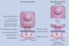
******What’s going on here?******

Separation of the stratum basale and basement membrane due to autoantibodies against hemidesmosomes = Bullous Pemphigoid; here it is shown on IF (linear pattern) and in clinic. Will form thicker blisters becausehe whole epidermis is the blister.

******What’s going on here?******

Sepration of stratum basale and stratum spinosum due to autoantibodies agaisnt desmosomes = Pemphigus Vulgaris; Shown here is it on IF and in Clinic
What is atopic Dermatitis associated with?
Genetic defects in filaggrin; Clinical examples shown here

What is the specific genetic mutation that causes Epidermolysis Bullosa Simplex?

Mutations in Keratin 5/14; the blister forms when the K5 and K14 keratin filamens no longer hold cell together as shown in picture
What is a nevus vs melanoma?
Nevus is a mole and is a benign collection of melanocytes whereas a melanoma is a growth of malignant melanocytes; melanocyte shown

What’s going on here?

Local thermoregulatory dysfunction a.k.a. Raynauds’ phenomenon and is chronic episodic attacks of digital ischemia; can be provoked by cold; painful and can ulcerate; can be assocaited with underlying AI dz
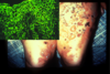
What’s going on here?

Small vessel vasculitis; dz’s that injure blood vessels + disrupt circulation = curatneous necrosis and/or ulceration; Here it is shown widespread due to Antiphospholipid antibody syndrome
