Diabetic Foot Charcot Neuropathy Flashcards
1
Q
What is Charcot foot?
A
- A CHRONIC AND PROGRESSIVE JOINT DISEASE after LOSS OF PROGRESSIVE SENSATION
2
Q
What does charcot foot lead too?
A
- Destruction of joints and surrounding bony structures
- May require Amputation if left untreated

3
Q
What is the epidemiology of Charcot foot?
A
- 0.1-1.4% of pt w diabetes
- 7.5% with diabetes and neuropathy
- Presents type 1 DM age 5th decade ( 20yrs from DX)
- Present type 2 - in 6th decade ( 5-10 yrs from DX)
location
-
Foot and ankle
- 9-35% bilateral disease
- Shoulder and elbow
- knee

4
Q
Which joints does Charcot effect?
A
- Foot and ankle 9-35% have bilat disease
- Shoulder and elbow
- Knee -> ligamentous laxity
5
Q
What are the risk factors for Charcot ?
A
- **Diabetic Neuropathy **
- xs ETOH
- Leprosy
- Myelomeningogcele
- Tabes Dorsalis/Syphilis
- Syringomyelia
6
Q
What is the pathophysiology of Charcot?
A
-
NEUROTRAUMATIC
- insensate joint subjected to REPETITIVE MICRO TRAUMA
- body unable to adopt mechanism to protect due to abnormal sensation
-
NEUROVASCULAR
- autonomic dysfunction increases blood flow thru Av shunting -> bone absorption and weakness
- ? INFLAMMATORY CYTOKINES IL10, TNF ALPHA
- lead to increased production of transcription factorKB, rank/rankl/opg triad pathway
7
Q
Do you know any classifications?
A
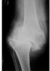
- EICHENHOLTZ
- Stage O= Joint oedema, radiographs are negative, bone scan positive in all stages
-
Stage 1 FRAGMENTATION= Joint oedema
- radiographs= osseous fragementation with joint dislocation - see pic
-
Stage 2= COALESCENCE
- decrease bone oedema
- xray- coalescence of fragments and absorption of bone debris
-
Stage 3= RECONSTRUCTION
- no local oedema
- xray- consolidation & remodelling of fracture fragments

8
Q
What are the PC of someone with a charcot joint?
A
- Swollen foot and ankle
- PAIN 50%,
- PAINLESS 50%
- Loss of function
9
Q
What do you find on examination?
A
- Acute
- Swollen
- Warm
- Erytherma- decrease with elevation cf infection
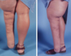
- Chronic
- Structurally deformed foot
- rocker bottom deformity- see pic
- pes planus
- bony prominence
- Lack of sensation- semmes- weinstein testing

10
Q
What do you see on X-rays ?
A
- Acute- degenerative changes may mimic OA
-
chronic-
- obliterated joint space
- Fragmentation of bony surface-> subluxation/dislocation
- HERETROPHIC OSSIFICATION
- scattered bone in soft tissue swelling
11
Q
Are bone scans helpful in charot ?
A
- Yes to identify presence of superimposed osteomyelitis
- Technetium- maybe positve for neuropathic joint and osteomyelitis
- Indium wc- cold neuropathic but hot for osteomyelitis
12
Q
Are MRI scans helpful?
A
- Yes identify abscess from soft tissue swelling
13
Q
Are biopsy useful in charot joints?
What investigations are also helpful?
A
- Yes
- Can guide antibiotic tx in cases of osteomyelitis or soft tissue abscess
- FBC/ESR- both elevated in infection/Charcot arthropathy
14
Q
What are the tx options for charcot arthropathy?
A
- TOTAL CONTACT CASTING- Cast changed 2-4 wks for 2-4 months
- Orthotics- charcot restraint walker boot used after contact casting
- SHOE WEAR MODIFICATIONS- double rocker reduce risk of ulceration
- Medication- bisphosphonates, topical anaesthetics, antidepressants
- Outcomes 75% success rate
Operative
-
Resection of bony prominence (exostectomy) & TAL
- Braceable foot w equinus deformity + focal bony prominenece= skin breakdown
- Joint stability good
- Aim achieve a plantigrade foot that allows ambulation
-
Arthrodesis and osteotomies
- severe defoemities, unstable joints that are non braceble
- v high complx rate 70%
-
Amputation
- failed surgery, infection
- goal is partial/limited amputation if vascularity allows

15
Q
Describe the surgical technique for arthrodesis in charcot foot?
A
-
Fixation technique
- Screws/plates & tibiocalcaneal nail
- ex fix - bone quality poor
- post op minimal weight bear 3 months
-
High complication rate up tp 70%
- Infection
- hardwear malposition
- recurrent ulceration
- fracture
20
Q
What is the pathophysiology of diabetic foot ulcers?

A
-
Neuropathy
- largest effect
- sensory dysfunction-> lack of protective sensation-> ulcer
- autonomic dysfunction-> drying of skin due to lack of normal glandular function
- increased mechanical & axial stress on skin more prone to injury due to drying
-
Angiopathy
- Iesser effect than neuropathy
- >60% of diabetic ulcers have decreased blood flow due to peripheral vascular disease
27
Q
What does a syme amputation include?
A
- Ankle disarticulation
- removal of malleoli
- anchoring heel pad to weight bearing surface- must have a viable heel pad- branches of posteriot tibial artery= NB important palpable POST TIBIAL PULSE