BB Anatomy 1 COPY Flashcards

1
Q

the central sulcus disects which lobes of the brain?

frontal lobe from the temporal lobe
frontal lobe from the parietal lobe
frontal lobe from the occipital lobe
frontal lobe from cerebellum
parietal lobe from temporal lobe

A

the central sulcus disects which lobes of the brain?

frontal lobe from the temporal lobe
frontal lobe from the parietal lobe
frontal lobe from the occipital lobe
frontal lobe from cerebellum
parietal lobe from temporal lobe

2
Q

label 1-3

A

1: midbrain
2: the pons
3: medulla oblongata

3
Q

what structure is this?

pons
medulla oblongata
midbrain
hypothalamus
fasciculus gracilis

A

what structure is this?

pons
medulla oblongata
midbrain
hypothalamus
fasciculus gracilis

4
Q

what structure is this?

pons
medulla oblongata
brainstem
hypothalamus
fasciculus gracilis

A

what structure is this?

pons
medulla oblongata
midbrain
hypothalamus
fasciculus gracilis

5
Q

Which of the following is A?

Pons
Medulla
Cerebral aquaduct
Fourth ventricle
Midbrain

A

Which of the following is A?

Pons
Medulla
Cerebral aquaduct
Fourth ventricle
Midbrain

6
Q

Which of the following is B?

Pons
Medulla
Cerebral aquaduct
Fourth ventricle
Midbrain

A

Which of the following is B?

Pons
Medulla
Cerebral aquaduct
Fourth ventricle
Midbrain

7
Q

Which of the following is C?

Pons
Medulla
Cerebral aquaduct
Fourth ventricle
Midbrain

A

Which of the following is C?

Pons
Medulla
Cerebral aquaduct
Fourth ventricle
Midbrain

8
Q

Which of the following is D?

Pons
Medulla
Cerebral aquaduct
Fourth ventricle
Midbrain

A

Which of the following is D?

Pons
Medulla
Cerebral aquaduct
Fourth ventricle
Midbrain

9
Q

Which of the following is E?

Pons
Medulla
Cerebral aquaduct
Fourth ventricle
Midbrain

A

Which of the following is E?

Pons
Medulla
Cerebral aquaduct
Fourth ventricle
Midbrain

10
Q

What are the two layers of the dura mater? [2]

A
  • *periosteal layer** (which lines the inner surface of the bones) [1]
  • *meningeal layer** which forms dural folds. [1]
11
Q

Lumbar puncture needle is inserted into:

  • space between arachnoid mater and pia mater
  • space between dura mater and arachnoid
  • space between arachnoid and pia mater
  • space between vertebrae and dura mater
  • into the spinal cord
A

Lumbar puncture with needle is inserted into:

  • *- space between arachnoid mater and pia mater**
  • space between dura mater and arachnoid
  • space between arachnoid and pia mater
  • space between vertebrae and dura mater
  • into the spinal cord
12
Q

Epidural needle is inserted into:

  • space between arachnoid mater and pia mater
  • space between dura mater and arachnoid
  • space between arachnoid and pia mater
  • space between vertebrae and dura mater
  • into the spinal cord
A

Epidural needle is inserted into:

- space between arachnoid mater and pia mater
- space between dura mater and arachnoid
- space between arachnoid and pia mater
- space between vertebrae and dura mater
- into the spinal cord

13
Q

which side of spinal cord carries motor fibres? [1]

which side of spinal cord carries efferent fibres? [1]

A

which side of spinal cord carries motor fibres? [1]
ventral root

which side of spinal cord carries efferent fibres? [1]
dorsal root

14
Q

which out of A & B is the dorsal root and ventral root?

A

A: dorsal root - look out for DRG. DORSAL IS BIGGER
B: ventral

15
Q

describe the overview of 1st order, 2nd order & 3rd order neuron pathways [3]

A

first order neurons: peripheral receptors –> spinal cord, where is will synapse with second order neurons in the spinal cord or brainstem.

second order neurons:spinal cord –> brain, usually the thalamus.

third order neurons: thalamus –> primary sensory cortex.

16
Q

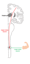
describe basic pathway of descending tracts: first order and second order neurons

A

first order neuron: (aka upper motor neuron): brain to the spinal cord or brainstem.It will synapse with the second order neuron,

second order neuron (aka lower motor neuron): spinal cord –> skelatal muscle

17
Q
A
18
Q

Which functional division of the nervous system would be responsible for the physiological changes seen during exercise (e.g., increased heart rate and sweating)?

Somatic
Central
Autonomic
Enteric

A

Which functional division of the nervous system would be responsible for the physiological changes seen during exercise (e.g., increased heart rate and sweating)?

Somatic
Central
Autonomic
​Enteric

19
Q

how many pairs of spinal nerves are there?

30
31
32
33
34

A

how many pairs of spinal nerves are there?

30
31
32
33
34

20
Q
A
21
Q

what is the verterbral breakdown of spinal nerves?

cervical =
thoracic =
lumbar =
sacral =
coccygeal =

A

what is the verterbral breakdown of spinal nerves?

cervical = **8**
thoracic = **12**
lumbar = **5**
sacral = **5** 
coccygeal = **1**
22
Q

the spinal cord terminates at the:

cauda equina
filum terminale
conus medullaris
foramen magnum
1st coccygeal vert

A

the spinal cord terminates at the:

cauda equina
filum terminale
conus medullaris
foramen magnum
1st coccygeal vert

23
Q

A patient is experiencing numbness across the lateral aspect of their shoulder, what spinal level do you think the injury is at?

T1
T2
C6
C5
C7

A

A patient is experiencing numbness across the lateral aspect of their shoulder, what spinal level do you think the injury is at?

T1
T2
C6
C5
C7

24
Q

which part of the spinal cord do the vertebral artierise travel in? [1]

A

which part of the spinal cord do the vertebral artierise travel in? [1]
transverse foramen

25
Q

which vertebrae has the dens?

C1
C2
C3
C4
C5

what is the role of the dens? [1]

A

which vertebrae has the dens?

C1
C2
C3
C4
​C5

what is the role of the dens? [1]
forms pivot joint with C1

26
Q

what are the type of joints found between adjacent verebral bodies?

synchondrosis
symphysis
synostosis
syndesmoses

A

what are the type of joints found between adjacent verebral bodies?

synchondrosis
symphysis
synostosis
syndesmoses

Symphysial joints are where the bones are united by a layer of fibrocartilage. They are slightly movable (amphiarthrosis).

27
Q

what type of joint is the articulation of the inferior articular facet of one vertebra with the superior acrticular facet of the vertebra immediately below it?

  • pivot joint
  • facet joint
  • condyloid
  • plane joint
A

what type of joint is the articulation of the inferior articular facet of one vertebra with the superior acrticular facet of the vertebra immediately below it?

  • pivot joint
  • *- facet joint**
  • condyloid
  • plane joint
28
Q

which of the following is the ligamaentum flavum?

A
B
C
D
E
F

A

which of the following is the ligamaentum flavum?

A
B
C
D
E
F

29
Q

which of the following is the interspinous ligament?

A
B
C
D
E
F

A

which of the following is the interspinous ligament?

A
B
C
D
E
​F

30
Q

which of the following is the posterior longitudinal ligament?

A
B
C
D
E
​F

A

which of the following is the posterior longitudinal ligament?

A
B
C
D
E
​F

31
Q

which of the following is the anterior longitudinal ligament?

A
B
C
D
E
​F

A

which of the following is the anterior longitudinal ligament?

A
B
C
D
E
​F

32
Q

where is the fracture?

base of skull
C1
C2
C3
C4

A

where is the fracture?

base of skull
C1
C2

C3
​C4

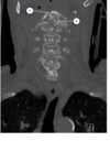
There is a fracture through the right anterior arch of C1 (B) seen on the coronal view. We can also see a fracture through the dens (D), which is the most common type of fracture of the cervical spine. It is angled posteriorly, which is common after hyperextension injury.

33
Q

Which structure is at risk of compression by a very posteriorly displaced dens? [1]

A

spinal cord

34
Q
A
35
Q

Which structure may be affected by cervical vertebral fractures? [1]

A

Which structure may be affected by cervical vertebral fractures? [1]
vertebral arteries

36
Q

what is A? [1]

A
37
Q

which verebtral level do u go below to do lumbar puncture?

A

L2

38
Q

Why might lumbar puncture be more challenging in a young child? [1]

A

Why might lumbar puncture be more challenging in a young child? [1]

Acceptable responses: spinal cord lower, the spinal cord ends lower down, the spinal cord travels further down the vertebral column

39
Q

Which meningeal space is targeted in lumbar puncture? [1]

A

Acceptable responses: subarachnoid space, subarachnoid, csf space, csf

40
Q

Louise’s friend, Rodrigo, is a third year medical student. He is worried about Louise having the lumbar puncture as he has learnt that the pressure change can start to pull the brain out of the skull base. What is Rodrigo referring to here? [1]

A

Louise’s friend, Rodrigo, is a third year medical student. He is worried about Louise having the lumbar puncture as he has learnt that the pressure change can start to pull the brain out of the skull base. What is Rodrigo referring to here? [1]

Rodrigo is worried about cerebellar tonsillar herniation, or ‘coning’. This is a risk of lumbar puncture in the context of obstructive hydrocephalus.

Normally, CSF will circulate freely around both brain and spinal cord as it is produced and reabsorbed.

Where there is obstructive hydrocephalus, however, there is a blockage in the cerebral ventricular system, effectively creating two closed CSF compartments. In this situation, when CSF is drawn off during lumbar puncture, a pressure differential is created, with spinal pressure much lower than cranial. This pulls the cerebellar tonsils out through the foramen magnum, compressing the brainstem - invariably an immediately fatal event.

If obstructive hydrocephalus is a possibility, imaging should always be undertaken to rule it out prior to lumbar puncture.

41
Q
A