10 - Prostate and Genital Tract Flashcards
What are the different ways you should inspect and examine any lump?
Inspect 6 S’s: Site, Size, Shape, Symmetry, Skin changes, Scars
Palpate CAMPFIRE: Tenderness, Temperature, Transillumination, Consistency, Attachments, Mobility, Pulsation, Fluctuation, Irreducibility, Regional lymph nodes, Edge
What investigations should be carried out with a testicular lump?
1st Line: US of scrotum
Blood tests: LDH, b-HCG, AFP

What are some differential diagnoses for scrotal lumps?
ALWAYS CONSIDER MALIGNANCY

Extra-testicular:
- Hydrocele (transilluminates)
- Varicocele
- Epididymal cyst (transilluminates)
- Epididymitis
- Inguinal Hernia
Testicular:
- Tumours
- Torsion
- Benign tumours (Leydig, Sertoli, Lipomas, Fibromas)
- Orchitis
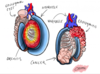
How do hydroceles present and how are they investigated and managed?
Abnormal collection of peritoneal fluid between parietal and visceral layers of tunica vaginalis. Not separate from testes
Symptoms: painless fluctuant swelling that will transilluminate, either unilateral or bilateral. If big can be painful
Causes: primary or secondary (infection, malignancy, trauma), patent processus vaginalis in infants
Ix: Urgent US if aged 20-40 and cannot palpate testes
Mx: if congential often regress spontaneously, ligation if patent processus vaginalis, if large can do surgical management

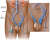
What is a varicocele and what are the complications of this pathology?
Abnormal dilatation of the pampiniform venous plexus within the spermatic cord. Like a bag of worms and disappears on lying down. Examine patient lying and standing whilst doing valsalva
Often on left due to testicular vein going to left renal vein before IVC
Complications: testicular atrophy and infertility due to increased intra-scrotal temperature so sent for semen analysis

How are varicoceles investigated and managed?
- If asymptomatic and no red flags no investigation and treatment
- If red flags (right sided, doesn’t disappear on lying, acute onset) needs urgent US investigation and then embolisation to ligate spermatic veins either laparoscopically or open
What are epididymal cysts (spermatoceles) and how are they treated?
Benign fluid-filled sacs arising from the epididymis. Smooth fluctuant nodule, above and separate from the testis that will transilluminates, often they are multiple
Very common and not associated with malignancy
No treatment unless large and painful can remove with surgery but avoid in young men as can cause infertility

What is epididymitis, how does it present and how is it managed?
- Unilateral acute onset scrotal pain with associated swelling, overlying erythematous skin, systemic symptoms like fever
- On examination testis is tender and pain relieved on elevation (Prehn’s sign)
- Often due to STI bacteria in young men and enterococcus bacteria in older men
- Antibiotics and analgesia

How can you tell if a lump in the scrotum is an inguinal hernia?
- Cannot get ‘above’ it - cannot get to the superior surface
- Cough will exacerbate and lying down will make it disappear
How do malignant testicular tumours present and how are they managed vaguely?
Painless lumps that are firm, irregular and do not transilluminate. 5% of men have pain so delays diagnosis
Urgent US for diagnosis and then tumour markers
Mx: radical inguinal orchidectomy then chemotherapy
What is orchitis and how is it treated?
- Inflammation of the testis that is often due to mumps virus (history of parotid swelling)
- Rest and analgesia
- If intra-testicular abscess forms then drainage and sometimes orchidectomy

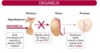
What is the pathophysiology of BPH?
- Most common cause of bladder outlet obstruction and LUTs in men aged over 60
- Prostate converts testosterone to dihydrotestosterone (DHT) using the enzyme 5α-reductase which is more potent. It is the only tissue that always responds to testosterone so DHT remains high and enlarges the prostate
- Occurs in the transitional zone
What are some risk factors for developing BPH?
- Age
- Family history
- Afro-Carribbean ethnicity
- Obesity
What are the presenting features of BPH?
- Voiding and Storage LUTS (e.g terminal dribbling, hesitancy, weak stream)
- Haematuria and haematospermia
- DRE: firm, smooth, symmetrical enlarged prostate. (more than two finger width)

What is the IPSS?
Can be used when men have LUTS to quantify how bad it is
0-7 is mild
8-19 is moderate
20+ is severe

What are some differential diagnoses with BPH?
- Prostate cancer
- UTI
- Bladder cancer

What investigations should you do if you suspect a patient has BPH?

- Urinary frequency and volume chart for all with LUTS
- Urinalysis to rule out infection
- Post-void bladder scan for chronic retention
- PSA
- US to assess prostate and look for any hydronephrosis/retention. Enlarged if prostate>30ml
- Urodynamic studies with BOOI to look for obstruction

How is BPH managed conservatively and medically?
Conservatively
- If asymptomatic found incidentally reassure
- Symptom diary
- Review drugs to see if iatrogenic LUTs, lifestyle advice (e.g cut down caffeine, urethral milking)
Medically
- 1st line: alpha-blockers like Tamsulosin to relax prostate smooth muscle. 4 point improvement in IPSS in few days if works
- 2nd line: if above doesn’t work give 5a-reductase inhibitors like finasteride to decrease prostatic volume by decreasing amount of DHT but can take up to 6 months to take effect
What are some side effects of alpha-blockers used to treat BPH?
- Postural hypotension
- Retrograde ejaculation
- Floppy Iris Syndrome (during cataracts surgery)

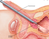
BPH is managed surgically if a patient doesn’t respond to medical management or if they are having complications like high pressure retention. How is BPH managed surgically?

1st Line: TURP
Endoscopic removal of obstructive prostate tissue using a diathermy loop to increase the urethral lumen size
Holmium Laser Enucleation of the Prostate (HoLEP)
Holmium:YAG laser used to heat and dissect sections of prostate into the bladder
Others: PVP (Photoselective Vaporization of the Prostate), TUVP (Transurethral Vapourization of the Prostate), and TUMT (Transurethral Microwave Thermotherapy), Prostaectomy
What are some complications of BPH itself and some complications of the surgical treatment for BPH (TURP)?
BPH: high pressure retention with chronic retention so post-renal kidney injury, UTI, haematuria
TURP: haemorrhage, sexual dysfunction, retrograde ejaculation, urethral stricture, TURP syndrome
What is TURP syndrome?
TURP uses hypoosmolar irrigation during the procedure which can result in significant fluid overload and hyponatremia as the fluid enters the circulation through the exposed venous beds
Symptoms: confusion, nausea, agitation, or visual changes due to hyponatraemia and fluid overload

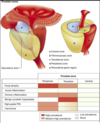
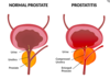
What is the pathophysiology and histology of prostate cancer?
- Most common cancer in men
- Influenced by androgens (testosterone and DHT)
- Mostly acinar adenocarcinomas found in the peripheral zone that are multifocal
Acinar adenocarcinoma: from the glandular cells. Most common form of prostate cancer
Ductal adenocarcinoma: from the cells that line the ducts of the prostate gland. They grow and metastasise faster than acinar

What are the main risk factors for developing prostate cancer?
- Age, Ethnicity, FHx
- BRCA1/2
- Obesity, diabetes mellitus, smoking, little exercise

What are some of the symptoms of prostate cancer and what are some differential diagnoses for this?
- LUTS: weak urinary stream, increased urinary frequency, and urgency
- Advanced localised disease: haematuria, dysuria, incontinence, haematospermia, suprapubic pain, loin pain, rectal tenesmus
- Metastatic: bone pain, lethargy, anorexia, and unexplained weight loss.
- DRE: asymmetry, nodularity, or a fixed irregular mass
DD: BPH, prostatitis, bladder cancer

What are some investigations (not including imaging) if you suspect prostate cancer?
- PSA: however may be artifically raised
- Free:total PSA ratio
- PSA density: which is the serum PSA divided by prostate volume determined by imaging (i.e. TRUS, CT, or MRI)
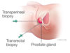
- Transperineal (Template) biopsy: general anaesthetic and less infection
- TRUS biopsy: local anaesthetic, 12 cores, risk of sepsis
When should men have a repeat prostate biopsy?
- Repeat if negative biopsy but rising PSA or suspicious DRE
- Repeat if atypical small acinar proliferation (ASAP) or >3 sites of high-grade prostatic intraepithelial neoplasia (HGPIN),
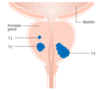
How is prostate cancer graded once biopsied?
Low grade: 3+3 = 6
Intermediate: 3+4 = 7
High grade: 7,8,9, 10

What imaging is used to diagnose and stage prostate cancer?
- Multi-parametric MRI can identify abnormal prostate so you can biopsy
- Abdomino-pelvic CT imaging and bone scan
How is prostate cancer managed?
Risk stratification based on PSA levels, Gleason score, and T staging (from TNM).
Low risk: active surveillance, if signs of progression then radical prostatectomy
Intermediate and high risk disease: radical prostatectomy, some intermediate can have surveillance
Metastatic disease: chemotherapy agents and anti-hormonal agents
Castrate–resistant disease: further chemotherapy agents such as Docetaxel. Corticosteroids are used as third line if androgen deprivation therapy and anti-androgen therapy doesn’t work

What are the mainstay treatments of localised or locally advanced prostate cancer?
- Radical prostatectomy: removal of the prostate gland, resection of the seminal vesicles, along with the surrounding tissue +/- dissection of the pelvic lymph nodes
- External-beam radiotherapy: focused radiotherapy just to target prostate gland
- Brachytherapy: transperineal implantation of radioactive seeds

What are some side effects of a radical prostatectomy?
- Erectile dysfunction (affecting 60-90% of men)
- Stress incontinence
- Bladder neck stenosis
What are some examples of chemotherapy and anti-androgen therapy used in the treatment of metastatic prostate cancer?

Chemotherapy
- Used in metastatic diease
- Docetaxel (recommended in men with testosterone-resistant cancer) and Cabazitaxel (used with prednisolone, recommended for treating relapsed prostate cancer)
Anti-androgen Therapy
- LHRH agonists (e.g. goserelin or triptorelin) stops LH production
- GnRH receptor antagonists (e.g. degarelix)
- Orchidectomy for androgen deprivation
- Enzalutamide and Abiraterone lower testosterone in metastatic disease
What are the different types of prostatitis?
- Acute bacterial prostatitis
- Chronic bacterial prostatitis
- Nonbacterial prostatitis
- Prostatodynia
What are some risk factors for acute and chronic prostatitis?

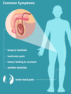
What are some symptoms of prostatitis?
Acute: LUTS, pyrexia, perineal or suprapubic pain, or urethral discharge, tender boggy prostate on DRE, inguinal lymphadenopathy
Chronic: pelvic pain for at least 3/12 (Prostatodynia) alongside LUTS with pain usually in perineum but can be in suprapubic, lower back and rectum

How is prostatitis managed?
Acute
- Prolonged abx (usually quinolone like ciprofloxacin as good prostate penetration)
- Analgesia (paracetamol and NSAIDs)
- Alpha blockers of 5a-reductase inhibitors for second line
Chronic
- 6 week abx if present for less than 6/12
- Symptom control with analgesia, stool softeners, alpha blockers
- If pain persists chronic pain specialist

What is the pathophysiology of epididymo-orchitis? (inflammation of epididymis and testes)
- Bimodal common age distribution aged 15-30 then >60 years
- Extension of infection from lower urinary tract (bladder and urethra), either via enteric (i.e. classic UTI) or non-enteric (i.e. sexually transmitted) organisms.
- <35 usually due to sexual transmission of N. gonorrhoeae and C. trachomatis (if anal intercourse then E.Coli)
- >35 usually due to enteric bacteria like E. coli, Proteus spp, Klebsiella pneumoniae, Pseudomonas aeruginosa due to bladder outflow obstruction due to enlarged prostate so allows organisms to travel retrograde

What are some risk factors for developing epididymo-orchitis?
Non-enteric: males who have sex with males, multiple sexual partners, sex with known contact of gonorrhea
Enteric: recent instrumentation or catheterisation, bladder outlet obstructuon (e.g prostate enlargement or urethral stricture) or immunocompromised state

How does epididymitis present and what are some important differentials to consider?
Symptoms

- Unilateral scrotal pain and swelling with possible fever and rigors
- May have LUTS due to underlying cause e.g dysuria, urethral discharge
- Find out sexual history
Examination
- Red and swollen
- Tender on palpation with possible associated hydrocele
- Cremasteric reflex in tact
- Prehn’s sign
Differentials
- Testicular torsion
- Testicular abscess or tumour
- Epididymal cyst
- Hydrocele
How is a case of suspected epididymitis investigated?
- Urine dipstick and possible MSU (if non-enteric suspected take first void urine then NAAT for G,C and M.Genitalium)
- Further STI screening
- Routine bloods (CRP and FBC) and blood cultures if signs of systemic infection
- US Doppler to confirm and rule out complications. If suspect torsion from this urgent scrotal exploration

How is epididymitis managed?

- Abx (see image)
- Analgesia
- Bed rest and scrotal support
- Abstain from sex until abx done
- If chronic or persisting with pain then may warrant orchidectomy

What is the pathophysiology of testicular torsion and what are some risk factors for this?
- Mobile testis rotates on spermatic cord, reduced arterial flow, impaired venous return so oedema and infarction to testes
- Men with Bell-Clapper deformity more likely to twist as testis lacks normal attachment to tunica vaginalis so more mobile
- Risk Factors: Age (12-25), previous testicular torsion, FHx, undescended testes

How does testicular torsion present?
Symptoms

- Sudden onset severe unilateral testicular pain
- Nausea and vomiting due to pain
- Referred abdominal pain
Examination
- Testis will have a high position
- Swollen and tender to touch
- Cremasteric reflex absent
- Prehn’s sign negative
What are some differentials to consider with acute scrotal pain apart from torsion?
- Epididymo-orchitis (gradual onset of pain, LUTs, Pyrexia)
- Trauma
- Incarcerated inguinal hernia
- Renal colic
- Hydrocele
- Torsion of hyatid of Morgagni

How is suspected testicular torsion investigated and managed?
Ix

- If suspected take straight for scrotal exploration with strong analgesia, anti-emetics and NBM
- If not enough evidence Doppler US to see if compromised blood flow
- Urine dipstick
Mx
- Surgical emergency with 4-6 hour window
- Bilateral orchidopexy once confirmed intra-operatively to prevent further torsion
- If tissue non-viable orchidectomy and possible prosthesis
What are the different classifications of testicular cancer?
- Most common in men aged 20-40 years or that are Caucasian
- Germ Cell (malignant) or Non-Germ Cell (Benign)
- Seminoma (better prognosis) or Non-Seminoma

What are some risk factors for developing testicular cancer?
- Cryptorchidism
- Previous testicular malignancy
- FHx
- Kleinfelter’s syndrome

What are some presenting signs of testicular cancer and what are some non-malignant differentials for a scrotal lump?
- Unilateral painless testicular lump
- Irregular, firm, fixed lump that does not transilluminate
- No lymphadenopathy as metastasises to paraaortic nodes
- Evidence of metastases: weight loss, back pain (retroperitoneal metastases), or dyspnoea (lung metastases)
Differentials: epididymal cyst, haematoma, epididymitis, or hydrocoele
How is testicular cancer diagnosed?
Tumour markers:
- b-hCG in NSGCTs and seminomas or AFP in non-seminomas
- LDH for tumour volume usually seminomas
Scrotal US for inital lump investigation then CT with contrast of chest/abdo/pelvis
Diagnosis purely on markers and imaging. Biopsy not done due to seeding!!!

How is testicular cancer managed?
Dependent on tumour subtype, disease stage, and risk scoring. Most will have inguinal radical orchidectomy (remove testes, spermatic cord and lymph)
NSGCTs
- Orchidectomy then if low risk (no vascular invasion) just surveillance. If high risk, give adjuvant chemotherapy before surveillance
- If metastatic give cycles of chemotherapy if intermediate prognosis but if poor prognosis just one cycle of chemo
Seminomas
- Orchidectomy and surveillance monitoring. If high chance of relapse consider chemo
- If metastatic chemo or radio

What should be offered to patients before an orchidectomy for testicular cancer?
- Pretreatment fertility assessment with semen analysis
- Cryopreservation
This is because cancer often causes sperm abnormalities and Leydig cell dysfunction and then chemo/radio can also reduce fertility

What causes urethritis?
Gonococcal urethritis: N.Gonorrhoeae
Non-Gonococcal urethritis: C.Trachomatis, M.Genitalium, T.Vaginalis
Risk factors: men aged <25, men who have sex with men, new sexual partner, more than one sexual partner in a year, previous STI

How does urethritis present?
Dysuria, penile irritation, discharge from urethral meatus
Can present with complications like reactive arthritis or epididymitis

How is suspected urethritis diagnosed?
Ix:
- Gold standard: First void urine sent for NAAT for N.Gonorrhoeae, C.Trachomatis, M.Genitalium
- Urethral gram stain from swab. Triple site testing if suspect gonorrhoeae
- MSU dipstick
- Further STI screening for HIV/syphillis

How is urethritis managed?
- Abx therapy
- Abstain from sexual activity for 7 days after antibiotics, symptoms have resolved and sexual partners are treated
- Test of cure if gonorrhoea
- Counsel on condom use and contact tracing

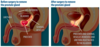
What is the pathophysiology of Fournier’s gangrene?
Necrotising fascitis of the perineum that is a urological emergency
Can be mono or polymicrobial (Group A Strep, C.Perfringes, E.Coli)
Anatomic barriers stop testes and epididymis being involved (Dartos fascia of the penis/scrotum, Colles fascia of perineum, Scarpa fascia of anterior abdominal wall)

How does Fourniers gangrene present and what are some risk factors for developing this?
- Initially pain out of proportion to findings or pyrexia and small amount of cellulitis
- As progresses crepitus, skin necrosis, haemorraghic bullae and sensory loss of overlying skin
- Patient rapidly deteriorates and can go into septic shock
- Risk factors: Type 2 DM, excess alcohol, poor nutritional state, steroid use, haemotological malignancies, recent trauma to area

How is Fournier’s gangrene diagnosed and managed?

Dx
- Clinical diagnosis
- Can take blood routine bloods, culturesand work outLRINECto see if6 or more
Mx
- Urgent surgical debridement
- Partial or total orchiectomy
- Broad spectrum abx
- Transfer to high dependency unit
- Further surgical debridement and skin grafts to recover scrotum

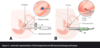
What is paraphimosis and what are some risk factors for developing this?
- Inability to pull retracted foreskin over glans penis which is an emergency
- Glans becomes oedamtous which can reduce venous return and lead to vascular engorgement so penile ischaemia, worsening infection, Fournier’s gangrene
- Risk factors: phimosis, indwelling urethral catheter, poor hygeine, previous paraphimosis

How does paraphimosis present and how is it managed?
Presentation
- Progressive pain and swelling with inability to retract foreskin
- May occur often if cause not managed
Management
- Reduce ASAP with penile block with local anaesthesia for analgesia
- Definitive management such as circumsion can occur as outpatient

What are the different classifications of priapism?

Painful erection not associated with sexual desire that lasts for more than 4 hours
High Flow (Non-ischaemic)
- Arterial blood rapidly enters the corpus cavernosum quicker than it can be drained. Associated with trauma and can be triggered by sexual stimulation
- PAINLESS AND NOT FULLY RIGID
Low Flow (Ischaemic)
- Blockage of the venous drainage of the corpus cavernosum so blood collects here, causes ischaemia and can lead to fibrosis and impotence
- Urological emergency
- Glans and Spongiosum not affected
- PAINFUL AND RIGID

What are some causes of priapism?
- Idiopathic in half of patients
- Non ischaemic: penile/perineal trauma, spinal cord injury
- Ischaemic: see image

What are some investigations carried out when a patient presents with priapism?
- Once initial management done obtain corporeal blood gas to determine if ischaemic or non-ischaemic
- Routine bloods to find underlying cause (FBC, CRP, ESR, coagulation screen, bone profile, haemoglobin electrophoresis)
- If non-ischaemic do work up for potential spinal cord injury

How is priapism managed and what is the prognosis?
Corporeal Aspiration!
If no response give intracavernosal injection of sympathomimetic agent like phenylephrine
If above not working promt surgical shunt between corpus cavernosa and glans. Can cause ED
- If priapism lasts >24 hours most people do not regain ability to have sexual intercourse so consider penile prosthesis

Penile cancer is very rare and has a 5 year survival of 70%. What is the pathophysiology of penile cancer and some risk factors for this?
- Most common in men>60
- Strong association with HPV 16, 6 and 18
- SCC from the epithelum of the inner prepuce or glans in 95% of cases
Risk factors: HPB, phimosis, smoking, lichen sclerosis, untreated HIV, previous PUVA for psoriasis
Protective factors: Circumsion

What are some of the clinical features of penile cancer?
- Palpable or ulcerating lesion on penis most commonly on glans
- Lesion is painless but may discharge or bleed
- Inguinal lymphadenopathy

How is penile cancer managed?
Combination of surgery, radio, chemo
- If superficial non-invasive can give topical chemo (e.g imiquimod or 5-FU) then follow up with repeat biopsy. Or can use laser treatment or glans resurfacing
- If invasive but confined to the glans can do organ sparing treatment with 5mm tumour margins. Local excision, partial glansectomy or total glansectomy with reconstruction. Radical circumsion if confined to foreskin
- If invasive partial amputation or total penectomy with perineal urethrostomy. Afterwards phallic reconstruction with forearm phalloplasty May need neoadjuvant chemo or radio
- If in inguinal nodes may need radical inguinal lymphadenectomy, neoadjuvant chemo or radio
What is the pathophysiology of a penile fracture?

Urological emergency common in 30-40 year olds and more commone to occur damage on the right side
Rupture of corpus cavernosa and the tubica albuginea in an erect penis
Due to blunt trauma up to pressure of 1500mmHg where penis is violently deviated from axis e.g forceful masturbation, falling from bed with erect penis, partner on top

What are the clinical features of a penile fracture?
- Popping sensation with immediate pain, swelling, detumescence after forceful thrusting into pubic bone of partner
- Aubergine Sign: penile swelling and discolouration (due to haematoma) with deviation to opposite side of lesion
- Rolling Sign: firm immobile haematoma palpated in the shaft
May be butterfly shaped haematoma in the perineum may suggest a urethral injury

How is a penile fracture diagnosed and managed?
Ix/Dx
- Diagnosed clinically
- Cavernosography can be used to identify rupture site but associated with complications like priapism and fibrosis of corpus cavernosum
- Retrograde urethography if patient has symptoms suggesting urethral injury e.g voiding difficulties or blood at meatus
Mx:
- Analgesia and anti-emetics before urgent surgical exploration and repair
- Surgical exploration involves circumferential incision and penile skin degloved proximally up to base, haematoma evacuated then tear identified and repaired with absorbable sutures
- Abstinence from all sexual activities for 6-8 weeks post op

What are some complicatgions following a penile fracture repaire?
- Penile curvature during erection
- Penile paraesthesia
- Dyspareunia/Painful erectiom