4 - Uroradiology and Histology Flashcards
What are the different ways of imaging the kidneys?
- CT
- IVU
- Renal Scintigraphy (Tc)
- Ultrasound
What is an IVP? (pyelogram)
Form of imaging of the renal pelvis and ureter. Urogram includes bladder, kidneys and ureter.
intravenous pyelogram
What is the abnormality in this CT scan?

Horseshoe kidney - can lead to risk of sepsis as with duplex kidneys
Why is ultrasound the image of choice for the kidneys?
- Doppler effect around calcified areas and abscesses as increased blood flow around peripheries. Also blood flow for stenosis
- No radiation
- Can see calcium as white
- Can do guided biopsy

What is hydronephrosis?
Kidney with a dilated pelvis and collecting system. It can be caused by obstruction of the ureters or bladder outlet

What is adult polycystic kidney disease?
Autosomal dominant diseases where small fluid filled renal cysts replace renal parenchyma
The renal parenchyma is the functional part of the kidney that includes the renal cortex (the outermost part of the kidney) and the renal medulla

What is the medical term for kidney stones, what are the symptoms and how would you image to confirm the diagnosis?
Method of treatment?
- Urolithiasis
- Loin to groin pain, blood in urine, pain
- CT gold standard after ultrasound
- Most pass on their own n few months, over 6mm need to intervene with surgery or ESWL

What is cystinuria?
Autosomal recessive disease.
High concentrations of the amino acid cysteine in the urine, leading to the formation of cystine stones in the kidneys, ureter, and bladder
How do we investigate suspected urological malignancies?
- CT unless kid or pregnant
- Cystoscopy (Inserted into urethra)
- Ultrasound

What are the different types of epithelia throughout the course of the nephron?
Everything cuboidal except for DESCENDING LOOP
- PCT: (Cuboidal with microvilli) lots of mitochondria for active reabsorption
- Descending Loop: (simple squamous) only water reabsorption
- Ascending Loop: (simple cuboidal) Cl and Na ions
- DCT: (simple cuboidal no microvilli) reabsorbs water, Ca and Na
- Collecting Duct: (simple cuboidal)

What is the minor calyx lined with epithelia wise?
Transitional - like ureters, bladder and urethras.
Label this picture of a Bowman’s capsule.


What is the macula densa? Give an example of use
Collection of specialized epithelial cells in DCT that detect sodium.
Elevated sodium, the cells trigger contraction of the afferent arteriole, reducing flow of blood to the glomerulus and the GFR

What is the function of the podocytes and the mesangial cells?
- Podocytes attached to glomerular capillarys and mesangial cells to provide support

How do you recognise the Loop of Henle histologically?
- Association of thin walled tubules (T) and parallel capillaries vesa recta (VR).
- Will also be visible collecting ducts (CD) which is a much wider section of the uriniferous tube. Proximal tubules (PT) which belong to other nephrons will also be visible.

What do renal pyramids look like histiologically?
- Collecting ducts merge to form ducts of Bellini
- Major calyces merge to form renal pelvis from papilla of pyramid

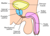
What epithelia lines the bladder and ureters?
- Transitional
- Longitudinal inner muscles and circular outer
- In distal ureter and bladder, outer longitudinal muscle

What epithelia lines the urethra?
- Firstly transitional
- In males, eplaced by a pseudostratified columnar below the openings of the ejaculatory ducts into the urethra
- Distal urethra stratified squamous

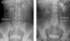
Label this diagram and decide what vertebral level this CT is at?


Label this CT scan.


At what vertebral level do you find each kidney?
Right pushed down by liver.
Right: T12 - L3
Left: T11 - L2

From the hilum, what is the course of the ureter?
- Anterior to psoas major
- At sacroiliac joint goes inwards to pelvis and crosses bifurcation of common iliac and under gonadal arteries
- At ischial spine goes into a transverse plane into the bladder at oblique angle
- 25cm

Where are renal stones most likely to lodge?
- VUJ
- Pelvicoureteric J
- Crossing of iliac arteries
Due to non-uniformity of the lumen of the ureter

How long is the urethra and where is the membranous part?
- F: 4cm
- M: 20cm
As it passes through urogenital diaphragm
What is this showing?

Vasa Recta

A renal biopsy is taken from someone with sickle cell nephropathy. There is an infarct in an area supplied by vessels from the efferent arteriole of large glomeruli close to the medullary border - what vessel is most likely to have been occluded?
Vasa Recta as in the medullary part

What would be the cause of these symptoms and why would they not be due to renal vein entrapment?

- Ureteric entrapment by something like an AAA.
- This is causing hydronephrosis which is leading to referred flank pain
- Renal vein entrapment is associated with haematuria (as blood can’t drain), proteinuria and nausea (usually due to SMA over renal vein)

An 8 year old girl presents to the GP following 2 day history of pain on passing urine, she is otherwise generally fit and well. What is unusual and what tests would you do to investigate?

- Atypical culture
- Dipstick, Ultrasound now and 6 weeks, DMSA

What is a DMSA?
Radioactive technetium-99m and DMSA injected in. 2-3 hour later gamma camera can help form renal morphology image

What is an MCUG?
Test how well child’s bladder works, any abnormalities in urinary sytem and diagnoses why child may have a UTI

These are images from the 8 year girl Jenna’s investigations, what do they show and how can this be dealt with? She also had a DMSA showing reduced tracker uptake in upper left moeity.

- Duplex left kidney with ectopic ureter and uterocoele. Has lead to hydronephrosis and a risk of sepsis
- Could replant the ectopic ureter but kidney not working well with it so left upper pole heminephroureterectomy

What is the issue with a uterocoele?
- Blocks flow of urine so goes back up ureter causing swelling and possible hydronephrosis
- Can lead to a lot of UTIs
Uterocoele - Swelling at the bottom of one of the ureters
