8 - Ophthalmology Anatomy, History and Examination Flashcards
What are some examples of opthalmic pathologies that need urgent review?
- Sudden blurring or loss of vision e.g GCA, CRAO, retinal detachment, TIA
- New onset diplopia e.g first sign of GCA, PCA aneurysm
- Headache with eye pain: e.g Anterior uveitis, Acute angle closure glaucoma
- Orbital cellulitis
- Chemical trauma
What is the definition of the following ophthalmological words:
- Agnosia
- Amblyopia
- Aniridia
- Anopthalmos
Agnosia: Inability to recognise common objects despite an intact visual apparatus e.g dementia
Amblyopia: Reduced vision due to disuse of the eye with no organic defect
Aniridia: Congenital absence of an iris
Anopthalmos: Absence of a true eyeball

What is the definition of the following opthalmological terms:
- Aphakia
- Binocular vision
- Blindness
Aphakia: Absence of the lens
Binocular vision: Ability of the eye to focus on one object and fuse the two images together
Blindness: In the UK it is defined as the level of visul impairment such as to prevent any work for which vision is essential, usually 3/60 or worse
What is the definition of the following opthalmological terms?
- Bupthalmos
- Chemosis
- Coloboma
-
Bupthalmos: large eyeball in infantile glaucoma
Chemosis: Conjunctival swelling of any cause
Coloboma: Congenital cleft due to the failure of some portion of the eye or the lids to complete growth

What is the definition of the following opthalmological terms:
- Keratoplasty
- Cycloplegic
- Ectropion
- Entropion
Keratoplasty: Surgery where opaque cornea is replaced with transparent cornea
Cycloplegic: Drug that relaxes the cilliary muscle, paralysing accomodation and dilating the pupil
Ectropion: Turning out of the eyelid so the conjunctival lining the inside of the lid is exposed to the exterior
Entropion: Opposite of above, eyelashes may come into contact with cornea. Can be caused by trachomatous conjunctivitis causing scarring and contraction of conjunctivae. Common cause of blindness in Africa

What is the definition of the following opthalmological terms:
- Enucleation
- Esophoria
- Esotropia
- Exenteration
Enucleation: Complete surgical removal of the eyeball
Esophoria: Tendency of the eyes to turn inwards when binocular reflexes are disrupted by covering one eye
Esotropia: Inward deviation of the eye
Exenteration: Removal of all of the orbit including the eyeball and the eyelids
What is the definition of the following opthalmological terms:
- Exophoria
- Exotropia
- Macula Lutea
- Miotic
- Mydriatic
Exophoria: Tendency of the eyes to turn outwards when binocular reflexes are disrupted by covering one eye
Exotropia: Outward deviation of the eyes
Macula Lutea: Avascular area of the retina surrounding the fovea
Miotic: A drug causing pupillary constriction
Mydriatic: An agent causing pupil dilatation
What is the definition of the following opthalmological terms:
- Myopia
- Ophtalmia Neonatorum
- Optic Atrophy
- Pannus
Myopia: Near-sighted. Refractive error where the focal point for light rays from a distant object are anterior to the retina
Opthalmia Neonatorum: Conjunctivitis in the newborn usually due to gonococcus or chlamydia
Optic Atrophy: Optic nerve damage characterised by optic disc pallor
Pannus: Infiltration of the cornea with blood vessels
What is the definition of the following opthalmological terms:
- Photocoagulation
- Pterygium
- Strabismus/Squint/Tropia
- Sympathetic Opthalmia
Photocoagulation: Method of causing multiple areas of localised destruction of the retina for treatment of certain retinal disorders e.g diabetic retinopathy
Pterygium: Pathological fold of triangular tissue that extends from the conjunctiva over the cornea
Strabismus: Misalignment of the eyes
Sympathetic Opthalmia: Inflammation in one eye following traumatic inflammation in the other eye

What is the definition of the following opthalmological terms:
- Synechia
- Trachoma
- Uvea
- Zonule
- Tarsorrhaphy
- Tonometer

Synechia: Adhesion of the iris to the cornea (anterior synechia) or lens (posterior synechia)
Trachoma: Infectious keratoconjunctivitis due to repeated chlamydial infections in hot dry climates where flies are present. WORLDWIDE COMMONEST CAUSE OF BLINDNESS!!!!!!
Uvea: Iris, Cilliary body, Choroid
Zonule: Numerous fine tissue strands that stretch from the ciliary muscles to the lens and hold the lens in place
Tarsorrhaphy: A surgical procedure for uniting upper and lower lids
Tonometer: A device for measuring intraocular pressure.

What are mydriatic and cyclopegic drops and what are they used for?
Mydriatic: Dilate the pupil
Cycloplegic: Paralyse the cilliary muscles so can no longer accomodate and focus. All cycloplegics are mydriatics
Uses: dilate pupil to look at retina, management of amblyopia, refraction of children for the prescription of glasses

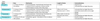
What are four examples of mydriatics/cycloplegics, how long do they act for and what are the contraindications of these?
- Atropine
- Cyclopentolate
- Tropicamide
- Phenylephirine
ALL DROPS SHOULD BE AVOIDED IN UNTREATED NARROW ANGLE GLAUCOMA
What are the side effects of mydriatics/cycloplegics?

- Cannot drive until blurring effect has warn off
- Mydriatics will sting for a few seoconds after instillation
- Atropine can cause redness and warmness of the face
- Whitening of eyelids due to vasoconstriction
Think about side effects of anticholinergics e.g dry mouth

How do you apply mydriatic/cycloplegic drops?

What are fluorescein drops used for and whay are the contraindications of these?
Uses:
- Detect defects in the corneal epithelium
- Assess tear drainage in children with congenital nasolacrimal duct obstruction
- Helps in tonometry to measure intraocular pressure
- Can be administered in a solution with local anaesthetic

What are the side effects of fluoroscein drops?
- Staining of skin or clothes. Lasts for 6-12 hours
- Can discolour contacts so take them out and don’t put them in for another hour after the stain

How do you apply fluorescein drops?

What are miotic eye drops?
They cause pupil constriction, used in the treatment of acute angle closure glaucoma as they increase aqueous drainage

What are steroid and NSAID eye drops used for and why do they need to be used with caution?
Used for the treatment of allergy, episcleritis, scleritis, or iritis
They can increase the IOP so can precipitate glaucoma and can cause progression of dendritic ulcers.
Must look in slit lamp before application as can miss ulcers on opthalmoscopy

What are some topical antibiotics used for the eye?
- Fusidic acid
- Chloramphenicol
- Neomycin
Some systemic drugs can cause opthalmological side effects. What drugs cause the following opthalmological side effects:
- Dry eyes
- Corneal deposits
- Lens Opacities
- Glaucoma
- Papilloedema
- Retinopathy
Dry eyes: B-blockers, anticholinergics
Corneal Deposits: amiodarone, chloroquine, chlorpromazine
Glauvoma
How do you take an opthalmological history?
(important card)

PC (see further cards): onset and duration, has it happened before, discomfort?, visual loss?, double vision?, colour changes?, any trauma?
PMHx: any inflammatory conditions (AS,RA), previous eye surgery/injections, contact lenses, smoker, thyroid issues, hypertension
DHx: any toxic drugs
FHx: of eye disease
ICE

What are some symptoms of eye discomfort and examples of differentials associated with this?
Foreign Body Sensation: foreign body, conjunctivitis, blepharitis, eyelid cysts
Photophobia: Anterior uveitis, Corneal ulcer, Corneal abrasion
Eye pain: Scleritis, Acute angle closure glaucoma
Headache: GCA, Migraine, Meningitis, Raised ICP, cluster headache
Lid irritation: Blepharitis, Stye, Chalazion, Eczema
If a patient presents with visual disturbance, what questions do you need to ask in the history and what differentials could each symptom be?
(Important card)
•Generalized blurring? Refractive error, cataracts, steroids
•Central blurring? Macula or Optic disc pathology e.g AMD, Diabetic maculopathy, optic disc swelling
•Black spots, blob or curtain? Amaurosis Fugax, Retinal detachment, Vitreous detachment
•Photopsia? Migraine, Retinal detachment
•Double vision- monocular or binocular? vertical or horizontal?
•Colour changes? Cataracts, Optic nerve pathology
What is the difference between monocular and binocular double vision?
Binocular double vision only occurs if both eyes are open, can correct by closing one eye
Monocular double vision is due to pathology of the affected eye e.g cataracts, cornea abrasion. Binocular is due to pathology of nerves, neuromuscular junction or muscles

What are some important co-morbidities to acquire about in opthalmological histories?
- Any vascular risk factors

What tests can you do to test visual function?
(IMPORTANT CARD)

- Visual acuity with Pinhole test added on (Snellen Chart)
- Near visual acuity
- Visual Fields: Confrontation test, Perimetery
- Colour Vision: Ishari chart
- Macula: Amsler Grid

How do you perform an OSCE eye examination?
https://geekymedics.com/wp-content/uploads/2021/04/OSCE-Checklist-Eye-Examination.pdf (watch video, important card)
1. Intro and Consent
2. Visual Acuity, Colour Vision, Visual fields
3. Inspect eye (make sure to flip eyelids)
4. Eye movements
5. Cover test
6. Pupil reflexes (direct, consensual, accomodation, swinging light test for RAPD)
7. Anterior segment of eye: cornea reflexes, corneal opacities, hypopyon (pus), hyphema (blood)
8. Dilate the pupil: mydriatics, turn lights down
9. Direct Opthalmoscopy: red reflex, optic disc, retina, macula
10. Thank patient and further tests: Slit lamp, Tonometry, Ishari, Amsler

What is a corneal light reflex?
Light from opthalmoscope should shine in the same place of the pupil, if it doesn’t then the eyes are not aligned so strabismus
Can have second light reflection after cataracts surgery

How do you measure visual acuity and how do you record it?
Test
- Place Snellen chart at 6m and ask patient to wear glasses if they wear them
- Cover each eye and read the lowest line you can with each eye
- Pinhole test
- Near visual acuity test with glasses
Documentation
- If read from 6m and can read to 12 line then 6/12
- Can bring patient to 5, 4, 3, 2, 1m
- If still can’t read do cout fingers, hand movements, light. If no perception this is blind

What cranial nerves are involved in the direct/consensual/accomodation pupillary reflexes?

Brainstem reflex
Direct/Consensual Light Reflex: CNII afferent, CNIII efferent with EDW nucleus in midbrain
Accomodation: Focusing on object causes constriction of pupil. Same nerves as above but includes the LGN and visual cortex

What is a relative afferent pupillary defect and how do you assess for it?
Swinging light test
- Due to retinal or optic nerve disease
- Light shone in bad eye both pupils will constrict but not fully, then when shone into the good eye both pupils constrict further. Then back to bad eye both pupils will dilate a bit
- Do not swing across bridge of nose

What are some causes of an RAPD?

- Optic neuritis (e.g MS)
- Optic nerve tumour
- Unilateral glaucoma (often bilateral so not relevant)
- Optic nerve inflammation (e.g SLE, Sarcoidosis)

What are some things that can affect the red reflex?
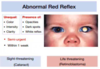
ALWAYS BE THINKING CATARACTS (SIGHT THREATENING) AND RETINOBLASTOMA (LIFE-THREATENING)
Dense black sillhouette: opacity in the media e.g mucous on tear film, cataracts, corneal opacities, retinoblastoma
Absent red reflex (white): complete opacity e.g cataracts, retinoblastoma, vitreous haemorraghe
Less bright: room not dim enough, heavily pigmented eyes

How do you use the opthalmoscope to examine the fundus?

Examine right fundus:
- Dim lights and give mydriatic drops
- Adjust opthalmoscope to largest patch of light and set to 0 (or others if you wear glasses)
- Stand to the right of the patient 20 degrees, hold the opthalmoscope in your right hand, hold their forehead with your left hand. Use focusing dial to get red reflex
- As you get closer you should see a retinal vessel, follow this vessel to the optic disc. Examine for outline, colour, cupping, haemorraghes, abnormal blood vessels
- Examine periphery of retina by following each of the blood vessels
- Examine macula by asking patient to look straight at the light. Difficult to see without mydriatics

How do you test a patients visual fields very basically?

Confrontation test

- Sit face to face at the same eye level
- Ask patient to cover their eye and you cover the opposite eye
How do you test extraocular movements and what muscles is each movement testing?
H test

What tests do you do to assess for squint?
- Corneal Light Reflex (previous flashcard)
- Cover Test: picks up latent squints (squint when eye not being used) as well as manifest squints

How does the cover test work?
https://www.youtube.com/watch?v=Wf8DGL7WE8U

Manifest Squint: when covering the normal eye the squinty eye moves to come to fixed position
Latent Squint: when covering the unknowingly abnormal eye it moves out of position (image on reverse of flashcard)
Alternate cover test allows prisms to be put in until movement stopped to know which prism to use

What are some investigations you can order for eye pathology after you have done an eye examination in clinic?
- Perimeter visual field testing
- Fluorescein angiography: IV inject, can view retinal vessels and see if any leakage
- Orthoptic assessment
- OCT
- MRI
- US

What bones make up the wall of the orbit and which ones fracture with trauma?
Floor and medial walls are thinnest so fracture on trauma. Orbital blow-out fracture
Floor: maxillary, palatine bone, orbital plate of the zygomatic bone, maxillary sinys

What is the role of eyelids and what muscles move them?

- Protect the cornea from injury
- Keep the cornea moist by covering it with a tear film
- Palpebral conjunctiva line inner eyelid and is continuous with bulbar conjunctivae
- Orbicularis Oculi (CN7) and Levator Palpebrae Superioris (CN3)

What is the passage a tear takes from gland to the end destination?
Gland –> Lacrimal Ducts –> Lacrimal Lake (medial angle of eye) –> Lacrimal Sac –> Nasolacrimal duct –> Inferior meatus
No anastomotic pathway so if a blockage anywhere will be overflow of tears (epiphora). Stagnant tears can lead to infection

Why do we need a tear film and what is it made up of?
Need a smooth ocular surface for light rays to be refracted uniformly and to provide lubrication to prevent friction when the lids close.
Also has antibacterial properties and prevents cornea drying out and getting abrasions which are uncomfortable and gritty

What are the different layers of the eyeball?

Outer protective layer: Sclera and Cornea (Transparent bit). Allows attachment of extra-ocular muscles
Middle layer: Choroid, Ciliary Body, Iris. Blood vessels layer. Ciliary body connects choroid and iris
Inner layer: Retina

What are the functions of the cornea?
Transparent avascular structure that separates the tear film and anterior chamber. Made of 5 layers (see image)
- Maintaining transparency
- Ocular protection (corneal reflex)
- Refraction of light

How long does it take for trauma to heal in the corneal epithelium layer and Bowman’s layer?
Corneal Epithelial: 3 to 14 days, longer for central defects. Cells need to migrate from basal layer to the surface. Stem cells found at limbus on the periphery of the cornea which is why central defects take longer
Bowman’s Layer: will never fully heal if trauma this deep, will cause corneal scarring as this layer is acellular

Aqueous humour maintains the intraocular pressure of the eye. What is the route that aqeous humour travels through the eye?
Made in the cilliary processes of cilliary body –> Between iris and lens to get to the pupil –> Trabecular meshwork in iridocorneal angle –> Canal of Schlemm –> Episcleral Vessels –> Systemic Venous Circulation
Can also drain through uvoscleral route (into root of iris or ciliary muscle and drain into scleral vascular system)

How do you measure inraocular pressure and what are the normal ranges for this?
Tonometry
Normal: 11-21
Over 21 is ocular hypertension and an increased risk of developing glaucoma. However can have glaucoma with normal IOP so beware

What are some classes and examples of drugs that can be used to lower the IOP?
Important card

What part of the nervous system controls pupil movements?
Mydriasis (dilate): sympathetic
Miosis (constrict): parasympathetic

What is the functions of the choroid?
Choroid is connected to retina by Bruch’s membrane

- Allows nerves and vessels to get to anterior eye
- Removes waste product from outer retina
- Supplies nutrients to outer retinsa
- Absorbs any light travelling through the retina so it does not reflect back and interfere with vision
What structures make up the uveal tract?
- Iris
- Choroid
- Cilliary body

What is the blood supply to the retina?
- 2/3 supplied by central retinal artery, 1/3 by choroidal vessels
- Central retinal artery splits into four, the superior and inferior arteries cross at the posterior pole
- Macula has a dense capillary network but the fovea does not, it relies on the choroid

Draw the visual pathway.

What is the path of the pupil reflexes?
Light travels with normal visual pathway until just before the LGN where it goes into the pre-tectal area and synapses at BOTH EDW nuclei.
Efferent fibres travel to ciliary ganglion via occulo motor nerve and then to sphincter pupillae
