Pictures Flashcards
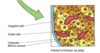
What do these pictures show?

The right pic is normal histology of the thyroid gland. The left pic shows the decrease in colloid volume with diffuse goiter, the hypertrophy & hyperplasia of the thyroid gland.
The symptom shown on this picture is an indication of what disease?

Graves’ Disease
myxedema (not always present)
What symptom does this picture show? Which disease is this consistent with?

exophthalmos
Graves’ Disease
**pic shows before & after decompression surgery
**Remember: autoimmune rxn causes edema of retroorbital tissues & degeneration of extra-ocular muscles
What symptom does this picture show? What disease is this consistent with?

Thyroid Stare
Graves’ Disease
What does this picture show? Which disease is this consistent with?
Hashimoto’s Disease
hypothyroidism
**lymphocyte infiltration of the thyroid
What condition does this picture show?

Cretinism or congenital hypothyroidism
sketal muscle development is slowed more than soft tissue growth, thus the short & stocky appearance…
What structure is this stain taken from? What are the main features of this slide? What are cell types of A & B? What is the cell type found in b/w them?

Adenohypophysis–note the extensive sinusoidal system
A: acidophil
B: basophil
The other cell type in b/w that is clear is a chromophobe.
Which cells are being pointed to? What gland is this?

Parathyroid gland
larger, lighter cells–>oxyphils
more compact, darker cells–>chief cells
Describe what is shown in this picture.

Lysosomal enzymes are packaged in the Golgi. Lysosomes & H+ from inside the osteoclast are both released into the microenvironment. The acidic environment allows for the dissolution of the calcium phosphate of the bone…bone resorption begins taking place. The products of the bone breakdown are taken up into the osteoclast cytoplasm, further broken down & released into capillaries.
What do the 2 unlabeled arrows indicate?

