Cerebral Cortex Flashcards
Cerebral Cortex:
Anatomical organization
- Embryologically derived from the telencephalon
- Can be subdivided into:
-
Archicortex (3 layers)
- Hippocampus and Dentate gyrus
-
Paleocortex
- Olfcatory cortex
- Neocortex (6 layers)
-
Archicortex (3 layers)
- Made up of 3 poles (frontal, occipital, and temporal) and 6 lobes
6 Anatomical Lobes:
- Frontal lobe
- Parietal lobe
- Temporal lobe
- Occipital lobe
-
Insular lobe
- The Insula
-
Limbic lobe:
- includes Cingulate gyrus, Parahippocampal gyrus, and Hippocampus
Blood supply to the cerebral cortex:

- Anterior cerebral
- Middle cerebral
- Posterior cerebral
- Anterior communicating
- Posterior communicating arteries
- all of which form the Circle of Willis
Drainage of the cerebral cortex:
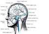
- Superior Sagittal sinus
- Inferior sagittal sinus
- Straight sinus
- Transverse sinuses
- Sigmoid sinuses
- which drain into the Internal jugular veins
6 Layers of the Neocortex:
- Layer I: Molecular layer
- Layer II: External granular layer
- Layer III: External pyramidal layer
-
Layer IV: Internal granular layer
- well-developed in sensory areas
-
Layer V: Internal pyramidal layer
- well-developed in motor areas
-
Layer VI: Multiform layer
- project back to the thalamus
How are myelinated axons organized in the cortex?
horizontal bands and vertical bundles
What are the major cell types of the cortex?
Pyramidal and Non-pyramidal
Functional units: Columns and Modules
- Each column extends through the 6 layers that share similar functions
- Functional columns form modules in various cortical areas
- especially the primary somatosensory, visual, and auditory cortices
- Columns of cortical neurons are interconnected within the same hemisphere and between the two hemispheres
Areas 3, 1, 2 (Postcentral gyrus):
Primary somatosensory cortex
- Somatotopic organization (contralateral)
- Sensory homunculus
- Greater representation of the face and hand
- Lower limb representation is medial
-
Lesion:
- Contralateral loss of somesthetic sensation
Area 4 (Precentral gyrus):
Primary motor cortex
- Somatotopic organization (contralateral)
- Motor homunculus
- Greater representation of the face and hand
- Lower limb representation is medial
-
Lesion:
- Contralateral spastic paralysis
Area 17 (Cuneus and Lingual gyri):
Primary visual cortex
-
Visuotopic organization
- Central visual field: Most posterior
- Peripheral visual field: most anterior
- Vertical meridian: border of areas 17 and 18
- Horizontal meridian: bisect horizontally
-
Lesions:
- Contralateral hemianopia
- If restricted to upper or lower banks of the Calcarine fissure ⇒ contralateral inferior or superior quadrantanopia
Areas 41 and 42: Transverse gyri (of Heschl)
Primary auditory cortex
- Tonotopic organization
- Biaural representation
-
Lesions:
- Bilateral lesions lead to loss of hearing
Areas 44 and 45: part of the **Inferior frontal gyrus **
Motor area of speech (Broca’s area)
- mostly dominant in the left hemisphere
-
Lesions:
- Dominant side (left) ⇒ motor aphasia, Broca’s aphasia, or expressive aphasia
- Non-dominant (right) ⇒ difficulty in expressing emotional aspect of language
Main functional areas of the frontal cortex:
- Primary motor cortex: area 4
- Premotor cortex: area 6
- Supplementary motor cortex
- Frontal eye field
- Broca’s area
- Prefrontal cortex:
- Dorsolateral prefrontal cortex: working memory
- Ventromedial prefrontal cortex (orbitofronal cortex): limbic
Association cortical areas of the parietal lobe:
- Posterior parietal lobe: polymodal convergence
- Superior parietal lobule: areas 5 and 7
- Inferior parietal lobule: Supramarginal gyrus (area 40) and Angular gyrus (area 39)
Lesions:
- **Dominant (usually left) hemisphere: **
- astereognosis (area 40)
- lose the meaning of touch
- aphasia, alexia and agraphia (area 39)
- inability to read and write
- astereognosis (area 40)
-
Non-dominant (usually right) hemisphere:
- spatial distortion
- contralateral neglect
Cortico-cortical connections within the same hemisphere and between the two hemispheres:
- Short association fibers
- Long association fibers
- Callosal fibers
