LEC 24: Abdomen I Flashcards
What are the organs of the digestive tube
- mouth
- pharynx and esophagus
- stomach
- small intestine (duodenum, jejunum, ileum)
- large intestine (cecum, colon, rectum, anal canal, anus)
What are the accessory organs of the digestive tract
- teeth
- salivary glands
- liver
- pancreas
When does the primordial gut begin
Week 4
- FGF signal A-P axial patterning
- signals for induction of endoderm
- FGF4 and activins
- Endoderm: specifies temporal and positional information
- stomodeum, proctodeum

What are the 5 classes of foregut derivatives
- primordial pharynx and derivatives
- lower respiratory system
- esophagus and stomach
- duodenum–PROXIMAL to bile duct
- liver, billiary apparatus, pancreas
Where does the esophagus and stomach derive from
foregut
Where do the oral cavity, pharynx, tongue, tonsils, salivary glands, upper respiratory system derive from
foregut (primordial pharynx and derivatives)
Where does the duodenum derive from
foregut
Where do the liver, billiary apparatus, and pancreas derive from
foregut
Development of esophagus
- T-E suptum separates esophagus
- epithelium obliterates lumen and then it is recanalized
- Upper 1/3: striated muscles from pharyngeal arches
- move food down
- Lower 1/3: smooth muscle from surrounding splanchnic mesenchyme
- Cranial Nerve X (VAGUS)
What innervates the esophagus and what is the main supplier of blood flow
- Cranial Nerve X (Vagus) innervates the esophagus
- Celiac trunk is the primary blood supply to the esophagus
esophageal stenosis
- incomplete recanalization of esophagus during 8th week
OR
- failure of esophageal blood vessels to develop (atropy–lack of developing tissue causes dying)
When does the development of the stomach occur
Stomach development happens in week 4
Stomach development

- dilation of tubular structure
- fusiform enlargment
- initially oriented in median plane
- during weeks 4-6, broadens ventro-dorsally
- dorsal boder grows faster, greater curvature forms
- greater curvature toward vertebral column
- lesser curvature towards anterior abdominal wall
- surfaces are still left/right oriented

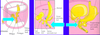
Rotation of stomach

- Stomach, situated in median plane with greater curvature towards dorsal surface, lesser curvature toward ventral surface
-
rotates 90 degrees in a clockwise direction
- lesser curvature (ventral border) moves right
- greater curvature (dorsal border) moves left
- original left side now ventral surface
- original right side now dorsal surface

What nerves supply the walls of the stomach
LEFT VAGUS
- supplies anterior wall
RIGHT VAGUS
- supplies posterior wall
How is the stomach suspended in the abdomen

The stomach is suspended by dorsal mesogastrium (originally in median plane)
- mesentary carried to the left during rotating
- this forms omental bursa

omental bursa
- clefts in dorsal mesogastrium coalesce to form omental bursa
- with rotation, it is pulled to the left and stretched
- allows free movement of stomach

Congenital Pyloric Stenosis
- 3/1000
- males>females
- stenosis of pyloric canal
- obstruction of food
- projectile vomiting

What is the duodenum derived from
Duodenum has a dual origin and dual blood supply
FOREGUT
- proximal to level of bile duct
- blood supplied by celiac trunk
MIDGUT
- blood supplied by superior mesenteric artery

How does the duodenum rotate
- duodenal loop projects ventral
- with rotation, moves rightward
- the lumen becomes obliterated and then recanalizes during weeks 5-6
duodenal stenosis
- partial occlusion of the lumen
- due to incomplete recanalization from defective vacuolization
- stomach contents (with bile) are vomited

liver, gallbladder, and biliary apparatus
- during week 4, outgrowth in foregut
- Fibroblast growth factors (FGF) from heart –> bipotential cells –> hepatic diverticulum
- hepatic diverticulum extends into septum transversum (from early embryo folding)
- hepatic diverticulum divides into 2 parts
- endodermal cells –> hepatic cords –> sinusoids
- mesenchyme in septum transversum –> kupffer cells and hematopoetic tissue

What is the gallbladder formed from
small part of hepatic diverticulum forms the gallbladder

What is the cystic duct formed from
stalk of diverticulum

ventral mesentary
- lesser omentum
- hepatogastric ligament
- heaptoduodenal ligament
- falciform ligament
- forms visceral peritoneum of liver

development of pancreas

- caudal foregut endodermal cells–>pancreatic buds (dorsal to ventral)
- rotation of stomach, duodenum leads to pancreas lying on dorsal abdominal wall
- ducts fuse to form main pancreatic duct
- exocrine: endodermal tubules branch to form acinar cells and ducts
- endocrine: clumps of cells from exocrine part form islets

annular pancreas
- growth of bifid ventral pancreatic bud around duodenum
- can cause duodenal obstruction

Where does the spleen develop from
- spleen develops from mesenchyme in dorsal mesogastrium
- thickening of mesoderm in dorsal mesogastrium
- mesenchymal cells differentiate
- capsule, connective tissue, parenchyma
- center for hematopoiesis
