Muscles of the Back Flashcards
What is the organization of back muscles according to innervation?
Which one is intrinsic? which one is extrinsic?
- Hypaxial mucles:
- extrinsic back musles
- innervated by cranial nerves and ventral rami of spinal nerves.
- superficial muscles.
- Epaxial muscles:
- intrinsic back muscles
- innervated by dorsal rami of spinal nerves.
- What constitute the superficial group?
- To what muscle group do they belong?
- Superficial muscles: muscles realted to and involved in movements of appendicular elements of skeleton
* Clavicle, scapula, humerus - They belong to the hypaxial muscle group.
- What constitute the intermediate muscle group?
- To what muscle group to they belong?
- intermediate group: muscles attached to costal elements and with possible respiratory functions.
- they belong to the hypaxial muscle.
1.Name the group of muscles that constitute the epaxial musles?
- spinotransversales group
- erector spinae group
- transversospinales group
- segmental group
- suboccipital group
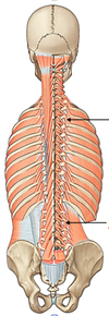
What muscles are part of the superficial group of the hypaxial muscles?

Superficial group:
- Trapezius
- Latissims dorsi
- levator scapulae
- rhomboid minor
- rhomboid major
State the following for the Trapezius:
- Origin
- Insertion
- Function
- Innervation
Trapezius:
- Origin:
- Superior nuchal line
- ligamentum nuchae
- external occiipal protuberance
- thoracic vertebral spines.
- Insertion: lateral 1/3 clavicle + acromion and spinae of scapula
- Funtion:
- Upper fibers: elevation of shoulder girdle (as in shrug and during arm elevation)
- Middle fibers: retraction of scapula
- Lower fibers: depression of scapula + participation in cranial rotation of glenoid during arm abduction
- Innervation:
- Motor: Accessory n. (CN XI)
- (Motion sensor) Proprioception: C3-C4 ventral rami
State the following for the Latissimus Dorsi:
- Origin
- Insertion
- Function
- Innervation
- Origin: Iliac Crest + spinous process of T6-sacrum
- Insertion: Floor of intertubercular sulcus of humerus.
- Function:
- Extension
- adduction
- medial rotation of humerus
- Innervation: Middle subscapular nerve ( C6-C8)
State the following about Levator Scapulae:
- Origin
- Insertion
- Function
- Innervation
- Origin: Transverse processes of C1-C4
- Insertion: Upper portion of vertebral border of scapula
- Function: Upward and anterior movement of superior angle of scapula –> as in reaching forward or extending arm.
- Innervation: Dorsal scapular nerve (C4-C5)
State the following about the Rhomboid minor:
- Origin
- Insertion
- Functions
- Innervations
- Origin: Lower part of ligamentum nuchae + C7-T1 spines
- Insertion: Vertebral border of scapula at root of spine
- Function:
- Retraction (adduction)
- elevation
- caudal rotation of glenoid fossa of scapula
- Innervation: Dorsal scapular nerve (C4-C5)
State the following about the Rhomboid Major:
- Origin
- Insertion
- Function
- Innervation
- Origin: T2-T5 spines
- Insertion: Vertebral border of scapula between spine and inferior angle.
- Function:
- Retraction (adduction)
- elevation
- caudal rotation of glenoid fossa of scapula
- Innervation: Dorsal Scapular nerve (C4-C5)
Which mucles are part of the intermediate group of the Hypaxial Mucles?

- Serratus posterior inferior.
- Serratus posterior superior.
State the following about the Serratus Posterior Superior:
- Origin
- Insertion
- Function
- Innervation
- Origin:
- Lower part of ligamentum nuchae
- C7-T3
- subraspinous
- Insertion: Upper border of ribs 2-5 just lateral to their angles
- Function: elevate rib 2-5
- Innervation: ventral rami of spinal nn.
State the following about the Serratus posterior inferior:
- Origin
- Insertion
- Function
- Innervation
- Origin:
- T11-L3 spines
- supraspinous ligament
- Insertion: lower border of ribs 9-12 lateral to their ribs angles.
- Functions:
- Depression of ribs 9-12
- possible prevention of elevation of lower ribs during contraction of diaphragm
- Innervation: Dorsal rami of spinal nn.
- Where is this located?
- To what is this attached to?

- The Thoracolumbar fascia is the separartion between the hypaxial and epaxial muscles in the thoracic region.
What does the posterior layer of the Thoracolumbar fascia covers?
Erector Spinae

What does the anterior layer of the Thoracolumbar fascia covers?
Quadratus lumborum

- Which muscles are part of the Spinotransversales group?
- To which group of muscles do they belong?
- Are the intrinsic or extrinsic?
- Splenius capitis and Splenius cervisis
- They belong to the epaxial muscle group
- They are intrinsic muscles of the back
State the following about Splenius capitis:
- Origin
- Insertion
- Functions
- Innervation
- Origin:
- Ligamentum nuchae
- C7-T4 spines
- Insertion:
- Mastoid process
- skull bellow superior nuchal line
- Functions:
- Bilateral activation: Extension of head and neck. Extend the head to one side when activated.
- Unilateral activation: Rotation of head toward ipsilateral side
- Innervation: Dorsal Rami spinal nn.
State the following about the Splenius Cervicis:
- Origin
- Insertion
- Functions
- Innervation
- Origin: T3-T6
- Insertion: Transverse processes of C1-C3
- Function:
- Bilateral activation: Extension of head and neck. Extend the head to one side when activated.
- Unilateral activation: Rotation of head toward ipsilateral side
- Innervation: Dorsal rami spinal nn.
- Which muscles are part of the Erector spinae group?
- To what muscle groups do they belong?
- Are they intrinsic or extrinsic?

State the following about Spinalis:
- Origin
- Insertion
- Function
- Innervation
- Origin: Vertebral spines
- Insertion: vertebral spines
- Function:
- Bilateral activation: Extension of vertebral column and head. Rate head to active side and the spine.
- Unilateral activation: Lateral flexion of vertebral column + rotation of head to ipsilateral side. Rotate the spine
- Innervation:
- Dorsal Rami of spinal nerves
State the following about the Longissimus:
- Origin
- Insertion
- Function
- Innervation
- Origin: Aponeurotically from sacrum. More cranial fibers originate from transverse processes of pre-sacral vertebrae
- Insertion: transverse and costal elements of vertebrae near their junctions. Most cranial fibers insert onto mastoid process, and are called longissimus capitis.
- Function:
- Bilateral activation: Extension of vertebral column and head. Rate head to active side and the spine.
- Unilateral activation: Lateral flexion of vertebral column + rotation of head to ipsilateral side. Rotate the spine
- Innervation: Dorsal rami of spinal nerves
State the following for the Iliocostalis muscle:
- Origin
- Insertion
- Function
- Innervation
- Origin: Aponeurotically from sacrum and iliac crest. More cranial fibers originate near costal angles
- Insertion: ribs near their angles. Mores cranial fibers insert onto posterior tubercles of cervical vertebrae.
- Functions:
- Bilateral activation: Extension of vertebral column and head. Rate head to active side and the spine.
- Unilateral activation: Lateral flexion of vertebral column + rotation of head to ipsilateral side. Rotate the spine
- Innervation: Dorsal rami of spinal nerve.
- What muscles are part of the Transversospinales muscle group?
- To what group do they belong?
- Are they intrinsic or extrinsic muscles?

- Semispinalis capitis, semispinalis, rotatores, multifidus
- They belong to the epaxial muscles
- They are intrinsic muscles.
State the following for Semispinalis muscle group:
- Origin
- Insertion
- Function
- Innervation
- Origin: transverse process of vertebrae
- Insertion: vertebral spines
- Functions:
- Bilateral activation: Contribution to extension of vertebral column
- Unilateral activation: Lateral flexion and (if intervertebral joints permit) axial rotation of vertebral column toward contralateral side
- Innervation: Dorsal rami spinal nerve.
State the following for the Semispinalis capitis:
- Origin
- Insertion
- Function
- Innervation
- Origin: Transverse process of upper thoracic and lower cervial vertebrae.
- Insertion: Nuchal plane of occipital bone near midline.
- Function:
- Bilateral activation: Powerful extension of head/skull
- Unilateral activation: Rotation of head to ipsilateral side
- Innervation: Dorsal rami spinal nerves.
State the following for the Multifidus muscle:
- Origin
- Insertion
- Function
- Innervation
- Origin:
- Transverse processes of vertebrae
- Iliac crest
- Insertion: vertebral spines
- Functions: same as semispinalis
- Innervation: dorsal rami spinal nerves
State the following for the Rotatores muscles:
- Origin
- Insertion
- Function
- Innervation
***SAME AS SEMISPINALIS MUSCLES***
- Origin
- Insertion
- Function
- Innervation
What muscles are part of the segmental group?
To what muscle group do they belong to?
Are the intrinsic or extrinsic?
- Levatores costarum, interspinales and intertransversarius
- They belong to the epaxial muscle group
- They are intrinsic muscle.
State the following for the Levator Costarum:
- Origin
- Insertion
- Function
- Innervation
- Origin: Transverse processes of C7-T11
- Insertion: Rib below vertebra of origin near costal tubercle
- Function: Elevate of rib
- Innervation: Dorsal rami spinal nerve
State the following about the Interspinales:
- Origin
- Insertion
- Function
- Innervation
- Origin: Spinous process of a more caudally located vertebra.
- Insertion: Spinous process of the vertebra located immediately cranial to the vertebra of origin
- Funtion: Stabilization of adjacent vertebrae during trunk movements
- Innervation: Dorsal Rami spinal nerve
State the following about the Intertransversari muscle:
- Origin
- Insertion
- Function
- Innervation
- Origin: Transverse process of a more caudally located vertebra
- Insertion: Transverse process of the vertebra located immediately cranial to the vertebra of origin
- Funtion: Stabilization of adjacent vertebrae during trunk movements
- Innervation: Dorsal Rami spinal nerve
What muscles are located in the Suboccipital muscle group?
To what muscle group do they belong?
Are they intrinsic or extrinsic?

- Rectus capitis posterior major, rectus capitis posterior minor, obliquus capitis superior, obliquus capitis inferior.
- They belong to the epaxial muscle
- They are intrinsic muscles
- What are the boundaries of the Suboccipital triangle?
- What are the contents of the suboccipital triangle?

- Boundaries:
- Medial → Rectus capitis posterior major
- Lateral → Obliquus capitis superior
- Inferior → Obliquus capitis inferior
- Contents:
- Suboccipital n. (dorsal ramus of C1)
- Vertebral a.
Name the external features of the spinal cord? (3)

- Anterior median fissure on anterior surface
- Posterior median sulcus on posterior surface
- Posterolateral sulcus on each side of posterior surface. Entrance for rootlets.
How many longitudinal arteries does the spinal cord contains?
Where are these arteries branching from?
Along what structure of the spinal cord do they descend?

- Anterior spinal artery
- originates from union of branches of vertebral aa.
- passes through the anterior median surface of spinal cor
- Posterior spinal artery (R/L)
- branches of vertebral (posterior inferior cerebellar) aa.
- Descends along the spinal cord
Where does the segmental medullary arteries originate from?
The segmental medullary arteries entry into the vertebral cana through __________

- Segmental arteries originate from:
* segmental spinal aa. ( spinal branches of ascending cervical, deep cervical, vertebral, posterior intercostal, and lumbar aa.) - The segmental medullary arrteries enter through the intervertebral foramina at entry level.
- What is the largest segmental medullary artery?
- Where does it originate from?
- True or False: These arteries reinforce the anterior and posterior spinal aa.

- The largest segmental medullary arteries are the Arteria radicularis magna (of Adamkiewics)
- They originate in lower thoracic or upper lumbar region from intercostal/lumbar aa. Usually on left side (65%)
- False, this arteries reinforce the arterial supply of 2/3 of spinal cord.
****The segmental medullary aa are the ones that reinforce the anterior and posterior spinal aa.
A. Where does the Radicular aa branch from?
B. This artery provides blood supply to where?

- The radicular artery is a branch of the segmental spinal artery.
- This provides blood supplu to the ventral and dorsal roots o spinal nn. + superficial part of gray matter.
How is the spinal cord positioned in the center of the subarachnoid space?
Denticulate ligament
