Vertebral Column Flashcards
Vertebral Column
Skeleton of neck and back. Functions to support weight, protect spinal cord, serve as axis and pivot, aid posture/movement

What are the differentiated segments of the vertebral column?
7 Cervical vertebrae, 12 thoracic vertebrae, 5 lumbar vertebrae, 5 sacral vertebrae, variable coccygeal vertebrae (typically 4)
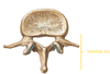
Vertebral Body
Anterior most structure of the vertebral column. Major weight bearing component of the structure. Size of vertebrae varies (increases) as you go down the vertebral column (weight borne on the vertebra significantly increases as you go down the spine)

Vertebral (Neural) Arch
Paired pedicle and lamina of a vertebra.
Pedicle
Paired structures on either side of vertebral foramen. joins vertebral arch and body
Lamina
Paired structures on either side. Flat plates contacting pedicles and spinous process, bound the vertebral foramen.

Vertebral foramen (Canal)
Midline of vertebral column. Houses the spinal cord. Formed by pedicles and lamina.

Superior vertebral notch
Sit above the vertebral arch, immediately superior to the pedicle just posterior to the body. When the vertebrae are articulated, these notches correspond with their superjacent inferior counterpart to form the intervertebral foramen for the spinal nerve

Inferior vertebral notch
deep incisure placed inferior to the more superiorly situated pedicle. Its anterior border is the back of the vertebral body and its posterior wall is the inferior articular process. When the vertebrae are articulated, these notches correspond with their subjacent superior counterpart to form the intervertebral foramen for the spinal nerve

Intervertebral foramen
Formed by superior and inferior vertebral notch; where the spinal nerve roots exit

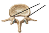
Transverse processes
Processes (2 per vertebrae) projecting horizontally bilaterally from intersection of lamina and pedicles. Serve as a point of attachment for muscles and ligaments.

Spinous process
Process (1 per vertebra EXCEPT C1) projecting posteriorly and inferiorly from the junction of the laminae. Serves as an attachment for muscles and ligaments.

Zygapophysis/Articular process
4 per vertebra, 2 superior (pre-zygapophyses) and 2 inferior (post-zygapophoses). Act to link vertebrae adjacent together. Spring from junction of pedicles and lamina. superior processes or prezygapophysis project upward from a lower vertebra, and their articular surfaces are directed more or less backward. inferior processes or postzygapophysis project downward from a higher vertebra, and their articular surfaces are directed more or less forward and outward

Typical Cervical Vertebra: Transverse Foramen
Located on either side of the body. Vertebral arteries run C2-C6, enter through foramen magnum to reach the brain (become basillar artery). C7 has transverse foramen, but the artery does not run through it! (Runs along it)

Typical Cervical Vertebra: Anterior and Posterior tubercles
The transverse processes are divided into an anterior and posterior portion, termed the anterior and posterior tubercles. Anterior portion is analagous to the rib, posterior portion is more analagous to “true” transverse process

Carotid Tubercle
Anterior tubercle of C6. Carotid artery runs alongside it. Carotid artery can be compressed at this point easily to occlude bloodflow.
Typical Cervical Vertebra: Vertebral Foramina
large for C3 – C7 due to cervical enlargement of spinal cord
Typical Cervical Vertebra: Uncinate Process/Uncovertebral joint
Raised margins of superior border of body. Form saddle-shaped joint on C3-C7 and T1. Prevents a vertebra from sliding backwards off the vertebra below

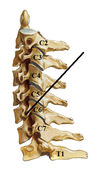
Spinous Process of C7
Vertebra prominens; very long and prominent.

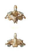
Atlas
C1. Articulating surfaces for articulation with the occipital condyles (superior articulating surfaces) and Axis (Inferior articulating surface). LACKS A VERTEBRAL BODY; instead has an anterior and posterior arch (appears as a ring). Groove for vertebral artery on superior surface

Axis
C2. Superior articulating surface (articulation with C1). Dens/odontoid process: former body of C1, allows pivoting for skull
Dens
Odontoid process. Allows for pivoting of skull. Enlarged spinous process of C2. Articulates with C1.

Typical Thoracic Vertebrae: Superior Costal Facet
Location where rib forms articulation with the top of a vertebra. Superior costal facet is located on the inferior thoracic vertebrae; inferior costal facet is located on the superior vertebrae. While these terms may be confusing, it helps to know that the costal facets are named for their position on the vertebral body itself, NOT for the part of the rib that they articulate with

Typical Thoracic Vertebrae: Inferior Costal Facet
Location where rib forms articulation with the top of a vertebra. Inferior costal facet is located on the superior vertebrae; superior costal facet is located on the inferior thoracic vertebrae;. While these terms may be confusing, it helps to know that the costal facets are named for their position on the vertebral body itself, NOT for the part of the rib that they articulate with

Typical Thoracic Vertebrae: Transverse Costal Facet
Point where rib tubercle articulates with transverse process of vertebra.

Typical Thoracic Vertebrae: Apperance
More superior ones look more like cervical vertebra; more inferior ones look more like lumbar vertebra

T11 & T12
Differ from the other thoracic vertebrae in lacking facets for the ribs on their transverse processes, which additionally are shorter here. More similar and size and function similarly to lumbar vertebrae.
Lumbar vertebra
Weight bearing vertebrae of the body. Largest vertebral bodies.

Accessory process
Inferior tubercles, found posteriorly on each transverse process. Attachment for intertransverse lumborum muscles. Specialized tubercles for muscular attachments.

Lumbar vertebra: Spinous process
Short, rectangular shaped

Mammillary process
Superior tubercle, attachments for muscles/articulation: attachments for multifidus and median intertransverse muscles

L5 Vertebra
largest vertebra in column. Significant contributor to lumbosacral angle

Lumbosacral angle
angle between lumbar and sacral vertebrae (duh). Also known as sacrovertebral angle. ~120 degrees. Changes significantly during pregnancy.

Anterior edge of L5
Larger than posterior, results in wedge shaped structure/attachment. Results in the existence of the lumbosacral angle.

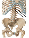
Sacrum
Fused vertebra. Discs not visible. functions to transfer body weight to pelvis and contribute to strength and stability.

Sacral Canal
sacral continuation of vertebral canal
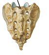
Sacral foramina
Four paired foramina descending, diminishing in size as they descend. Sacral nerves exit out of these foramina.

Sacral promonotory
Elevation at base of sacrum. anterosuperior projecting edge of S1

Median crest of sacrum
Formed by fusion of spinous processes

Medial crest
Fusion of articular processes (WHY ARE THEY NAMED SO CONFUSINGLY SIMILAR [MEDIAN AND MEDIAL?!] WHY IS THE MEDIAN CREST MORE MEDIAL??)

Lateral crest
Fusion of transverse processes.

Sacral hiatus
Gap in lamina of 5th (someimes 4th as well) sacral vertebrae

Sacral cornua (Horns)
former inferior articular processes of S5. Most caudal parts of the sacral crest. Projected downwards, articulate with the coccygeal cornua.

Auricular surface
Large articulating surfaces on sides of sacrum. Transfer weight bearing from vertebral column; part of sacroiliac joint.

Coccyx
Rudimentary tailbone. Fusion of variable # of vertebrae (typically 4)

Coccygeal cornuas
Part of Co1. Found on the posterior supperior portion of the coccyx, articulate with sacral horns

Primary curvatures of the spine
Present at birth due to differences in anterior and posterior size/shape of vertebrae

Cervical vertebral column curvature
Anteriorly convex, secondary curvature

Thoracic vertebral column curvature
Anteriorly concave, primary curvature

Lumbar vertebral column curvature
Anteriorly convex, secondary curvature

Sacral vertebral column curvature
Anteriorly concave, primary curvature

Movement of spine
flexion/extension, lateral bending, & rotation (GO READ OMM IF STILL CONFUSED). Restricted by: IV discs, orientation of zygapophysial joints, articular capsules, muscles, & ligaments. Range of motion results primarily from elasticity of IV discs.

Degeneration of vertebrae
Osteoporosis, osteoarthritis. Progressive bone diseases that result in decrease in bone mass and density which can lead to an increased risk of fracture

Kyphosis
Convex curvature of spine. Abnormal if excessive

Lordosis
Concave curvature of spine. Abnormal if excessive

Scoliosis
Lateral curvature of spine

Intervertebral Joints
Secondary cartilaginous joints. Allow the vertebral column to articulate via interposed intervertebral discs. IV disc comprised of anulus fibrosis and nucleus pulposus.

Nucleus pulosus
Ring in the “center”; slightly posteriorly positioned gelatinous, elastic, avascular core

Anulus Fibrosis
Hard ring around the outside; outer portion of concentric fibrocartilaginous circles

Uncovertebral Joints
joints between uncinate process of C3-C6 and superjacent body

Vertebral Arch Joints (Zygapophyses)
synovial joints between superior and inferior articular processes. allow gliding between vertebrae. variable orientation limits movements

Anterior longitudinal ligament
strong band extending across anterolateral vertebral bodies and IV discs. Prevents hyperextension of vertebral column. Runs entire course of vertebral column (sacrum to occiput & C1’s anterior tubercle). stabilizes vertebral body joints.

Posterior longitudinal ligament
weaker band extending along anterior aspect of vertebral canal. Prevents hyperflexion of vertebral body and herniation/protrusion of discs

Accessory Ligaments
stabilize laminae, transverse processes, and spinous processes. Include ligamentum flava, interspinous, supraspinous, and intertransverse ligaments

Ligamentum flava (flavum)
Broad yellowish ligaments. Connect adjacent lamina. prevents abrupt flexion of vertebral column, preventing injury to IV discs. Extends to the posterior wall.

Interspinous ligaments
Ligaments connecting spinous processes

Supraspinous ligaments
Connect tips of spinous processes, extends from C7-sacrum. Merge with the ligamentum nuchae.

Tectorial ligament
Extends from C2 through foramen magnum. Superior continuation of posterior longitudinal ligament

Nuchal Ligament/Ligamentum nuchae
extends from external occipital protuberance /posterior foramen magnum to spinous processes of cervical vertebrae

Inter-transverse ligaments
Connect ADJACENT transverse processes. obvious in thorax

Craniovertebral Joints
Articulations between the vertebral column and the cranium and their associated ligaments
Cruciate ligaments
Aka cruciform ligaments. Shaped like a cross. Formed by superior and inferior longitudinal bands + transverse ligament of atlas

Transverse ligament of atlas
Holds the dens of C2 with arch of C1. Found on the posterior wall of dens’ socket

Longitudinal Bands
extend from transverse ligament to occiput and C2 body

Alar ligaments
extend from sides of dens to C1 & foramen magnum. Limit head rotation

Vertebral arteries
Converge on brainstem, give off various spinal arteries.

Herniation of nucleus pulposus
Nucleus pulposis is posteriorly oriented; trauma or age-related degeneration of anulus fibrosis can result in nucleus pulposis herniating or “pushing out” of the posterior aspect of the IV disc and bulging out beyond the damaged ring. Usually occurs in lumbar discs due to weight bearing nature of the lumbar vertebrae, can result in paralysis, paresthesia, muscle weakness.

Internal Decapitation
Skull separates from spinal column as the result of severe head injury. Usually fatal due to nerve damage or severing of spinal cord. Frequently the result of a car accident; usually the cause of death in hanging. CAN BE SURVIVED.

Terminal radicular arteries
Branch off of spinal arteries after they enter the IV foramen. Terminal radicular arteries supply ventral and dorsal roots

Medullary segmental arteries
Branch off of spinal arteries after they enter the IV foramen. Provide blood flow to the surface and inside the spinal canal at each segmental level. Anastomose with spinal cord’s arteries

Vertebral venous supply
Internal and external venous plexuses. Anterior and posterior components for both internal and external venous plexuses.

Initial vertebral development
involves sclerotomes, paired mesenchymal condensations around notochord. Part moves cranially to form IV disc, part forms mesenchymal centrum/future vertebral body. each centrum forms from 2 adjacent sclerotomes and becomes intersegmental. Notochord degenerates where surrounded by vertebral bodies, forming nucleus pulposus between vertebral bodies. Portion around the neural tube forms neural arch. Mesenchymal cells in body wall form ribs

Chondrification
development of cartilaginous vertebral column. Occurs at several chondrification centers: 2 fuse to form cartilaginous centrum –> 2 centers fuse to form neural arch, then arch fuses with body.

Ossification
development of bony vertebral column.

secondary ossification centers
tip of spinous process (1 per vertebra). tip of transverse processes (2 per vertebra). annular epiphyses, on superior and inferior rims of body (2 per vertebra)

Spina bifida
Incomplete closing of the embryonic neural tube. Has multiple forms BUT WE DON’T NEED TO KNOW THEM FOR THIS TEST WOOOOOO
