2 Normal Histology of the Parenchyma, Airways, & Blood Vessels Flashcards
Conducting vs. respiratory airways
- Conducting airways
- Include…
- Main functions
- Respiratory airways
- Include…
- Main function
- As air moves distally into the lung, the following is observed
- Conducting airways
- Include the nasal cavities, pharynx, larynx, trachea, bronchi, and bronchioles
-
Main functions
- Deliver atmospheric air to the site of gas exchange
- Clear the air of pollutants
- Normalize its temperature and humidity
- Respiratory airways
- Include the respiratory bronchioles, alveolar ducts, and alveolar sacs
- Main function: exchange oxygen and carbon dioxide between atmospheric air and blood
- As air moves distally into the lung, the following is observed
- Air flow velocity decreases
- Air turbulence increases (enhances circulation of air contacting the airways walls and later the alveolar walls)
- Humidity & warmth increase
How trachea & bronchi are alike (components seen on cross-section)
- Mucosa
- Submucosa
- Muscularis & cartilage
- Adventitia

Trachea & bronchi:
Mucosa
- Mucosa consists of…
- The trachea and bronchi are lined by…
- Mucosa consists of…
- The epithelium and the underlying connective tissue, the lamina propria
- The trachea and bronchi are lined by…
-
A classic “respiratory epithelium”
- Epithelium
- Basement membrane
- Lamina propria
- Pseudostratified columnar epithelium consisting of ciliated columnar cells and goblet cells
- Each of these cells is attached by a fine, deep cytoplasmic foot process to the underlying basement membrane
- Situated between the deep attachments of these cells, lie the basal or germinal cells, which are also attached to the basement membrane
- Ciliated cells are found throughout the lining epithelium of the bronchial tree as far distally as the terminal bronchioles
-
A classic “respiratory epithelium”

Trachea & bronchi:
Pseudostratified columnar epithelium:
Ciliated cells
- Joined to each other by tight junctions near their luminal surfaces and contain numerous mitochondria
- The cilium is a cytoplasmic extension from the surface of the cell, being covered externally by the same cell membrane
- The ciliary rippling movement beats in one direction, returning to the original position by a slower sweeping movement

Trachea & bronchi:
Pseudostratified columnar epithelium:
Goblet cells
- Normally interposed between ciliated cells
- Non-ciliated
-
Narrow at the base & bulge apically
- Looks like a wine glass
-
Produce mucus, which is an acid glycoprotein (sialomucin)
- Makes the walls sticky
- Ultramicroscopically
- Goblet cells contain distinctive round or oval cytoplasmic vesicles filled with mucin
- Their cytoplasm is rich in endoplasmic reticulum

Trachea & bronchi:
Pseudostratified columnar epithelium:
Kulchitsky cells & basal layer of cells
- Kulchitsky cells
- Specialized secretory cells located at the base of the epithelium
- Endocrine cells in a class referred to as APUD cells (Amine Precursor Uptake & Decarboxylase)
- Concentrated granules at the basal aspect of the Kulchitsky cells contain a variety of pharmacologically active peptides
- Produce seratonin, ADH, calcitonin, somatostatin, and others
- When stimulated, the secretions are released and carried away by the blood stream
- These local agents act as paracrine and endocrine factors that respond to hypoxia and help to regulate the respiratory tract
- Kulchitsky cells are small and pale staining and have long dendritic-like cytoplasmic processes situated between the bronchial epithelial cells
- Basal layer of cells
- Progenitors of the other varieties of bronchial epithelial cells

Trachea & bronchi:
Basement membrane
- Respiratory epithelium
- Bronchial basement membrane
- Lamina propria
- Respiratory epithelium
- Mucociliary escalator for the trachea and bronchi
- Mucus secreted by the goblet cells and submucosal glands is carried by the action of cilia up to the junction with the esophagus, where it is swallowed
- Bronchial basement membrane
- Laminated structure about 5 μm thick
-
Neutrophils & other inflammatory cells can migrate to the surface
- Open-network structure through which nerve fibers and leucocytes and other inflammatory cells may migrate to the surface from the underlying connective tissues
-
Porous: allows fluids to pass freely to nourish the overlying epithelium
- Tissue fluid also passes freely through the basement membrane to nourish the overlying epithelium, passing into the intercellular canals between the epithelial cells
- Image: abnormal in asthma
- Lamina propria
- Beneath the basement membrane
- Normally contains a few lymphocytes, numerous mast cells, and an occasional polymorphonuclear leukocyte, together with a rich capillary network surrounded by non-myelinated nerve fibers

Trachea & bronchi:
Metaplasia
- Metaplasia
- Squamous metaplasia
- Goblet cell metaplasia
- Smooth muscle cell metaplasia
- Metaplasia
- The change in the type of epithelial cells to a form which is not normal for the tissue/organ
- With persistent or chronic irritation or injury to the lung, the normal cellular constituents may change shape or proliferate such that the normal mucosa or mesenchyme of a site will be altered
- Usually serves a protective function
- Squamous metaplasia
- With chronic irritation of the airways, the pseudostratified epithelium of airways will change to a squamous epithelium
- Goblet cell metaplasia
- With lesser degrees of inflammation the respiratory epithelium will develop an abnormal number of goblet cells, which secrete more mucus to provide a more substantial protective surface gel
- Smooth muscle cell metaplasia
- Almost all scars of the lung will elicit hyperplasia and metaplasia of the smooth muscle of the airways and vessel walls and stromal fibroblasts

Trachea & bronchi:
Submucosa
- Loose area of connective tissue outside of the lamina propria
-
Characterized by blood vessels and glands of mixed salivary type that secrete both mucous and serous components
- Glands occur in bronchi down to a diameter of about 1 mm and reach their greatest development in the second to the fifth generation of bronchi, where they are distributed about one gland per square millimeter of mucosal surface
- The gland acini are arranged in lobules surrounded by loose connective tissue containing scattered mast cells, and their draining ducts open on the surface of the bronchi

Trachea & bronchi:
Muscularis/cartilage layer
- In the trachea
- Contains C-shaped rings of hyaline cartilage
- The posterior gap in the cartilage is spanned by smooth muscle, the trachealis muscle
- The trachealis muscle acts to keep the trachea open during the pressure swings of inspiration and expiration
- In the lobar bronchi the cartilage plates are continuous
- As bronchi become smaller, they are reinforced by progressively fewer islands of cartilage
- With decreasing cartilage, the smooth muscle slowly increases to form a complete ring in the smaller bronchi
Trachea & bronchi:
Adventitia
- Layer of dense connective tissue containing large nerves, lymphatic vessels, fat, and occasional lymph nodules
- This layer blends into the general connective tissue of the central mediastinum
Bronchioles
Within lung segments
Lined by simple columnar epithelium
Decreasing cilia and goblet cells
Clara cells
Full circle of smooth muscle
Decreasing submucosal seromucous glands
No cartilage
- Within lung segments
- Lined by simple columnar epithelium
- Decreasing number of goblet cells & cilia
- Clara cells
- Full circle of smooth muscle
- Decreasing submucosal seromucous glands
- No cartilage

Terminal bronchioles
- Terminal bronchioles: main points
- Clara cells
- Bronchi divide to become bronchioles, which further divide to become…
- As the bronchioles become smaller, they…
- Each terminal bronchiole supplies a unit of the lung called…
- Each respiratory bronchiole and its surrounding structures is referred to as…
- Terminal bronchioles: main points
- Within lung lobule
- Lined by simple cuboidal epithelium
- No goblet cells or cilia
- Increasing number of Clara cells (80% of epithelial cells)
- Patches of smooth muscle
- No cartilage or submucosal glands
- Clara cells
- Non-ciliated peg-shaped cells
- Have numerous small microvilli
- Secrete surfactant proteins & lysozyme
- Serve as main progenitor cells after bronchiolar injury
- Bronchi divide to become bronchioles, which further divide to become terminal bronchioles
- These finally develop single alveolar pockets along their walls and are then referred to as respiratory bronchioles
- As the bronchioles become smaller, they progressively lose goblet cells, cilia, and seromucous glands
- The lining epithelium becomes less complex and changes from simple columnar to simple cuboidal and then low cuboidal
- Goblet cells are replaced by Clara cells that have a domed-shaped appearance
- Clara cells have numerous small microvilli projecting from their free surfaces, and the bases of the cells contain numerous mitochondria
- Their most characteristic feature is the osmiophilic lamellar bodies containing CC10, a Clara cell specific surfactant protein
- The surfactant decreases surface tension, allowing the bronchiolar lumen to remain patent
- Each terminal bronchiole supplies a unit of the lung called a lobule
- Each respiratory bronchiole and its surrounding structures is referred to as anacinus

Respiratory bronchioles
- Within lung acini
- Low cuboidal epithelium
- Interrupted with outpockets of alveoli along wall
- First respiratory gas exchange
- Clara cells
- Patches of smooth muscle
- Lead into the alveolar duct

Lung acinus
- Lung acinus
- Respiratory bronchioles
- Alveolar ducts
- Alveoli
- Lung acinus
- Functional unit of the lung
- Consists of a respiratory bronchiole, alveolar ducts, and alveolar sacs
- Respiratory bronchioles
- Only partly lined by a single layer of cuboidal epithelium, which covers the fixed portion of the wall adjacent to the accompanying branch of the pulmonary artery
- From the remainder of the wall arise numerous alveoli
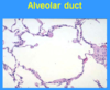
- Alveolar ducts
- About 14 million in the adult lung
- Form the final alveolated canals before reaching the air sacs
- About thirty alveoli arise in a spiral manner down the length of the alveolar duct
- Alveoli
- The average number of alveoli at birth is 25 million and in an adult lung is 375 million
- The surface area formed by the alveoli of a single normal adult lung is estimated to be between 600 and 800 square feet
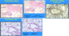
Pulmonary parenchyma
- If you scan this slide & try to estimate what is the amt of air vs. tissue on this slide
- 99% air
- 1% tissue
- Lung interstitium
- Tissue in these thin lines in the alveolar walls
- So thin in normal lungs that it basically doesn’t exist
- This image: normal
- Little tissue
- Little to no inflammatory cells
- No increased interstitium
- Unfortunately, there are interstitial lung diseases
- This thickness becomes significantly greater
- Thick walls separate atmospheric air from blood
- Gas exchange is significantly impaired

Lung acinus:
Alveolar wall
- The alveolar epithelium consists of two different types of cells
- Type I pneumocytes
- Type II pneumocytes
- Other characteristics of the alveolar wall
- The alveolar epithelium consists of two different types of cells
- Type I pneumocytes
- Cover 95% of the alveolar surface
- Flattened cells
- Ideally suited for gas exchange
- Type II pneumocytes
- Cover 5% of the alveolar surface
- Rounded cells that occupy the niches and corners of the alveolar walls
- Their most characteristic feature, the so-called “osmiophilic lamellar bodies,” represent surfactant material synthesized within the cells
- Surfactant has strong hydrophobic properties, decreases surface tension forces, and consists of surfactant proteins and lipids (dipalmityl lecithin, and dipalmityl phosphatidyl ethanolamine)
- Ability to proliferate, regenerate, & turn into type I pneumocytes (ex. if there’s damage to the alveolar structure)
- Type I pneumocytes
- Other characteristics of the alveolar wall
- Shared basement membrane
- Pulmonary capillary endothelium

Lung acinus:
Alveolar macrophages
- Large phagocytic cells that are present within alveoli
- Often they appear to be free in the lumen but are actually loosely attached to the epithelium
- They are derived from peripheral blood monocytes
- Like other macrophages, these cells ingest material, in this case dust and debris that gain access to the alveoli
- The lungs of smokers appear black because of the ingested carbon contained within the cytoplasm of macrophages
- Image: not normal b/c too many macrophages

Visceral pleura
-
Covers the surface of the lungs
- Also divides the underlying lung into lobules
- Consist of vascularized connective tissue, an elastic lamina, and an outer mesothelial layer
- Lined by mesothelial cells (simple squamous epithelium)
- Mesothelial cells are flattened and inconspicuous, but rapidly become rounded, cuboidal, and columnar in shape in response to damage

Vasculature, lymphatics, lymphoid tissue, & pleura:
Pulmonary vasculature
- The pulmonary vasculature includes…
- The lungs receive..
- The low-pressure pulmonary arteries run…
- The pulmonary circuit…
- The high-pressure bronchial arteries…
- The pulmonary vasculature includes…
- Pulmonary arteries
- Bronchial arteries
- Pulmonary veins
- Bronchial veins
- Alveolar microvasculature
- The lungs receive a dual arterial blood supply
- The low-pressure pulmonary arteries run alongside bronchi and bronchioles
- The pulmonary circuit continues to divide into the capillary networks of the alveolar walls
- The high-pressure bronchial arteries arise from the aorta and accompany the airways only to the terminal bronchioles

Vasculature, lymphatics, & lymphoid tissue
- Pulmonary arteries vs. veins
- Endothelium-lined lymphatic channels
- Lymphoid tissue
- The distal branches of the pulmonary arteries pass through the center of the
pulmonary lobules, whereas the pulmonary veins are situated at the edge of the
lobules- The pulmonary arterial system is designed to function as a low-resistance
maximum-flow system of vessels
- The pulmonary arterial system is designed to function as a low-resistance
- Endothelium-lined lymphatic channels are present around terminal bronchioles and the muscular branches of the pulmonary arteries
- Lymphoid tissue is distributed throughout the entire lung
- It can be divided into the central lymph nodes, lymphoid nodules, and aggregates of lymphocytes in the periphery of the lung, including the pleura
- Lymph nodes and aggregates consist mainly of T-lymphocytes and B-lymphocytes and constitute the part of the immune system designated as bronchus-associated lymphoid tissue (BALT)