2nd week 1 Flashcards
(44 cards)
Resting membrane potential:
Voltage difference across the membrane of a neuron when it is at rest (non-signalling)
Intracellular -70 mVs, (compared to extracellular 0mV)
Concentration gradient:
Positive or negative ions is higher/lower in one area than another.
Depolarisation:
A change in a neurons membrane potential that makes it more positive (less negative).
Hyperpolarization:
A change in a neurons membrane potential that makes it more negative. It is the opposite of depolarization.
Ionotropic receptors:
Transmembrane proteins that form a channel allowing ions to travel in/out of a cell.
These channels are opened when the receptor binds a ligand, like a neurotransmitter.
Glutamate receptors and GABA A receptors are examples of ionotropic receptors.
Voltage-gated ion channels:
Transmembrane proteins that form ion channels whose opening and closing is regulated by the membrane potential near the channel.
What types of electric activity:
hints
- up to 700-600volts (one 120mV)
- -70 mV (-80 to -60mV) present in most neurons
- 1 - 40 mV small variable changes
- 100 mv, fast, all or nothing
Large voltages generated by animals
(electric eels or rays: electroplaque)
Negative resting membrane potential
(most neurons)
Postsynaptic potentials
(small variable changes in membrane potential)
Action potentials
(large, fast, all or none fashion)
Explain functioning of an electroplaque
An electroplaque has Na/K pump maintaining membrane potential, operates on ATP.
When acetylcholine binds (ionotropic ligand gated) to nicotinic Ach receptor nAchR, sodium (Na+) flows in. depolarisation 120 mV.
Has electroplaques piled up, can create a shock up to 700V (volts)
concentration/electrical grade
draw directions


EPSPs are generated by activation of ion channels that let ….. ions into the cell –> …polarise neurons.
IPSPs are generated by activation of ion channels that let …. ions into the cell –> …polarise neurons.
EPSPs are generated by activation of ion channels that let positive ions into the cell –> depolarise neurons.
IPSPs are generated by activation of ion channels that let negative ions into the cell –> hyperpolarise neurons.
EPSPs and IPSPs are:
- graded in amplitude due to the ….. of neurotransmitter and …. the neurotransmitter is in the synaptic cleft
- additive, but decay in …. as they move around the neuron
concentration and length of time the neurotransmitter is in the synaptic cleft
amplitude
describe what happens to Nav and Kv (sodium and potassium gates) in
a) resting Vm
b) upstroke
c) peak
d) downstroke
e) resting Vm

Nav and Kv
a) closed, closed
b) open, closed
c) closed inactive, starts open
d) closed inactive, open
e) closed, closed
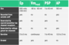
Electroplaque
Vm rest
PSP
AP
size
recorded from outside the cell
generated by ionotropic ion channel receptors (NT)
generated by voltage-gated ion channels
duration
graded
Field potential:
Electric potential in the extracellular space around neurons.
Nerve:
Nerve: a bundle of axons.
Compound axon potential:
Compound axon potential: the sum of the activity in a number of nerve fibers [axons].
Extracellular recording (ER):
(5 types)
- field potentials
- whole nerve activity
- multi-unit activity
- single unit activity
- multi-electrode arrays (MEAs)
Intracellular recording (IR):
(3 types)
- activity within single cells
- sharp electrodes
- patch suction electrodes
Single channel recording (SCR):
(2 types)
- recording activity of single ion channels
- patch clamp-type electrode
Extracellular recording (ER)
- the electrode is xxxside but close to the xxxx
- the electrodes pick up only xxxx potentials and xxx frequency filtered action potentials
- it is not possible to record xxx or post-synaptic potentials
- the electrode is outside but close to the neurons
- the electrodes pick up only field potentials and low frequency filtered action potentials
- it is not possible to record Vm rest or post-synaptic potentials
Example of recording field potential in mouse hippocampus, tri circuit
Stimulating electrode in tissue, Schaffer collaterals.
When stimulus given, activates Sch collaterals –> release of NT onto purkinje neurons in the area of CA1.
One electrode records the fEPSP and another sum of many AP of CA1 neurons = somatic population spike.
O’keefe & Nadel (1978); The Scripps Research Institute (2008)

Give an example of recording compound AP
Whole nerve recording
Frog sciatic nerve
Maximum capacity can be recording, adding voltage over hat won’t change the curve of an AP.
Mark CNS end, place on a dish over stimulating an recording electrodes. Apply olive oil at the ends, ringer in the middle (ions). Silicon grease between containers [conductance all the way through]
Lilley & Robbins (1998)
Rattus rattus has been used to separate different axons in the vagus nerve by measuring xxxx of the stimulus and their xxx xxxx.
Dochery et al. 2005
Rattus rattus has been used to separate different axons in the vagus nerve by measuring the intensity of the stimulus and their conducting velocity.
Multi-unit extracellular recording
- can de done in vivo?
Give an example
- rat LGN
yes
Electrode in rat brain, lateral geniculate nucleus.
Flash of light
Measures the neuron closest by but a neuron further away. Simultaneously measuring two neurons.
