Ophthalomolgy Flashcards

1
Q

What is Non Arteritic anterior ischemic optic neuropathy (AION)?

A

Occlusion of the short posterior ciliary arteries resulting in infarction of the optic nerve head.

2
Q

What are the risk factors for non arteritic AION?

A

Male Systemic ateriopathy 40-60yrs Small hypermetropic optic discs Hypertension Diabetes

3
Q

What are the symptoms and signs of non ateritic AION?

A

Painless, monocular sudden loss of vision

Can be asymptomatic if good vision in other eye Moderate to severely reduced visual acuity in the affected eye

Swollen optic nerve with splinter or flame shaped haemorrhages

Relative afferent pupillary defect

Visual field defect - most common inferior altitudinal defect

4
Q

How do you manage no arteritic AION and what is the prognosis?

A

Refer to ophthalmology - needs to exlcude GCA

R/V and treat all cardiovascular risk factors

Consider asprin

30% make substantial improvment

15-50% risk in other eye

Optic disc swellign resolves to leave optic disk pallor (loss of neural tissue)

5
Q

What is another name for giant cell arteritis?

A

Arteritic anterior ischemic optic neuropathy - optic nerve is damaged as the posterior ciliary arteries are blocked by inflammation

6
Q

How does GCA present?

A

Sudden loss of vision associated with headache

jaw and tongue claudication

tender scalp and temporal arteries

Loss of appetite and weight loss

symptoms of polymyalgia rheumatica

O/E: swollen optic disc and RAPD, flame shaped haemorrhages and cotton wool spots - retinal nerve fibre layer infarction

7
Q

How do you manage giant cell arteritis?

A

Ophthalomology referal

bloods for ESR (>47)

High dose steroids - prednisolone 80mg/24hrs ASAP

Temporal artery biopsy

8
Q

Describe the anatomy and physiology of the aqueous production of the eye

A

The CILIARY BODY - joins the iris to the choroid, it produces the aqueous humor and controls lens convexity

The ANTERIOR CHAMBER - the space between the cornea and the iris (shallower in the hypermetropic eyes as the eyes or smaller)

TRABECULAR MESHWORK - sieve like structure through which aqueous drains into the SCHLEMMS CANAL and out of the eye

The aqueous humour is produced and drained to maintain an appropriate intraoccular pressure.

9
Q

What is the mechanism in acute angle glaucoma?

A

Failure of the aqueous humour to pass through the pupil and drain out through the trabecular meshwork.

Occurs when:

  1. the iris is pushed forward and blocks the meshwork
  2. the aqueous fails to pass through the pupil (a pupil block)

The result is raised intraoccular pressure

10
Q

What are the risk factors for acute closed angle glaucoma?

A

Hypermetropia - eyes are normally smaller than usual and therefore have a shawllow anterior chamber which a narrower angle that is more prone to closure

Family history

Narrow anterior chamber angles

>30yrs - the lens thickens with age

Race - shallower angles in asians and eskimos

Women

11
Q

What are the symptoms and signs of acute closed angle glaucoma?

A

Unilateral sudden loss of vision

Painful red eye

Halos around lights

Nausea and vomiting

O/E: corneal oedema, oval unreactive pupil, raised IOP

12
Q

What is gonioscopy?

A

Uses prisms and a slit lamp to visualise the iridocorneal angle in 4 quadrants simultaneously

13
Q

How do you treat acute angle glaucoma?

A

acetazolamide 500mg IV STAT - decreases the aqeous formation

timolol 0.5% and apraclonidine 1% eye drops - misosis opens the blocked drainage angle.

Laser - YAG peripheral iridotomy to create an alternative outflow tract for the aqueous to drain through

14
Q

What is the anatomy of the retina?

A

Two main layers:

Neurosensory retina - in contact with the vitreous

The pigment layer - in contact with the choroid

The centre = the macula (5.5mm) and the fovea is the central area of the macula (high density of cones)

15
Q

What are the different types of retinal detachment?

A

rhegmatogenous - a seperation of the vitreous layer causing a tear in the retina and detachment. The sensory retina separates from the retinal pigment epithelium

Tractional retinal detachment: secondary to intraocular problems - melanoma or fibrous bands in the vitroeous, seen in DM

post cataract surgery or trauma

16
Q

What are the risk factors for retinal detachment?

A

MYOPIA - the higher the degree the higher the risk

previous retinal detachment in other eye

Trauma

17
Q

What are the symptoms of retinal detachment?

A

Floaters, Flashes, Field loss, Fall in acuity

Usually field loss unless the macula is affected and then there will be a central loss of vision

18
Q

How do you manage a retinal detachment?

A

REFER TO OPHTHALMOLOGY

the aim is to treat before the macula detaches.

Cyrobuckle surgery - silicome explant which indents the sclera to push back the retinal pigment epithelium against the retina and a cryotherapy probe to create a scar that seals the retinal break

Pars Plana vitrectomy - threet port pars plana cetrectomy involves removal of the vitreous, cryotherapy or laser to seal the break and then a bubble of gas to push the retina back against the retinal pigment epithelium.

19
Q

Describe the visual pathway:

A

Nerve fibres from each retina leave the eye at the optic disc and travel along the optic nerve

The optic nerves of each eye join at the optic chiasm

Nerve fibres from the nasal retina of each eye cross over at the chiasm and travel down the contralateral optic tract

Nerve fibres from the temporl retina do NOT cross over so travel down the ipsilteral tract

The optic tracts connect with the lateral geniculate body. At this point synapses are formed with the neurones passing through the optic radiations to reach the visual cortex.

20
Q

Describe the postion of lesions and what type of visual field defects they produce:

A
21
Q

What is a homonymous hemianopia?

A

Field loss affecting the corresponding half fields of each eye. eg left homonymous hemianopia, the left side of the visual field is affected which means the right side of the visual pathway is damaged.

Must be posterior to the chiasm as the loss is bilateral.

Stroke is most common cause, others include tumours

22
Q

What is a quandrantanopia?

A

Loss of a quarter of field of vision.

Bilateral to posterior to the chaism

Normally due to damage to the optic radiations.

A right homonymous inferior quadrantopia is due to lesion in the upper optic radiation in the left parietal lobe

23
Q

What causes a bitemporal hemianopia?

A

Lesions in the optic chiasm

eg pituitary tumours - usually cause bi-temporal superior quadrantopia.

24
Q

What causes central visual field defects?

A

Commonest cause = age related macula degeneration

Can be caused by optic nerve disease, often associated colour vision defects and visual acuity reduction

25
Q

When a patient presents with visual field defect what are the differentials?

A

Retinal detachment

Central retinal vein occulsion

Optic nerve disease

Visual pathway pathology

Glaucoma

Stroke

Pituitary tumour

26
Q

What causes diplopia?

A

When the visual axes of the two eyes deviate from fixating straight ahead or become misaligned during eye movement.

Displacement of globe in socket: tumours, trauma, infection

Cranial nerve palsies III, IV, VI

Cranial nerve palsies caused by DM and HTN

Decompensation of a latent squint

Extraocular muscle disease: MG

Thyroid eye disease

27
Q

What is the difference between monocular and binocluar diplopia?

A

Monocular - doube vision persists when one eye is covered. Caused by corneal abnormalities, uncorrected refractive error, cataractcs

Binocular - occurs when the two eyes become misaligned,

28
Q

Whic nerves supply the extraoccular muscles?

A

III:

Superior rectus, inferior rectus, medial rectus and inferior oblique

IV:

Superior oblique

VI:

Lateral rectus

29
Q

What would you see in a III nerve palsy?

A

exo hypo deviation - down and out postion

Due to superior oblique and lateral rectus working unoppsed.

Superior obiques main function is to internally rotate the eye, and depress and abduct it

30
Q

What are the causes of a IV nerve palsy?

A

DM, HT, demylinating disease, GCA, tumours, aneurysms

31
Q

What are the signs of a IV nerve palsy?

A

abnormal head posture - head tilts away from the lesion

Eye deviating upwards as it moves towards the nose

32
Q

What are the signs of a VI nerve palsy?

A

Patient suffereing with double vision especially when looking to the affected side.

Eye fails to abduct

33
Q

What are the causes of VI nerve palsy?

A

Adults: HTN, DM, MS, neoplasm, head trauma, infection, raised ICP

Children: transient in neonates, benign causes may occur 1-3 weks after viral infection

34
Q

How do you assess optic nerve function?

A

Visual acuity

Visual fields

Colour vision

Pupil reactions

35
Q

What are the causes of optic disc swelling?

A

Unilateral:

Central retinal vein occlusion, AION, papillits (inflammation of the optic nerve head), neuroretinitis

Bilateral:

Intracranial mass causing raises ICP, malignant hypertension, optic disc drusen

36
Q

What is the upper limit of size difference between the pupils that is physiological?

A

Up to 0.5mm of aniscoria (difference in size)

Physiological anisocria is the same in all levels of light where as pathological varies between light and dark conditions

37
Q

What determines pupil size?

A

The iris sphincter muscle is innervated by the parasympathetic division of cIII (occulmotor n). It constricts the pupil, miosis. Neurotransmitter is acetlycholine.

The pupillary reflex is a 4 neurone arc:

1st neurone passes from retina to the pretectal nucleus. 2nd neurone passes from the pretectal nucleus to the Edinger-Westphal nucleus. 3rd neurone passes from the EW nucleus to the cilary gangilon via the cIII. 4th neurone passes from the ciliary ganglion to the sphincter mucles via the ciliary nerves.

The dilator muscle is innervated by the sympathetic nervous system. Dilates the pupil, mydriasis.

Pupil mydrasis is a 3 neurone arc:

1st neruone passes from the posterior hypothalamus to the ciliospinal centre at the Budge at C8-T2. 2nd neruone passes from the spinal cord to the superior cervical ganglion. 3rd neurone passes from the superior cervical ganglion along the internal carotid artery through the cavernous sinus to the short posterior ciliary nerves and the dilator muscle.

38
Q

What is RADP and was does it signify?

A

Relative afferent pupillary defect.

Swing a light from one eye to the other, allow 3secs of illumination per eye. When the light is swung to the healthy eye both pupils will contstrict, but when it is moved to the defected eye both pupils constrict less and may appear to dilate.

It indicates asymmetrical optic nerve or retinal disease, the afferent signal is reduced. The efferent pathway is in tact as the defected eye has a consensual relfex when the light is shone in the other eye.

39
Q

What are the causes for an abnormally large pupil?

A

Holmes Adie pupil: poor reaction to light but constricts on accomodation. Part of Holmes Adie syndrome if there are reduced deep tendon relfexes.

Occulomotor palsy

Dilating eye drops

Traumatic mydriasis

Iris rubeosis: new blood vessel growth on the surface of the iris

In a bright room the larger pupil is likely to be the defect one.

40
Q

What are the causes of an abnormally small pupil?

A

Horner’s syndrome: disruption of the sympathetic supply to the eye. Associated with ptosis, and anhidrosis.

Pilocarpine eye drops

Uveitis/ posterior synechiae: inflammation causes adhesions between the pupil margin and the lens. The pupil is small and stuck down so cant dilate.

Physiological

In a dark room the small pupil is likely to be the defect

41
Q

What are the causes of Horner’s syndrome?

A

Posterior inferior ceebller artery or basilar artery occlusion

MS

Cavernous sinus thrombosis

Pancoast’s tumour

Hypothalamic lesions

Cervical adenopathy

Mediatinal masses

Pontine syringomelia

Kulmpke’s palsy

Aortic aneurysm

42
Q

What is an Argyll Robertson pupil?

A

Bilateral miosis, pupil irregularity and no response to light but there is a response to accomodation.

Seen in neurosyphilis and diabetes

43
Q

What is light near dissociation and what are the causes of it?

A

Where the pupil doesn’t respond to light but does respond to accomodation.

Causes: Argyll Robertson pupil. Holmes- Adie syndome, meningitis, alcoholism, tectal lesions, mesencephalic or thalamic lesions

44
Q

What is ptosis?

A

Drooping of the upper eyelid.

The eye lid is intervated ny occulmotor nerve.

45
Q

What presentations can give the false impression of ptosis?

A

Enophthalmous - posterior displacement of the eye makes the upper lid sulcus look deeper.

Dematochalsis: excess skin of the upper eyelid

Contralateral eyelid retraction

Contralateral proptosis

46
Q

What are the causes of ptosis?

A

Congential: Levator dystropy, Horner’s syndrome

Levator disinsertion: Age or trauma

Mechanical due to eye lid trauma

Occulomotor palsy: usually has an divergent squint, eye is down and out, and pupil may be dilates and unreactive

Myasthenia gravis: becomes worse throughout the day

Horner’s syndrome

Crhonic progressive external ophthalmoplegia (mitochonrial myopathy)

47
Q

How does light get focused onto the retina?

A

The cornea is the most refractive surface

The lens has variable focusing power: when near objects are focused on the ciliary body contratcs, relaxing the lens zonules so the lens becomes more convex and powerful.

The axial length helps determine focusing power.

48
Q

What is Emmetropia?

A

When there is no refractive error

49
Q

What is presbyopia?

A

Where with increasing age the lens looses its ablity to change shape and the point of closest focus moves further away for the eye.

Reading glasses are convex, providing extra power to focus the light on to the retina.

50
Q

What is hypermetropia?

A

Long sightedness - when the refractive power of the eye is too weak and light from distant objects falls behind the retina.

51
Q

What is myopia?

A

Short sightedness….the image falls in front of the retina

52
Q

What is astigmatism?

A

Where the eye has different refractive power in different meridians so there are mutlipe focal points on the retina.

The cornea is shaped liked a rugby ball rather than a football.

53
Q

What is keratoconus?

A

corneal collagen disorder where the cornea things and distorts.

Increase in corneal astigmatism and myopia, abnormal retinoscopy reflex (looks like a oil droplet), retinoscope reflex shows scissors movement.

Central corneal thinning and conical shape.

Bulging of the lower lif on dow gaze

Vertical lines in the corneal stroma

54
Q

What syndromes are associated with keratoconus?

A

Downs syndrome

Ehlers Danlos syndrome

Marfans syndrome

55
Q

How do you manage keratoconus?

A

Contact lens fit with Rigid gas permeable

Photorefractive surgery is contraindicated

Corneal graft may be needed in later stages if scaring is present

56
Q

What three bacteria ,ay cause neonatal conjuctivitis and how can you differentiate between them?

A

Chalmydia - creamy white discharge 5-12 days after birth

Gonorrhoea - 2-5days after birth, rapidly progressing, can lead to corneal ulceration.

Staphlococcus - yellow discharge

57
Q

In a baby with recurrent conjunctivits what must you consider?

A

Nasolacrimal duct obstruction - where the nasolacrimal duct fails to canalise by birth. Often resolves by its self in the first year of life but if not children may bee to be given a GA and have saline injected into the punctum to see if the canuli are open, and if not have a metal probe inserted to open them up.

58
Q

Describe some of the ocular presentations of non accidental injury:

A

Swelling or buising of the external eye

Lid swelling, bruising or cuts

Infestation of eyelashes with lice

Conjunctiva haemorrhages

Scaring or perforation of the cornea

Caracts

Vitreous haemorrhages

Retinal haemorrhages (normally in the posterior pole) or detachment

Papilloedema

59
Q

What is leucocoria?

A

White pupil

60
Q

In children what are the causes of leucocoria?

A

Congenital cataract - needs ophth referral ASAP and TORCH screen

Coats disease - rare idiopathic unilateral retinal telangiectasia. more common in boys around 5 yrs.

Persistent hyperplastic primary vitreous - when the embryonic vitreous fails to regress, usually unilateral, and vision is related to the extent of posterior retinal involvement.

Toxocariasis - rare worm infection, usually unilateral. Causes posterior or peripheral retinal granuloma. May be associated with chronic intraocular inflammation

Retinoblastoma - rare but most common intraocular malignanacy in children. Can be bilateral and hereditary. Needs urgent ophth referral.

61
Q

What is the inheritence pattern of retinoblastoma?

A

Hereditary retinoblastoma is different from non hereditary.

There is mutation of the RB gene on 13q14.

Autosomal dominant with 80% penetrance.

Those with the hereditary condition have only 1 allele in each cell and if the other allele develops a mutation then the conditiona arises

The RB is a tumour suppressor gene.

62
Q

What is the difference between a latent and a manifest squint?

A

Latent squint (phoria) is when there is an underlying tendency for the eyes to be misaligned but this can be controlled through subconscious effort.

Manifest squint (tropia) is when the eyes can not be controlled and they deviate

63
Q

How do you describe squints?

A

If the misaligned eye diverges in (towards the nose) when supposed to be looking straight ahead - convergent, this is called an ESO deviation. Esophoria or esotropia

If the misaligned eye diverges out (towards the temporal) when supposed to be looking straight ahead - divergent, this is an EXO deviation. Exophoria or exotropia.

If the visual axis is higher (the eye turns upwards) this is called a hypertropia.

64
Q

Describe the cover test:

A

The cover test is used to show squints.

When you cover the dominant eye the eye with the squint will move to correct itself. eg with a left exotropia when you cover the right eye the left eye will move inwards to correct the outward gaze.

65
Q

How do you treat squints?

A

Need to treat as soon as it is noticed.

Assess refractive state of the eyes using cyclopentolate 1% drops. and then corect this refractive error

Patch the good eye - helps to correct the loss of vision that occurs in the eye that is not aligning. the brain stops processing signals from that eye to reduce double vision in children, the process can be reversed if noticed soon enough.

Operations may be needed.

66
Q

What is retinopathy of prematurity?

A

Changes that happen to the retina in preterm babies.

The retina has no blood vessels until the 4th month of gestation, they grow from the optic disc towards to the peripheral retina.

Supplementary oxygen that is given to the preterm babies contributes to its development

>50% if babies weighing <1000g will have some ROP

67
Q

What are the stages of retinopathy of prematurity?

A
  1. demarcation line
  2. formation of three dimensional ridge
  3. fibrovascular proliferation at ridge
  4. partial retinal detachment (macula on v macula off)
  5. total retinal detachment
  6. agressive posterior retinopathy of prematurity
68
Q

What are the 3 parts of the lens?

A

Capsule (think outermost basement membrane)

Cortex (outer layer of lens epithelial cells)

Nucleus (central lens fibres

69
Q

What is a cataract?

A

Loss of transparency of the lens

70
Q

What are the symptoms and signs of cataracts?

A

Gradual reduction in visual acuity

Increasing myopia (short sightedness)

Glare

O/E - reduced visual acuity (usually in one eye)

diminished red relfex

difficult view of the fundus

71
Q

What are the risk factors for cataracts?

A

Most are age related - UV light and smoking increase risk

Cataracts occur earlier in DM, prolonged steriod use, trauma to eye, high myopia, dystrophia myotonica

72
Q

What causes congential cataracts?

A

1/3 are inherited, autosomal dominant

1/3 are caused by birht trauma, or maternal infection during pregnancy

73
Q

What different types of cataracts are there?

A

Nuclear

Cortical - spoke like from the edges

Posterior subcapsular - just deep to the lens capsule

74
Q

What are the risks with cataract surgery?

A

Development of posterior capsule opacification - can be treated by laser

Very rare

Rupture of the posterior lens capsule

Endophthalmitis (severe intraocular infection)

Suprachorodial haemorrhage ( severe)

75
Q

Descibe the procedure of cataract surgery:

A

Local anaestheisa

Self sealing corneal incision into the anterior chamber

Circular opening of the anterior lens capsule

Break up and aspiration of the lens material

Insertion of intraocular lens with chosen power to correct for any refractive error

76
Q

What are the different parts of the retina?

A

Macula - posterior pole of the eye, responsible for central vision

Fovea - in the centre of the macula, specialsed for high acuity vision

The rest of the retinal provides peripheral vision - only has rods

77
Q

What are the differences between the two types of Age related Macula Degeneration?

A

Both affect central vision, peripheral vision remains intact

Dry AMD-

Slowly progressing atrophy of the macular retinal pigment epitheial cells.

Drusen, mottled macular (hypo and hyper pigmentation) and atrophy on fundoscopy

Wet AMD-

New choroidal blood vessels grow into and under the retina. these then bleed or leak fluid causing macular oedema and scaring. Progresses quickly.

Green grey lesions consiting of new vessles, yellow exudates and retinal haemorrage on fundoscopy

78
Q

What is the main symptoms of age related macular degernation?

A

Loss of central vision

79
Q

What special tests can be done to confrim AMD?

A

Fluorescein angiography - identifies the areas of microvascular leakage

Optic coherence tomography - laser scanner is used to capture the cross section of the macular

80
Q

How do you manage AMD?

A

Use Amsler chart to monitor progress

Stop smoking

Counselling

Low vision aids

Dry AMD - no treatment

Wet AMD - regular intravetreal injections of anti-VEGF drugs

81
Q

What is primary open agle glaucoma?

A

Progressive optic neuropathy associated with optic nerve head damage (disc cupping) and visual field loss (usually peripheral) with raised IOP (21mmHg is the upper limit of normal)

Normal cup to disc ratio = 0.2/0.3, if above 0.6 needs investigating.

As the head of the optic nerve gets damaged the neuroretinal rim ges thinner so the cup gets bigger

82
Q

What is the pathogeneis of open angle glaucoma?

A

The trabecular meshwork may not drain as well with age so the IOP pressure rises.

This raised IOP can damage the nerve fibres of the optic nerve.

* can have increased IOP without optic nerve damage = ocular hypertension*

** can have normal IOP but optic nerve cupping and visual field loss = normal tension glaucoma**

83
Q

What are the risk factors for primary open angle glaucoma?

A

high IOP

family history

age

diabetes

myopia

black african or caribbean

* screening is very important as IOP is often raised asympotmatically, and when symptoms occur the damage is done*

84
Q

What is the treatment for primary open angle glaucoma?

A

Suppress the aqueous humour production: B blockers, carbonic anhydrase inhibitors and alpha agonist eye drops

OR

Increase the aqueous outflow: prostaglandin analogues

85
Q

What is Trachoma?

A

Bilateral follicular, non purulent conjunctivitis

Caused by Chlamydia trachomatis - spread by flys

Follicles develop on the superor tarsal conjuctiva and superior limbus after 2 -3 weeks

Repeated episodes leads to conjunctival scaring (cicatrisation), which causes entropian, trichiasis (eye lashes grow towards the cornea) and a dry eye.

86
Q

How do you diagnosis and trest Trachoma?

A

Diagnose using conjunctival scrapes and treat with oral antibiotics.

To reduce chronic development use tetracycline eye drops BD for 5 days each month for 6 months

87
Q

What is the other name for onchoceriasis and what causes it?

A

River Blindness

Caused by a naematode microfilaria (Onchocerca volvulus) transmitted by black flys of the Simulium species.

The flies bite and subcutaneous nodules form, the adult female worm sheds microfilariare which then migrate through the skin, eye involvement occurs when there is chronic infestation via the conjuctiva.

88
Q

What are the ocular signs of Onchoceriasis?

A

Lid nodules and depigmentation

Chronic conjunctivits and punctate keratitis

Corneal opacification

Iritis

Cataract

Gluacoma

Optic neuritis

89
Q

What caues xerophthalmia?

A

Vitamin A deficiency

It results in night blindess, corneal and conjunctival dryness and keratinisation. Corneal ulcers which can lead to perforation, and in its advance form - cornal necrosis.

Bitots spots form - small foamy plaques

90
Q

What signs might be present to suggest a person with a gradual loss of vision had choroiditis?

A

Acutely:

Blurred vision, grey white raised patch see on retina, vitreous opacities, cells in the anterior chamber.

Chronically:

Choroidoretinal scar - white patch with pigmentation around, will be symptomless unless they involve the retina

91
Q

What would you use fluorescein staining for?

A

To visualise any corneal abrasions

92
Q

What does linear vertical fluorescein staining on the superior cornea suggest?

A

Sub tarsal foreign body

93
Q

What is the difference between a penetrating eye injury and a perforating eye injury?

A

A penetrating describes an entrance wound only (need to assess for intraocular foreign body)

A perforating describes an entrance and exit wound to the eye

94
Q

Why is important to find out the history of what metal a person was working with when they injured their eye?

A

Ferrous metals can cause siderosis which leaks toxic ions into the eye, these ions are retinotoxic and can impair retinal function irreversibly.

95
Q

What are the signs of a ruptured globe?

A

Severe sub conjuctival haemorrhage

Hyphaema - blood in the anterior chamber

Restriction of eye movements, often with diplopia

Extruded intraocular contents

Collapsed globe

Irregular pupil and subluxed lens

Commotio retinae - retinal oedema

Traumatic optic neuropathy and retinal breaks

Vitreous haemorrhage

96
Q

What is the main treatment for chemical injury to the eye?

A

Irrigation - DO NOT DELAY

97
Q

What is traumatic optic neuropathy?

A

Damage often indirect to the optic nerve

Usually caused by a blow to the head face or orbit

98
Q

What is the most common orbital fracture?

A

A blow out of the orbital floor - can lead to herniation of inferior rectus into the maxillary sinus, restricting upward gaze

99
Q

Bacterial conjuctivitis:

Common causative organisms?

Signs and symptoms?

Management?

A

Staph aureus and strep pneumoniae are the most common

Patients complain of irritation and tearing, discharge, eyelids stuck together in the morning, red eye, often starts in one eye and spreads to the other.

O/E - mucopurulent discharge, lid erythema and oedema, conjunctival injection, papillae

Use chloramphenicol QDS for 1 week, ensure good higene

100
Q

Viral conjunctivitis:

Causative organisms

Symptoms and signs

A

Adenovirus type 3 = common

Patient complain of: acute red eye, watering, irritation and soreness, systemic viral symptoms, bilateral in 40%, intermittent blurred vision.

O/E: conjuctival injection, chemosis, watery discharge, eye lid swelling, follicules, preauricular adenopathy. May develope punctate keratitis.

101
Q

Allergic conjuctivitis:

Hx?

Symptoms and signs?

Management?

A

History of atopic disease, acute contact with allergen or seasonal

Patient complains of itching, tearing and intermitted bluring vision.

O/E: bilateral, eyelid oedema, normal cornea, conjuctival injection, papillae, watery/stringy mucoid discharge

Management: cold compresses, artificial tears, topical mast cell stabilisers Sodium cromoglicate

102
Q

When should a diagnosis of chlamydial conjuctivitis be considered and how is it confirmed?

A

If a viral or bacteral conjunctivitis has lasted over 3 weeks.

Fluorescent antibody stain, Giemsa stain: intracytoplasmis inclusion bodies in epithelial cells, polymorphonuclear leukocytes and lymphocytes.

103
Q

What is bacterial keratitis?

Risk factors?

Signs O/E?

Management?

A

Serious unilateral corneal infection

Risk factors: hx of trauma, corneal foreign body, contact lenses, corneal exposure

Signs: White corneal infiltrate with overlying epithelial defect, conjunctival injection, mucopurulent discharge, anterior chamber cells, hypopyon.

Refer to ophthalmology for: corneal scrapings, stop wearing contact lenses, hourly topical antibiotics

104
Q

What sign is diagnostic for herpes simplex in the eye?

A

Dendritic ulcers on staining

105
Q

What is the uvea?

A

The pigmented part of the eye.

The iris and cilary body are the anterior uvea and the choroid is the posterior component

106
Q

What are the symptoms of anterior uveitis?

What are the complications of anterior uveitis?

A

Painful red eye (circum corneal injection)

Photophobia

Normal/mildly decreased vision

O/E keratic precipitates, distorted or contricted pupil, hyponyon

Complications: posterior synechiae, secondary glaucoma, secondary cataract

107
Q

What are some of the causes of anterior uveitis?

A

Idiopathic - most common

Autoimmune HLA B27 - ank spond etc

Juvenile chronic arthritis

IBD

Non infectious systemic disease - sarcoidosis, MS

Infections - TB, syphilis, herpes zoster

Traumatic

Post op

108
Q

What is worrying with regards to a subconjuctiva haemorrhage?

A

When there is no posterior margin - may be associated with an intracranial bleed

109
Q

What are the signs and symptoms of episcleritis?

A

The episclera is the most superficial layer of the sclera.

Patients complain of localised redness in eye, associated with an ache or grittiness. They have normal acuity or mild bluring.

May nee NSAIDs to manage but normally self limiting

Usually idiopathic but may complicate rheumatic fever, SLE, polyarteritis nodosum

110
Q

What are the symptoms and signs of scleritis?

A

Gradual onset of pain and redness in the eye.

Very severe pain, assoicated with photophobia, but normal or only mildly blurred vision.

O/E: localised or diffuse scleral hyperaemia which is tender to palpation. If there are pale areas within the red zones this may indicate necrtoising scleritis

111
Q

What systemic illness’ are associated with scleritis?

A

Herpes zoster ophthalmicus

RA

Relapsing polychondritis

Wegner’s granulomatosis

Polyarteritis nodosa

SLE

112
Q

What is a chalazion?

A

Chronic condition caused by inflammation of the meibonium glands with secondary lipogranulomatous inflammtion.

Causes a swelling on the eyelid

113
Q

What is the difference between preseptal and orbital cellulitis?

A

Preseptal is an infection of the subcutaneous tissue anterior to the orbital septum

Orbital is inflamamtion of the soft tissue posterior to the obital septum

Both are medical emergencies

114
Q

What are the signs on fundoscopy of a central retinal artery occulusion?

A

White retina, cherry red spot at the macula.

Causes sudden loss of vision and afferent pupil defect.

115
Q

What are the signs of a retinal vein occulsion on fundoscopy?

A

ischemic retinal vein occulsion - cotton wool spots, swollen optic nerve, macular oedema, neovascuarlization

116
Q

What is retinitis pigmentosa?

A

Inherited degeneration of the retina.

Different forms of inheritence, results in reduced visual acuity

117
Q

What does toxoplasmosis look like in the eye?

A

Punched out pigmented chorioretinal scars

118
Q

What are the staged in thyroid eye disease?

A

No symptoms or signs

Only lid retraction +/- lid lag

Soft tissue involvement

Proptosis

Extraocular muscle involvement (diplopia)

Corneal disease (due to inability to close eye lids)

Sight threatening optic neuropathy

119
Q

What eye symptoms can patients with MS present with?

A

Optic neuritis - reduced visual acuity and colour vision (red desaturation), pain on moving eye

Visual field defects

Cranial nerve palsies - normall 6th

Nystagmus

120
Q

What does CMV retinitis present as?

A

HIV, low CD4 count

Paravascular retinal opacification from retinitis with associated haemorrhage

121
Q

What is HIV retinopathy?

A

Cotton wool spots

Retinal microhaemorrhages may occur

122
Q

What are the signs of hypertensive retinopathy?

A

Flame haemorrhages

Focal arteriolar narrowing

Cotton wool spots

Lipid exudates

Coper and silver wiring of the vessels

Normal optic disc

** often asymptomatic**

123
Q

How do you classify hypertensive retinopathy?

A

Grade 0 = normal

Grade 1 = arteriolar narrowing

Grade 2 = focal arteriolar narrowinf and AV crossing changes (AV nipping)

Grade 3 = haemorrhages, exudates and cotton wool spots

Grade 4 = grade 3 plus disc swelling

124
Q

What are the ocular manifestations of diabetes?

A

Diabetic retinopathy - due to microangiography in the capillaries, pre capillary arterioles and the venules leading to occulsion and leakage

Diabetic maculopathy - oedema of the macula from vessel leakage

Extraocular muscle palsy - an isolated 3rd, 4th or 6th palsy with paina round the eye. Usually if there is a 3rd it will spare the pupil

Retinal vein occulsions

Cataract - posterior subcapsular

125
Q

What is the likelihood of a diabetic patient getting eye problems?

A

20yrs after diagnosis - virtually ALL type 1 and 60% of type 2 will have retinopathy

30 yrs after diagnosis - 30% of type 1 and 3% of type 2 will have proliferative disease

126
Q

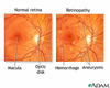
What are the retinal signs on fundoscopy of diabetic retinopathy?

A

Microaneurysms

Dot haemorrhages

Blot haemorrhages

Exudates

Cotton wool spots

Retinal oedema

Venous changes

Neovascularisation

127
Q

How is diabetic retinopathy classified?

A

Degree of retinopathy is taken into account

Presence of maculopathy is stated

If there is any evidence of previous photocoagulation this is stated

128
Q

What signs are seen at fundoscopy in diabetic maculopathy?

A

Macular odema, exudates, ischemia