The cranial cavity and face week 1 Flashcards
Identify the features of the roof of the cranial cavity.


What is the name of this part of the cranium?

anterior cranial fossa
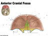
Identify the bones/foramina/prominences associated with the anterior cranial fossa.


What portion of the anterior cranial fossa does the frontal bone form? What does the frontal bone do for the orbit?
What portion of the anterior cranial fossa does the ethmoid bone form?
What is the function of the crista galli? What bone is it a part of?
What is the function of the foramen cecum?
What is the purpose of the foramina in the cribiform plate? What bone is the cribiform plate a part of?
What is the function of the anterior clinoid process?
Bones Associated with the Anterior Cranial Fossa:
- Frontal bone- is found anteriorly and forms the floor of the anterior cranial fossa and a roof over the orbit
- Ethmoid bone- is located centrally in the anterior cranial fossa forming part of the floor
- Sphenoid Bone- specifically the body and lesser wing of the sphenoid bone, are found posteriorly in the anterior cranial fossa
Foramina/Bony Prominences Associated with the Anterior Cranial Fossa:
- Crista Galli- part of the ethmoid bone which is the point of attachment for the falx cerebri (defined later in this lecture)
- Cribiform Plate of Ethmoid Bone- Contains foramina for passage of the olfactory nerves from the nasal cavities to the olfactory bulb of the brain
- Foramen Cecum- found between the frontal and ethmoid bones, this foramen allows passage of emissary veins which connect the nasal cavity with the superior sagittal sinus
- Anterior Clinoid Process- Part of lesser wing of sphenoid bone and site of attachment of tentorium cerebelli (defined later in this lecture)

Identify the bones/foramina/bony prominences of the middle cranial fossa.


What parts of the middle cranial fossa does the sphenoid bone contribute to?
What parts of the temporal bone contribute to the middle cranial fossa?
What part of the middle cranial fossa does the parietal bone contribute to?
Bones Associated with the Middle Cranial Fossa:
- Sphenoid Bone- Specifically the body and greater wings of the sphenoid bone contribute to the floor and lateral aspects of this fossa
- Temporal bone- Specifically the squamous part laterally and the petrous part posteriorly.
- Parietal Bone- Contributes to the lateral boundaries

What bone is the optic canal formed in? What is the purpose of the optic canal?
Where is the superior orbital fissure located? What does it transmit?
What does the foramen rotundum transmit?
What does the foramen ovale transmit?
What does the foramen spinosum transmit?
What organ is located in the hypophysial fossa?
Foramina/Bony Prominences Associated with the Middle Cranial Fossa:
- Optic canal: in the sphenoid bone allows passage of the optic nerve (CN II) from the orbit to the brain. Also transmits the opthalmic artery. This foramen may be considered part of the anterior or middle cranial fossa.
- Superior Orbital Fissure- is found between the greater and lesser wings of the sphenoid bone and transmits ophthalmic veins and nerves (CN III, CN IV, CN V1, CN VI, and sympathetic fibers entering the orbit).
- Foramen Rotundum- transmits V2 (maxillary nerve) to supply the skin, teeth, and mucosa related to the maxilla (upper jaw)
- Foramen Ovale- opens inferiorly to the infratemporal fossa and transmits CN V3 (mandibular nerve) and accessory meningeal artery
- Foramen Spinosum- also connects with the infratemporal fossa and transmits the middle meningeal artery (the groove for which can clearly be seen on the floor and lateral wall of the middle cranial fossa.
- Hypophysial Fossa: Note that this fossa houses the pituitary gland. You will learn later that the pituitary gland is surrounded by the Circle of Willis, a critical component of the vascular supply to the brain.

Identify the bones/foramina/bony prominences of the posterior cranial fossa.


What portion of the posterior cranial fossa does the occipital bone contribute to?
What is the contribution of the sphenoid bone to the posterior cranial fossa?
What parts of the temporal bone are in the posterior cranial fossa?
Bones Associated with the Posterior Cranial Fossa:
- Occipital bone- forms most of the posterior cranial cavity
- Sphenoid Bone- the dorsum sellae marks the anterior most aspect of this fossa
- Temporal Bone- the petrous and mastoid parts of the temporal bone contribute to the anterior-lateral walls of the fossa
- Parietal Bone

What is the internal occipital protuberance and grooves for transverse and sigmoid sinuses formed by?
What structures pass through the jugular foramen?
What does the internal acoustic meatus transmit?
What goes through the hypoglossal canal?
What lies against the clivus?
Foramina/Bony Prominences Associated with the Posterior Cranial Fossa:
- Internal occipital protuberance- is formed in relation to the confluence of sinuses (defined later in this lecture)
- Grooves for the transverse and sigmoid sinuses- are also created by the dural (cranial) sinuses (defined later in this lecture)
- Jugular foramen- transmits CN IX, X, and XI as well as the sigmoid sinus as it exits the skull as the internal jugular vein
- Internal acoustic meatus- transmits CN VII and VIII as well as the labyrinthine artery
- Hypoglossal canal- transmits the hypoglossal nerve (CN XII)
- The clivus: the brainstem, specifically the pons, lies against the clivus and extends from this region of bone to the foramen magnum, the large opening at the base of the skull. It is possible for the brainstem to “slide downward” along the clivus herniating into the foramen magnum. This typically would result from a pressure difference between the cranial and spinal cavities.

What is the difference between cranial dura and spinal dura?
Cranial dura mater is continuous with spinal dura mater, though the cranium the dura mater exists in 2 layers.
What 2 types of structures are created by separation of cranial dura mater?
The cranial dura mater exists in two layers which are fused together as the dura lines the cranial cavity. There are locations within the cranial cavity where the two layer of dura separate creating two types of structures:
1) Dural partitions: which incompletely separate parts of the brain
2) Dural venous sinuses (a.k.a. intracranial venous structures)
Where is the falx cerebri located? What does it do? What is the falx cerebri attahced to?
Where is the tentorium cerebelli located? What does it do? What is the tentorium cerebelli attached to?
Where is the falx cerebelli located? What does it do? What is the falx cerebelli attached to?
What does the diaphragma sellae do?
- Falx cerebri: is a crescent shaped structure that projects downward between the two cerebral hemispheres. It is attached anteriorly to the crista galli and the frontal crest (of the frontal bone) and blends posteriorly with the tentorium cerebelli. The falx is visible on CT imaging and should lie in the midline between the two cerebral hemispheres. Pathological processes inside the cranial cavity (e.g. a tumor or a bleed) may cause a deviation of the falx to one side.
- Tentorium cerebelli: is a horizontal projection of dura that covers the posterior cranial fossa. It attaches posteriorly to the occipital bone along the groove for the transverse sinuses, laterally to the petrous part of the temporal bone and anteriorly to the clinoid processes. This piece of dura separates the cerebellum (below) from the cerebrum (above).
- Falx cerebelli: is a small midline projection of dura within the posterior cranial fossa. It is attached superiorly to the tentorium cerebelli and posteriorly to the occipital bone. It separates the two halves of the cerebellum.
- Diaphragma Sellae: is a small horizontal shelf of dura that covers over the hypophysial fossa, which houses the pituitary gland.

meningiomas
Meningiomas are benign intracranial tumors which may arise from any part of the meninges and impact the surrounding brain tissue.
Where is the cerebrum located with respect to the tentorium cerebelli?
The corpus callosum (connects hemispheres of the brain) is situated where in relation to the falx cerebri?
What fossa does the frontal lobe of the brain lie in? What fossa does the temporal lobe lie in?
o The cerebrum (cerebral cortex & underlying structures) is situated above the tentorium cerebelli.
o The cerebrum is partitioned into right and left hemispheres by the falx cerebri in the midsagittal plane.
o The corpus callosum connecting the two hemispheres passes just beneath the falx cerebri.
o The frontal lobe of the brain lies in the anterior cranial fossa and the temporal lobes fill the middle fossa.

Between what menigeal layer(s) are dural venous sinuses located?
What do dural venous sinuses ultimately drain into?
What 2 types of veins drain into dural venous sinuses? Where do these veins drain blood from?
- Venous drainage of the brain ultimately empties into the dural venous sinuses.
- The dural sinuses are endothelial-lined spaces between the outer periosteal and inner meningeal layers of dura.
- This network of dural sinuses will drain into the internal jugular veins.
- Also draining into these sinuses are:
- Diploic veins: draining blood from the skull
- Emissary Veins: draining blood from outside the cranial cavity.

Identify the indicated veins.


What sinuses drain into the confluence of sinuses?
What sinuses drain blood from the confluence of sinuses?
The superior sagittal and straight sinuses as well as the occipital sinus (in the falx cerebelli) drain into the confluence of sinuses.
The paired transverse sinuses drain blood from the confluence of sinuses.

What drains into the paired transverse sinuses? What do the paired transverse sinuses become? What does this newly named sinus drain into?
The paired transverse sinuses drain blood from the confluence of sinuses and also receive the superior petrosal sinuses. The transverse sinuses become the sigmoid sinuses which receive blood from other regions of the brain/cranial cavity before ending at the beginning of the internal jugular veins.

What structures pass through the cavernous sinuses?
What structures pass through the lateral walls of the cavernous sinuses?
What structures do the cavernous sinuses receive blood from?
What is significant about cavernous sinuses as it pertains to spread of infection?
Why are structures passing through the wall of cavernous sinuses vulnerable to injury?
The cavernous sinuses are part of the system of dural venous sinuses. They are of great clinical importance because of their connections and the structures that pass through them.
Structures passing through each cavernous sinus:
- Internal carotid artery
- Abducent Nerve (CN VI)
Structures in the lateral wall of the cavernous sinus:
- Oculomotor nerve (CN III)
- Trochlear nerve (CN IV)
- Ophthalmic nerve (CN V1)
- Maxillary nerve (CN V2)
**The cavernous sinuses receive blood from cerebral veins but also from emissary veins AND the ophthalmic veins which drain the orbit.
**These extra-cranial connections are a potential pathway for spread of infections intracranially.
**Also, because structures are passing through the wall of the cavernous sinus they are vulnerable to injury due to inflammation or thrombosis in the sinus.

Identify the indicated muscles of the face (red boxes).


Where is the parotid gland located with respect to the ear?
What bony structures does the parotid gland extend from?
Landmarks:
- is anterior and slightly below the lower half of the ear
- extends from the zygomatic arch to the lower border of the mandible.

What structures does the main excretory duct of the parotid gland pierce to enter the mouth?
What nerve passes into the parotid gland and divides into its terminal branches within the parotid gland?
What artery enters or passes deep to the parotid gland?
Where is the retromandibular vein formed? What 2 veins come together to form this vein?
- The main excretory duct of the parotid gland crosses lateral to the masseter and pierces the buccal fat pad and the buccinator muscle to enter the mouth near the second upper molar.
- The facial nerve will pass into the parotid gland and typically divide into its five terminal branches within the substance of the gland. This relationship is clinically significant because it means that surgical removal of the parotid gland is quite difficult if all branches of the facial nerve are to be spared.
- The external carotid artery also enters or passes deep to the parotid gland.
- The retromandibular vein is formed within the parotid gland by the union of the superficial temporal and maxillary veins

Name the indicated nerves labeled in the attached picture.

Note that dorsal (posterior) rami innervate skin on the back of the head /scalp and the skin of the neck is innervated by ventral rami, specifically from branches of the cervical plexus (e.g. transverse cervical nerve). We first discussed these branches when we studied the posterior triangle of the neck.

Identify the indicated arteries of the face.


Identify the indicated branches of the maxillary artery.


What is the major vein that drains the face?
Why can infections in the face spread intracranially?
The facial vein is the major vein draining the face. Numerous connections exist between the facial vein and veins draining deeper regions of the face. These venous channels have interconnections with the cavernous sinus. There are no valves in any veins of the face so blood may flow in either direction. Because of these connections infections in the face can spread intracranially.

What lymph nodes drain the face?
State the specific parts of the face each node drains.
Lymphatic drainage of the face primarily moves towards three groups of lymph nodes:
1) Submental nodes: Drain the lower lip and chin bilaterally
2) Submandibular nodes: Drain part of the orbit, the external nose, part of the cheek, the upper lip, and part of the lower lip
3) Pre-auricular and Parotid nodes: Drain most of the eyelids, part of the external nose, and part of the cheek.
