Motor I Flashcards
Supplementary Motor Area (SMA) and Premotor cortex (PMC)
- Located in the frontal lobe (brodmann’s area 6)
- PROGRAM the “design” and “sequence” of complex movements involving groups of muscles
- TRANMITS the “program” of intedend movement to Primary motor cortex for execution
- Control axial (trunk) and prosimal limb (girdle) musculature of the upper and lower limbs (helps to orient the trunk and/or limbs towards the intended direction of movement)

Frontal Eye Field (FEF)
- Corresponds to Brodmann’s area 9
- Projects to brain stem centers that control ocular movements
- COORDINATES EYE MOVEMENT
- Plays a role in visual tracking

Posterior parietal cortex
- Broadmann’s area 7 (In superior parietal lobule)
- associated with VISUAL GUIDANCE OF MOVEMENT
- evaluates location or position of body/body parts, and forms a movement plan that would accomplish a task/reach a target

Primary motor cortex (M-I)
- Located in PRECENTRAL GYRUS (broadmann’s area)
- Function = EXECUTION of distinct, well defined, voluntary movement
- Gives rise to axons that descend to terminate in the brainstem and spinal cord
- CONTROLS MOVEMENTS OFF THE OPPOSITE SIDE OF BODY
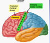
Primary Somatosensory (somesthetic) cortex (S-I)
- Corresponds to Brodmann’s area 3,1,2 (Postcentral gyrus)
- Gives rise to fibers that descend to terminate in the brain stem and spinal cord
- does NOT produce movement, but influences it instead
–> modulates the relay of sensory input from visceral and somatic structures to the spinal cord
–> acts as a filter or attenuated by descending fibers arising from somatosensory cortex
Internal pyramidal layer
- Layer V of cerebral cortex
- very prominent in the MOTOR cortex
- contains pyramidal cell bodies
Pyramidal cell bodies
- pyramidal cells are OUTPUT neurons of the motor cortex
- Pyramidal cells are UPPER MOTOR NEURONS (UMNs)
- give rise to axons that form DESCENDING MOTOR PATHWAYS
–> axon terminals synpase mostly with interneurons
–> interneurons in turn, synapse with motorneurons
- axon terminals synapse LESS OFTEN directly with motoneurons

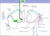
Upper Motor Neurons
- Cell bodies reside in the motor cortex or the brainstem
- INFLUENCE LOWER MOTOR NEURONS (LMN) located in the brainstem or spinal cord
- see descending axon of UMN and lower motor neuron in ventral horm of spinal cord
Examples of UMN
- corticonuclear tract
- anterior and lateral corticospinal tracts
- corticoreticular and reticulospinal tracts
- corticorubral and rubrospinal tracts
- vestibulospinal tracts
Lower Motor Neurons
- LMN that control movement of the body, reside in the ventral horn of the spinal cord
- their axons run in peripheral nerves that terminate in skeletal muscle
- innervate skeletal muscle with motor intervation
Describe the origin of the corticospinal tract
- 1/3 from brodmann’s area 4
- 1/3 from brodmann’s area 6
- 1/3 from bodmann’s area 5, 7 and 3, 1, 2
Describe the course of the corticospinal tract in Brain
- descends through the CORONA RADIATA, POSTERIOR LIMB OF THE INTERNAL CAPSULE, BASIS PEDUNCULI, PONS and MEDULLA
- In the medulla, the corticospinal tract fibers assemble to descend in the pyramid
- 85-90% of fibers decussate in the PYRAMIDAL DECUSSATION (CAUDAL MEDULLA) to the opposite side
- remaining 10-15% of fibers do NOT decussate, but instead, descend on the same side of Origin
describe the course of the lateral corticospinal tract in the spinal cord
- CROSS FIBERS descend in the LATERAL FUNICULUS of all spinal cord levels as the LATERAL CORTICOSPINAL TRACT
- Fibers synapse AT ALL SPINAL CORD LEVELS (concentrated in cervical and LS levels)
–> 55% of fibers terminate in Cervical cord
–> 20% of fibers terminate in thoracic cord
–> 25% of fibers terminate in lumbosacral cord
**Controls muscles of the upper and lower limbs, ESPECIALLY DISTAL MUSCLES OF UPPER LIMB**
** Involved in execution of distinct, skilled, well-defined manipulative, and independent voluntary movement of the fingers**
Describe the course of the Anterior corticospinal in the spinal cord
- UNCROSSED fibers DESCEND in the anterior funiculus of the cervical and upper thoracic spinal cord levels as the ANTERIOR CORTICOSPINAL TRACT
- Fibers DECUSSATE AT The level of their termination
- DESENDING AXONS OF THIS TRACT ARE THE ONLY UMN AXONS THAT DECUSSATE IN THE SPINAL CORD
**Controls the AXIAL MUSCLES (neck, shoulder, and trunk)**
Describe the signs of an upper motor neuron lesion
- Spastic paralysis
- hypereflexia
- mild muscle atrophy (wasting)
- babinski’s sign (extensor plantar response)
Blood supply to medial surface (hip, leg, and foot area) of the precentral gyrus
- anterior cerebral artery
- Occlusions result in motor deficits in contralateral leg and foot

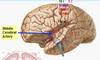
Blood supply to Lateral surface (trunk, upper limb, head area) of the precentral gyrus
- middle cerebral artery
- occlusion results in motor deficits in contralateral upper limb and face