cerebral hemisphere Flashcards

1
Q

Achicotex

A
  • part of allocortex (ancient center)
  • contains 3 layers of gray matter
  • involved in memory and emtions
  • HIPPOCAMPAL FORMATION
2
Q

Paleocortex

A
  • Part of allocortex (ancient cortex)
  • made up of 3-5 layers of gray matter
  • PARAHIPPOCAMPAL GYRUS, uncus and the lateral olfactory gyrus (olfactory system)
3
Q

Mesocortex

A
  • Part of allocrotex (ancient center)
  • made up of 3-6 layers of gray matter
  • cingulate gyrus and the insula
4
Q

describe Neocortex

A
  • is the evolutionarily newer cortex (makes up 90% of cerebral cortex)
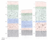
  • made up of 6-layers present with regional variation

–> HOMOTYPICAL CORTEX = association areas (all 6 layers are present)

–> HETEROTYPICAL CORTEX = PRIMARY AREAS

1) agranular = primary motor cortex
2) granular = primary sensory cortex

5
Q

define pyramidal cells

A
  • most abundant, 75% of cortical cells
  • Long axons FORM WHITE MATTER TRACTS
6
Q

Fusiform

A
  • Modified pyramidal
  • Located in the DEEPEST cortical layers
  • Axons PROJECT TO THALAMUS
7
Q

define Stellate (granule) cells

A
  • Involved in integration
  • Aspiny = INHIBITORY INTERNEURONS
  • Spiny = EXCITATORY interneurons, located in layer IV of the gray matter
8
Q

Horizontal cells of cajal

A
  • located in SUPERFICIAL most layer of gray matter
  • Seldom seen or completely lacking in adult brain
9
Q

Cells of MARTINOTTI

A
  • Interneurons
  • located in DEEP LAYERS of the gray matter
10
Q

Association fibers

A
  • Intracortical fibers = superior longitudinal fasciculus, uncinate fasciulus, occipitofrontal fasiculus
  • Send projections WITHIN SAME HEMISPHERE (IPSILATERAL)
11
Q

Commissural fibers

A
  • Commissural fibers = corpus callosum, anterior/posterior commissures
  • TO/FROM OPPOSITE HEMISPHERE (CONTRALATERAL)
12
Q

Projection fibers

A
  • Ex = crus cerebri, internal capsule
  • TO/FROM CORTEX and SPINAL CORD = corticobulbar, corticothalamic, corticopontine
  • INTERNAL CAPSULE
13
Q

Molecular layer (I)

A
  • Synaptic area = axons and MOSTLY DENDRITES
  • neuroglia and cells of cajal
  • INTEGRATION (sends axons horizontally)
14
Q

External granular layer (II)

A
  • Small pyramidal and STELLATE CELLS
  • Axons and dendrites from deeper layers
  • INTEGRATION (sends cells horizontally)
15
Q

External Pyramidal layer (III)

A
  • Moderate size PYRAMIDAL cells
  • EFFERENT LAYER (OUTPUT LAYER)
  • Corticocortical fibers = association and commissural fibers
16
Q

Internal granular layer (IV)

A
  • STELLATE CELLS DENSLY PACKED
  • outer band of baillarger = myelin staining
  • AFFERENT LAYER = CHIEF INPUT LAYER (EXPANDED IN SENSORY CORTEXS)
  • thalamic afferents = VPL, VPM-thalmocortical fibers
17
Q

Internal pyramidal layer (V)

A
  • GIANT PYRAMIDAL CELLS of betz, smaller number of stellate cells, cells of martinotti
  • Inner band of baillarger- myelin staining
  • EFFERENT LAYER (OUTPUT)
  • PROJECTION FIBERS = corticofugal (striate), corticobulbar, corticospinal fiber

**EXPANDED IN MOTOR CORTEX**

18
Q

polymorphic (multiform) layer (VI)

A
  • small pyramidal cells, stellate cells, CELLS OF MARTINOTTI, Fusiform cells
  • Efferent layer
  • corticothalamic fibers
19
Q

Precentral gyrus

A
  • Brodmanns 4 = primary motor cortex
  • Lesion = contralateral weakness, spastic paralysis, hyperreflexia (UMN lesion)
20
Q

Premotor and supplementary motor area

A
  • Brodmanns 6 = superior and middle frontal gyri
  • Premotor and supplementary motor area = programming necessary for movements
  • LESION = APRAXIA (inability to perform purposeful movements)
21
Q

frontal eye field

A
  • brodmanns 8 = superior and middle frontal gyri of pre-frontal cortex
  • LESION = disruption of some of the most complex aspecs of behavior (e.g Phinaeus gage)
22
Q

Brocas area

A
  • Opercular and triangular parts of inferior frontal gyrus
  • Brodmanns 44
  • motor programs necessary for generation of language
  • located in DOMINANT HEMISPHERE
  • LESION = Brocas aphasia
23
Q

define broca’s aphasia

A
  • nonfluent, motor, expressive
  • Comprehends language, difficult producing written or spoken words
24
Q

Primary somatosensory area

A
  • Brodmans 3,1,2 = post central gyrus
  • Lesion = loss of tactile discriminatory and proprioception on CONTRALATERAL SIDE
  • PAIN is not elicited, abolished or relieved
25
Q

Somatosensory association area

A
  • Brodmanns 5,7 = superior parietal lobule
  • Tactile and visual information, cognition of body itself and objects surroudning it
  • LESION = tactile agnosia, asterognosis, neglect syndrome
26
Q

Primary visual cortex

A
  • Brodmans = 17 = banks of alcarine sulcus
  • primary visual cortex:

–> forms on the walls of calcarine fissue

– initial processing from both eyes

–> distributes to association areas

27
Q

Visual association area

A
  • Brodmans 17, 18, 19
  • visual association area receives visual information from area 17
  • Motion, color, etc are analyzed
  • LESION = visual agnosia, achromatopsia, akinetopsia
28
Q

Primary auditory area

A
  • brodmanns 41 = SUPERIOR temporal gyrus
  • Granular cortex
29
Q

Auditory association area

A
  • Brodmans 42
  • Superior temporal gyrus
  • receives inputs from area 41
30
Q

Wernicke’s area

A
  • Brodmanns 39, 22
  • Superior temporal gyrus
  • Dominant side, the mechanisms for formualtion of language
  • LESION = wernicke’s aphasia
31
Q

define wernicke’s aphasia

A
  • Fluent, sensory, receptive
  • PATIENT is ABLE to PRODUCE WRITTEN or SPOKEN WORDS but they string together words and phrases in an order that conveys LITTLE OR NOT MEANING
32
Q

Limbic lobe

A
  • Horseshoe-shape cortex surroudning the junction of diencephalon and each cerebral cortex
  • drive related and emotional behavior functions
  • Contents = cingulate gyrus (24), Parahippocampus gyrus (27,28), Uncus (34), and Hippocampal formation
33
Q

define aphasia

A

loss of ability to understand or express speech, caused by brain damage

34
Q

define agnosia

A

inability to interpret sensations and hence to recognize things, typically as a result of brain damage