L4.2+4.3 Ab viscera Flashcards
1
Q
How is the abdominal quadrants divided?
A
- Horizontal line through umbilicus
- Vert line from sternum to pubic symphysis
2
Q
Esophagus
A
- Muscular tube, 25cm
- Conduct food from pharynx to stomhac via peristalsis
- Enters stomach from the L of side, into R side of somtach

3
Q
Esophogastric junction (Z-line)
A
- Changes from esophageal mucosa to gastric mucosa
- Stratified squamous epithelium → simple columnar epithelium)

4
Q
Esophageal narrowings
A
- Cervical: Upper esophageal sphincter
- Thoracic: Aortic arch & LMB
- Abdomen: Diaphragmatic orifice
5
Q
BS of the esophagus (ab part only)
A
- L gastric branch from aorta
6
Q
Venous drainage of the esophagus (ab part only)
A
- L gastric portal
7
Q
Herniation of the stomach
A
- Sliding hiatal hernia: Through esophagus
- Paraesophageal hernia: Next to esophagus

8
Q
Stomach
A
- LUQ, intraperitoneal
- J-shaped → have greater & lesser curvature
- Cardiac orifice (Prox opening of R border)
- Pyloric orifice (distal opening)
- Fundus (part that projects upwards above the cardio orifice - usually full of gas)
- Body
- Angular notch (on lesser surface where body ends - begins to funnel down)
- Pyloric antrum (funnel bit)
- Pylorus (converges on the most tubular & distal part)
- Has a pyloric sphincter - controls gastric outflow into duodenum
- Rugae (gastric folds in stomach - more predominant twd pylorus)

9
Q
Mesentery of the stomach
A
- Lesser omentum: Connected to under surface of liver on the lesser curvature of stomach
- Greater omentum: connects stomach to POS wall

10
Q
BS to the stomach
A
- Gastroepiploic vessels running along curvatures
11
Q
Duodenum
A
- Retroperitoneal (but 1st inch is intraperitoneal → hasn’t made it back to POS wall), 25cm
- C-shaped loop surrounding head of pancreas
- Site of digestion & absorption of digestive products
- Villi → ↑SA → ↑Abs

12
Q
Duodenum 1) Duodenal cap
A
- 5cm
- Upwards & backwards (adjacent to R.crus, overlying hilum of R. kidney)
- Ulcers tend to form (due to imbalance of gastric contents & acid)

13
Q
Duodenum 2) Descending vertical part
A
- 7.5cm
- Vertical descent on R.psoas next to head of pancreas
- Has transverse mesocolon (surrounds the transverse colon)
- Have pailla

14
Q
Duodenum 3) Horizontal part
A
- 10cm
- Has root of mesentery of SI
- R to L.psoas in front of IVC & aorta, at level of L3

15
Q
Duodenum 4) Ascending part
A
- DJ flexure
- Curves forward

16
Q
Duodenal papilla
A
- On P-M wall 1/2 down of 2nd part of duodenum
- Major: where common bile duct & pancreatic duct enters
- Minor: position is higher than maj, where accessory pancreatic duct enters

17
Q
Jejunum + Ileum
A
- 4-6m
- Starts at DJ flexure
- 2/5 jejenum, 3/5 ileum
19
Q
BS to SI
A
- Arcades: Mesenteric A arranged in loops
- Vasa recta: long projections twd intestines

20
Q
LI
A
- Frames the central coils of the SI
- Muscle coats:
- Inner circular coats
- Outer longitudinal muscle coats
- Forms 3 discrete muscle bands → Teniae Coli
- Bands are shorter than mucosal tubes → creates Haustra (sacs of LI)
- Epiploic appendices (fat tags) → unique to LI

23
Q
Appendix
A
- Hangs off base of caecum, where 3 teniae coli meet (fixed)
- Variable length
- Contains modules of lymphoids
- Tips of appendix is variable
- Pelvic appendix (~25%) → hangs twd pelvis
- Retrocaecal appendix (~65%) → tucked up behind A.colon

26
Q
Liver: Diaphragmatic surface
A
- Has sharp INF edge of liver
- Falciform ligament → divides into 2 functionally equal lobe (but R anatomically larger than L)

27
Q
Falciform ligament
A
- Double fold mesentery
- Connects to ANT wall
- Down to level of umbilicus → becomes the ligamentum teres (round ligament) → obliterated after birth → Remnant umbilical V in fetus

28
Q
Liver: Visceral surface
A
- Hilum of liver (creates H-shape fissure → creates 2 more lobes)
- Quadrate (INF)
- Caudate (SUP)
- Quadrate + Caudate + L lobe → functional L lobe
- Gallbladder b/w R lobe & quadrate lobe
- IVC embedded into V surface b/w R lobe & caudate lobe

30
Q
Hilum (porta hepatis)
A
- Structures all divide into R/L
- Left of hilum: Proper hepatic A
- Right of hilum: Hepatic duct → brings bile out (R+L = common hepatic duct)
- Back of hilum: Portal V → venous drainage from GI tract (all the products from GI presented to liver)

32
Q
Epiploic foramen
A
- Able to see hilum structures

33
Q
Gallbladder
A
- Stores & concentrate bile prod. From liver
- Sits in groove of visceral surface of liver
- Fundus of gallbladder hangs below INF margin of liver, corresponding with R.costal margin & with R. Rectus abdominus
- Able to palpate if gallbladder is infected (i.e. from gallstones etc…)
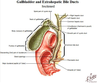
34
Q
Pathway of the gallbladder
A
- Fundus → Body (narrows) → Neck (narrowed further) → Cystic duct → joins common hepatic duct → becomes common bile duct → along edge of L.omentum → behind 1st of part duodenum → groove b/w head of pancreas of 2nd part of duodenum → maj papilla

35
Q
Hepatopancreatic sphincter
A
- At the terminal portion of pancreatic & bile duct
- Closed in resting state, relaxes only in the presence of fatty meals

38
Q
Pancreas
A
- Has exocrine functions (using ducts) & Endocrine functions (released into bloodstream ∴ rich BS)
- Head:
- Within C-shaped duodenum
- Uncinate process (landmark to identifying SUP mesenteric vessels)
- Neck
- Deep to pylorus of stomach
- Body
- Above DJ flexure
- Tail
- Leads directly to hilum of spleen

39
Q
Ducts of pancreas
A
- Begins at tails → joins common bile duct → maj duodenal papilla
- Accessory pancreatic duct:
- Drains uncinate process

41
Q
Spleen: Diaphragmatic surface
A
- Smooth & characterised by notches

42
Q
Spleen: Visceral surface
A
- Colic (colon impressions), Gastric, Renal surfaces
- Hilum → splenic A & V (VERY VASCULAR)
- A travels along SUP border of pancreas into hilum

43
Q
Position of the spleen
A
- LUQ, above L. splenic flexure
- Beneath diaphragm
