Chapter 18 –Circulatory Disorders :IMPAIRED BLOOD FLOW THROUGH THE LIVER Flashcards
What is the most common intrahepatic cause of blood flow obstruction?
cirrhosis
In addition, physical occlusion of the sinusoids occurs in a small but striking group of diseases.
What is the reason for the parenchymal necrosis of the liver in sicle cell disease?
In sickle cell disease the hepatic sinusoids may become packed with sickled erythrocytes, free
in the sinusoids or phagocytosed by Kupffer cells ( Fig. 18-37 ), leading to panlobular parenchymal necrosis.
Disseminated intravascular coagulation may occlude sinusoids.
This is usually inconsequential except for what?
for the periportal sinusoidal occlusion and parenchymal necrosis
that may arise in pregnancy as part of eclampsia (discussed later).
Wjhat can fill the hepatic sinusoids in
the absence of a mass lesion?
Finally, metastatic tumor
cells (e.g., breast carcinoma, lymphoma, malignant melanoma)
The attendant obstruction to blood flow and massive necrosis of hepatocytes can lead to fulminant hepatic failure.
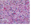
FIGURE 18-37 Sickle cell crisis in liver.
The photomicrograph shows several aggregates of
red blood cells, with some of them showing “sickle cell” appearance (arrow).
Why is Passive Congestion and Centrilobular Necrosishepatic manifestations of systemic circulatory compromise are considered together?
because they represent a morphologic continuum.
Both changes are commonly seen at
autopsybecause there is anelement of preterminal circulatory failurewithvirtually every
nontraumatic death.
What can lead to passive congestion of the liver?
Right-sided cardiac decompensation leads to passive congestion of the liver .
The liver is
slightly enlarged, tense, and cyanotic, with rounded edges.
Microscopically there is congestion
of centrilobular sinusoids.
With time, centrilobular hepatocytes become atrophic, resulting in markedly attenuated liver cell plates.
What can lead to hepatic
hypoperfusion and hypoxia, causing ischemic coagulative necrosis of hepatocytes in the central
region of the lobule (centrilobular necrosis) .
Left-sided cardiac failure or shock
In most instances the only clinical evidence of
centrilobular necrosis or its variants is transient elevation of serum aminotransferases, but the
parenchymal damage may be sufficient to induce mild to moderate jaundice.
The combination of hypoperfusion and retrograde congestion acts synergistically to cause what?
centrilobular hemorrhagic necrosis
The liver takes on a variegated mottled appearance,
reflecting hemorrhage and necrosis in the centrilobular regions, known as the nutmeg liver (
Fig. 18-38 ).
By microscopy there is a sharp demarcation of viable periportal and necrotic pericentral hepatocytes, with suffusion of blood through the centrilobular region.
What is cardiac sclerosis?
An uncommon complication of sustained chronic severe congestive heart failure is so-called cardiac sclerosis.
The pattern of liver fibrosis is distinctive, inasmuch as it is mostly centrilobular.
The damage
rarely fulfills the criteria for the diagnosis of cirrhosis, but the historically sanctified term cardiac
cirrhosis cannot easily be dislodged

FIGURE 18-38 Centrilobular hemorrhagic necrosis.
The cut liver section, in which major
blood vessels are visible, is notable for a variegated, mottled, red appearance (nutmeg
liver).
What is Peliosis
hepatis
Sinusoidal dilation occurs in any condition in which efflux of hepatic blood is impeded.
Peliosis hepatis is a rare condition in which the dilation is primary.
The liver contains blood-filled cystic
spaces, either unlined or lined with sinusoidal endothelial cells.
What is the pathogenesis of peliosis
hepatis?
The pathogenesis isunknown.
Focal apoptosis of hepatocytes or sinusoidal endothelial cells, and disruption of liver extracellular matrix seem to play a role in the pathogenesis.
Bartonella species have been seen in the sinusoidal endothelial cells in AIDS-associated peliosis. [65]
Clinically, peliosis hepatis is associated with many diseases, including what?
- cancer,
- tuberculosis,
- AIDS, or
- post-transplantation immunodeficiency.
- It is also associated with exposure to anabolic
- steroids and,
- rarely, oral contraceptives and danazol.
What are the clinical signs of peliosis?
Clinical signs are generally absent even
in advanced peliosis, but potentially fatal intra-abdominal hemorrhage or hepatic failure may
occur.
Peliotic lesions usually disappear after correction of the underlying causes.