Pediatric surgery Flashcards

1
Q

Fetal circulation

A

Blood from placenta via umbilical vein -> IVC -> right atrium. Venous return from body -> IVC, mixes with oxygenated blood from placenta. Most oxygenated blood shunted through foramen ovale to left heart -> aorta. Most oxygenated blood goes to brain. Distal to BCS origins, ductus arteriosus feeds deoxygenated blood from lungs into aorta -> mixed blood to body. Umbilical arteries return blood to placenta.

2
Q

Changes in circulation at birth

A

SVR increases, PVR drops. Ductus arteriosus closes in first 24 hours. Foramen ovale closes in first month.

3
Q

What drug keeps ductus arteriosus open?

A

Prostaglandin keeps open, indomethacin closes it.

4
Q

Cyanotic congenital heart disease (right to left shunting)

A

Tetralogy of Fallot, pulmonary stenosis/atresia, tricuspid atresia, Ebstein’s anomaly, transposition of great arteries (TGA)

5
Q

Definition of Tetralogy of Fallot (ToF)

A
  1. VSD 2. Pulmonary atresia 3. Overriding aorta 4. RVH (most common of the cyanotic heart defects). “boot shaped” heart
6
Q

Pulmonary stenosis/atresia with intact ventricular septum

A

Stenosis is more common than atresia, which is total obstruction, more severe. Must have PFO or ASD to survive. Tx: stenosis use balloon dilation. Atresia do surgical correction such as valvulotomy PLUS systemic-pulmonary shunt.

7
Q

Tricuspid atresia features

A

Tricuspid valve atresia PLUS 1) atresia/hypoplasia of right ventricle 2) ASD, PFO, or VSD. May have TGA or pulmonary stenosis. Most cases have a PDA as well.

8
Q

Treatment of tricuspid atresia

A

Emergent systemic-pulmonary shunt or enlargement of PDA/PFO in the newborn for initial palliation. Later, do bidirectional Glenn shunt followed by modified Fontan.

9
Q

Glenn shunt

A

SVC anastamosed to the pulmonary artery

10
Q

Fontan procedure

A

see attached image

11
Q

Ebstein’s anomaly definition

A

Malformed and downwardly displaced septal and posterior leaflets of tricuspid valve, leading to obstruction. PLUS PFO or ostium secundum defect –> shunting, cyanosis. Arrhythmias common.

12
Q

Risk factor for Ebsetin’s anomaly

A

Maternal lithium use during pregnancy, esp first trimester

13
Q

Transposition of Great Arteries (TGA)

A

Incompatible with life unless PDA, PFO, or VSD exists to allow mixing between the two separate circuits. If septum is intact, have to make emergent balloon septostomy of foramen ovale at birth.

14
Q

Jatene procedure

A

Transection of great vessels just superior to valves, then transfer of vessels to their correct locations. Used for kids with TGA with VSD, within first 2 weeks of life.

15
Q

Rastelli procedure

A

Aorta rerouted internally to left ventricle across VSD, pulmonary artery is attached to RV externally (see picture). Used for kids with TGA with VSD plus LVOT obstruction: do a palliative systemic-pulmonary shunt, then Rastelli at age 4-5.

16
Q

What are the three types of (at least initially) left to right shunt heart defects?

A

VSD, ASD, PDA

17
Q

Risk factors for PDA

A

Shorter gestational age, lower birth weight, female sex

18
Q

Treatment of PDA

A

Indomethacin is successful in 90%. Surgery in refractory cases. Unless CHF develops, can do surgery electively anytime between 6 months and 2 years

19
Q

What is the most common congenital heart defect overall?

A

VSD

20
Q

Do VSDs close on their own?

A

90% if small defects close by age 8. Treatment is to medically manage congestive symptoms. surgical closure is indicated if CHF is not controlled medically, or if it does not close by 9 months and pulmonic pressure has reached 2/3 of systemic pressure.

21
Q

ASD features

A

Usually asymptomatic until adult life unless large defect -> growth retardation, fatigue, recurrent PNA. Adults often present with arrhythmia or CHF.

22
Q

Three types of ASD

A

Secundum type (middle or lower defect, most common type), sinus venosus (high type assoc with partial anomalous pulmonary venous return), and ostium primum type

23
Q

Ostium primum type ASD

A

Endocardial cushion defect, cleft in mitral valve leaflet leading to mitral insufficiency. Uncommon except in Down syndrome

24
Q

Eisenmenger syndrome

A

When you have a left to right shunt, increased pulmonary blood flow, PVR increases to the point that shunt becomes right to left -> cyanosis. Only treatment is heart and lung transplant.

25
Q

What are the obstructive left sided congenital lesions?

A

Coarctation of the aorta, congenital aortic stenosis, hypoplastic left heart syndrome

26
Q

Where is aortic coarctation usually located?

A

Within four cm distal to left subclavian take off

27
Q

When is surgery done for aortic coarctation?

A

Neonates with CHF get it right away. Otherwise, ideal time is 3-4 years old

28
Q

What causes rib notching on CXR in aortic stenosis?

A

Collateral circulation distal to the coarctation causes marked dilation of intercostal arteries

29
Q

How do older children with aortic coarctation present?

A

Hypertensive but asymptomatic. Delayed femoral pulses

30
Q

What’s the deal with hypoplastic left heart syndrome?

A

Left side of heart is underdeveloped, with ineffective contractility and a valves less aortic remnant. Fetus survives because blood enters systemic circulation via PDA, so when that closes it is incompatible with life. Surgery is 80% survival.

31
Q

Norwood plan for hypoplastic left heart

A

Pulmonary artery anastamosed to aorta to establish flow from RV to systemic circulation. atrial septum resected to allow mixing of blood. Subclavian to pulmonary artery shunt created to control pulmonary blood flow, until 6+ months of age, when direct atrial to pulmonary connection is tolerated.

32
Q

What biomarkers indicate fetal lung maturity?

A

Lecithin/sphingomyelin ration > 2, 2.5

33
Q

What medication can help lungs reach maturity for babies born before 34 weeks?

A

Glucocorticoids given to mother before delivery

34
Q

Calculate TBW for neonate

A

Weight x 80%

35
Q

MIDLINE congenital neck lesions

A

Thyroglossal duct remnants, dermoid cyst, submental lymph node, goiter

36
Q

LATERAL congenital neck lesions

A

Cystic hygroma, branchial cyst, lymphadenitis, lymphoma

37
Q

Thyroglossal duct cyst

A

Most commonly presents as infection. Smooth, mobile, cystic midline mass arising from base of tongue. Treatment: complete excision necessary to prevent recurrence. Screen for hypothyroidism on follow up.

38
Q

Original of thyroglossal duct cyst

A

Develops from cells and tissues remaining after the formation of the thyroid gland during embryonic development

39
Q

Branchial cleft cyst

A

Remnants of the four paired branchial arches, clefts, and pouches. Most common = 2nd branchial cleft cyst. Presents as lateral neck mass at ant border of SCM, may present with drainage. Tx: complete excision of cyst and tract, abx if infected.

40
Q

Cystic hygroma

A

Congential lymphangioma; multiloculated lymphatic lesion that can arise anywhere, but is classically found in the left posterior triangle of the neck and armpits. Can cause upper airway obstruction. Tx: surgical incision (do not resolve spontaneously).

41
Q

Congenital lobar emphysema

A

Hyperexpansion of one or more lobes of lung -> respiratory distress. CXR shows hyperaerated lobe, hyperlucency (air trapping), mediastinal shift. Tx: resection of affected lobe

42
Q

Pulmonary sequestration

A

Nonfunctioning cystic pulmonary tissue that receives blood supply from systemic circulation and does not communicate with functional airways. Two types: intralobar (presents with infection) and extralobar (assoc with diaphragmatic hernia). Tx: surgical removal

43
Q

Congenital cystic adenomatoid malformation

A

Congenital pulm malformation involving varying degress of adenomatosis and cyst formation -> neonatal resp distress, recurrent respiratory infection. CXR: “Swiss cheese”

44
Q

Congenital cystic adenomatoid malformation present similarly to congenital diaphragmatic hernia. How to tell them apart?

A

CXR shows position of NG tube. NG tube in chest = CDH. NGT in abdomen = CCAM

45
Q

Hydrops fetalis

A

serious fetal condition defined as abnormal accumulation of fluid in 2 or more fetal compartments, including ascites, pleural effusion, pericardial effusion, and skin edema

46
Q

If a baby has a tracheoesophageal malformation, what other screening should be done?

A

Look for other anomalies, particularly cardiac and renal. Do a cardiac echo to define aortic arch

47
Q

How does TE malformation present?

A

Resp distress or choking following first feeding. Infant unable to swallow saliva. Cannot pass NG tube

48
Q

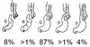
Types of TE malformations

A

Esophageal atresia + TE fistula is most common. See attached

49
Q

Gastroschisis

A

Full thickness abdominal wall defect with no covering over bowel, usually to the RIGHT of umbilicus. Tx: cover w/ saline-soaked sponges and plastic wrap, NG decompress, abx, TPN, surgical correction.

50
Q

Omphalocele

A

Herniation of abdominal contents into base of umbilical cord, CENTRAL, protective membrane is present. Assoc with syndromes (Beckwith-Wiedemann, trisomy 13, 18). Tx: if sac ruptured, treat emergently in same way as gastroschisis. If not ruptured, surgery can be planned, not urgent.

51
Q

Pyloric stenosis

A

Nonbilious, projective vomiting, 3rd-5th week of life. “Olive” shaped midepigastric mass, dx with US. Tx: give NS + D5W + K to fix electrolyte problems. Surgery is pyloromyotomy.

52
Q

Biliary atresia

A

Presents with neonatal jaundice, conjugated hyperbilirubinemia. Tx: correctible type (blind ending of hepatic duct) is fixed with direct anastamosis with RenY of loop of jejunum; noncorrectable type is treated with Kasai procedure

53
Q

Kasai procedure

A

Hepatoportoenterostomy. Sooner the better

54
Q

Most common cause of peds liver transplant

A

biliary atresia

55
Q

Malrotation and midgut volvulus

A

Acute onset of bilious vomiting, abdominal distention. Late sign = bloody stool. CXr shows double bubble sign with duodenal obstruction Tx: surgical emergent correction with Ladd procedure. Also do appendectomy (because cecum will remain in RUQ and future appendicitis will have confusing presentation).

56
Q

Ladd’s bands

A

Ladd’s bands, sometimes called bands of Ladd, are fibrous stalks of peritoneal tissue that attach the cecum to the abdominal wall and create an obstruction of the duodenum. This condition is found in malrotation of the intestine.

57
Q

Intestinal atresia is associated with..?

A

Down syndrome, esophageal atresia, imperforate anus

58
Q

What is the age with peak incidence of intussusception?

A

5-12 months (range 2-5 years)

59
Q

Causes of intussusception

A

Viral, lead point (Meckel’s, polyp, lymphoma, HSP, cystic fibrosis)

60
Q

Presentation of intussusception

A

Intermittent abdominal pain, biliious vomiting, currant jelly stool (later), sausage-sahped RUQ mass, target sign on imaging. Tx: barium enema unless perforation/peritonitis/shock (can try no more than 3 times)

61
Q

Meckel’s diverticulum

A

Persistence of the omphalomesenteric (vitelline) duct. Arises from antimesenteric border of ileum. Contains heterotopic epithelium (gastric, colonic, or pancreatic). TRUE diverticulum (contains all layers of bowel wall). Majority are asx but if presents it is usu in first 2 years, with intermittent painless rectal bleeding, intestinal obstruction, and/or diverticulitis. Most common GI congential abnormality.

62
Q

Inguinal hernia in peds

A

Males > females, premature infants more often.

63
Q

Imperforate anus tx and prognosis

A

If high (rectum ends above the puborectalis sling) -> colostomy.

If low, perineal anoplasty or dilation of fistula.

Higher up = worse prognosis

64
Q

Necrotizing enterocolitis

A

Mostly seen in premature infants. AXR shows pneumatosis intestinalis (air in bowel wall). Presents with sepsis, hematochezia, abd pain and distention. Tx: NPO, NGT, abx, TPN; surgical resection for perf or full-thickness necrosis of bowel. Overall 95% survival but also high morbidity.

65
Q

Wilms tumor

A

Nephroblastoma. Assoc with hemihypertrophy, aniridia, GU abnormalities. Triad = flank mass, HTN, hematuria. Most occur by age 6 years.

66
Q

Neuroblastoma

A

Arises from NC cells. Can arise in adrenal gland, sympathetic ganglia. Majority are abdominal. Dx: elevated VMA, HVA catecholamines, CT with contrast. Better prognosis if presents < 1 yr old. Majority have mets at time of presentation.