10/13- Metabolic Bone Disease Flashcards

1
Q

What are the main types of bone?

A
  • Cortical
  • Trabecular (cancellous)
2
Q

What are the different cell types in bone?

A
  • Osteoblast (4-6%)
  • Osteoclast (1-2%)
  • Osteocyte (90-95%)
  • Possibly old osteoblasts (?)
3
Q

Describe the process of bone turnover (remodeling).

How long does it take?

A
  • Activation
  • Reversal: osteoclasts start process of resorption
  • Formation: ostebolasts lay down bone matrix
  • Mineralization: osteoid mineralized by deposition of Ca and other minerals

Process is typically 4-8 mo duration

4
Q

Describe osteoclast features/characteristics?

A
  • CAII produces bicarb and H
  • H-ATPase pump sends H out into bone to break up material
5
Q

Describe osteoblast/osteoclast communication and feedback

A

RANKL = receptor activator of nuclear factor Kappa-B ligand

  • Expressed by osteoblast
  • Osteoblast stimulates monocytes to differentiate into osteoclasts

RANK = receptor activator of nuclear factor Kappa

  • Expressed by osteoclast

Osteoprotogerin (OPG) can bind RANKL and stop resorption process

  • OPG is a decoy receptor that prevents osteoclast activation
  • Expressed by osteoblast (?)
6
Q

T/F: Optimal bone strength occurs within physiologic window of bone turnover?

A

True

  • Weaker if too much or too little turnover
7
Q

What are high bone turnover diseases?

A
  • Osteoporosis
  • Paget’s disease
8
Q

What is osteoporosis?

A

A systemic skeletal disease characterized by:

  • Low bone mass and microarchitectural deterioration
  • Compromised bone strength with a consequent increase in bone fragility and susceptibility to fracture
9
Q

What are common fracture sites of osteoporosis?

A
  • Spine: wedge compression deformity
  • Hip: fracture typically at neck of femur
  • Wrist: typically distal 1/3 of the radius
  • Osteoporotic Fracture Syndrome (Dowager’s hump)
  • Multiple compression deformities leading to significant kyphosis
10
Q

Describe the gain, maintenance, and loss of bone throughout life

A
  • Gain of bone until age 20-30
  • Slight loss in 30s-40s (but not much)
  • Estrogen deficiency in post-menopausal women corresponds to steep decline in bone density
11
Q

Fracture risk most strongly corresponds to ____

A

Fracture risk most strongly corresponds to bone mass

12
Q

What are risk factors for fracture?

  • Measurable
  • Lifestyle
  • Medical Hx
  • Meds
  • 2ndary causes
A

Age (biggest risk)

Measurable

  • Low BMD
  • High bone turnover
  • Low body weight

Lifestyle

  • Risk of falls
  • Smoking
  • Excessive alcohol consumption

Medical History

  • Prior fracture
  • Family history

Medication use:

  • Corticosteroids

Some secondary causes of osteoporosis

Lack of estrogens or testosterone (sex hormones)

  • Rapid decline of bone mass in women within 1st 5 yrs following menopause
13
Q

How is osteoporosis diagnosed? Treatment?

A

Diagnosed by bone density scan

  • Fracture risk assessment model (FRAX) determines probability of major osteoporotic fractures
  • Only cost effective to treat osteoporosis if high FRAX risk (would go ahead and treat someone who has fragile bones, not just this early decreased bone density)
14
Q

Factors leading to fracture

A

Low bone density

  • Low peak bone mass
  • Increased bone loss
  • Aging
  • Menopause
  • Other risk factors

Also:

  • Propensity to fall
  • Poor bone quality
15
Q

Describe the incidence of osteoporotic fractures by age in men vs. women?

A

Men: really picks up around age 65

Women: starts picking up around 45 yo

  • Get big spike in Colles’ fracture at 60 yo
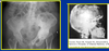
16
Q

T/F: there is increased remodeling after menopause?

A

True (measure of how bone turnover/osteoclatic activity)

17
Q

What is seen here?

A

Increased porosity in micro-architecture of osteoporosis (right pic)

18
Q

Diagnosis and evaluation of Osteoporosis

A
  • Diagnosis obvious if fragility fractures present (e.g. falling from stool)
  • BMD (bone mineral density) assessment in high risk patients; DEXA (dual x-ray absorption)
  • Lab tests to rule out secondary causes
  • Labs usually normal in primary osteoporosis
  • Biochemical markers of bone turnover?
19
Q

Describe the diagnostic categories for osteopenia and osteoporosis based upon bone density measurements

  • Normal
  • Low bone mass (osteopenia)
  • Osteoporosis
  • Severe (established) osteoporosis
A

Normal

  • BMD within 1 SD of young adult female reference mean

Low bone mass (osteopenia)

  • BMD 1-2.5 SDs below reference mean

Osteoporosis

  • BMD 2.5+ SDs below reference mean

Severe (established) osteoporosis

  • BMD > 2.5 SDs below reference mean in the presence of 1+ fragility fractures
20
Q

What is osteopenia?

A

Low bone mass (less severe than osteoporosis)

21
Q

What is T score? Z score?

A

T score

  • Compare pt’s BMD to young adult of same gender (comparing pt with optimal)
  • (Recall 20-30 yo adult has max bone mass; minimum fracture risk)

Z score

  • Compare pt’s BMD with age and gender matched average person
  • Low Z score means losing more bone than your peers
22
Q

Treatment for osteoporosis?

A
  • Calcium
  • Vitamin D

Reduction of non-vertebral fractures with Ca and Vitamin D by almost 50%

23
Q

What are anti-resorptive treatments for osteoporosis?

A

Bisphosphonates (first line!)*

  • Alendronate (oral)
  • Risedronate
  • Ibandronate (1/mo)
  • Zoledronic acid (15 min infusion once/yr)

SERMs (Raloxifene)

  • Selective estrogen receptor modulators
  • Estrogen agonist effect on the bone (and antagonist effect on the breast!)
  • Not as potent as bisphosphonates

HRT: (Estrogen) Hormone Replacement Therapy

  • Breast cancer risk; no longer used as treatment option for osteoporosis

Calcitonin

  • Not very potent
  • Good effect on pain (acute fracture scenario)

Densumab

  • Human monoclonal Ab against the RANKL (works like OPG)
24
Q

What is the mechanism of bisphosphonates?

A
  • Blocks GTPase binding; GTPase free from cell membrane
  • Prevents ruffled border formation of osteoclast (flattened border)
25
Q

What are concerns with bisphosphonates?

A

(Rare):

  • Osteonecrosis of jaw
  • Atypical femoral fractures
26
Q

What are anabolic agents that may be used to treat osteoporosis?

A
  • Teriparatide
27
Q

What is the effect of Teriparatide?

  • How best combined with bisphosphonates?
  • Uses
  • Duration of treatment
A
  • Reduces vertebral and non-vertebral fracture risk
  • Can NOT be used with bisphosphonates
  • Best data for Teriparatide for vertical issues (poor lumbar spine density or history of lumbar fracture)
  • Teriparatide can only be used ~ 2 yrs (once done, THEN start bisphosphonates)
28
Q

Do corticosteroids increase or decrease bone mass?

A

Corticosteroids (glucocorticoids) decrease bone mass

  • Induce osteoporosis
29
Q

Describe glucocorticoid induced osteoporosis

  • Rate of bone loss
  • Route of administration
  • Dosing
  • Pathophysiology
  • Treatment
A
  • Bone loss rapid during the early months of therapy
  • Any route of administration: risk
  • Alternate day delivery: does not decrease risk
  • Pathophysiology: Multiple mechanisms
  • Bisphosphonates for treatment
30
Q

Describe Paget’s disease

  • Cells involved
  • Extent (systemic vs. local)
  • Symptoms
  • Lab markers
A
  • Osteoclasts are extremely hyperactive
  • Localized bone disorder (unlike osteoporosis)
  • Overactive osteoclastic bone resorption increase in osteoblastic new bone formation
  • Usually asymptomatic

Labs:

  • Alkaline phosphatase (bone formation marker) is increased
  • Urinary and serum N-telopeptide (markers of bone resorption) are increased
31
Q

What is seen here?

A

Paget’s Disease

32
Q

What is treatment for Paget’s disease?

A

Bisphosphonates

33
Q

What is a disease of low bone turnover?

A
  • Osteopetrosis: osteoclasts do not “eat”
34
Q

Describe osteopetrosis

  • Osteoclast/blast function
  • Elevated levels of what
  • Results
A
  • Loss of osteoclastic bone resorption; normal osteoblastic bone formation
  • Osteoclast-derived tartrate-resistant acid phosphatase (TRAP) is elevated
  • Generalized symmetric increase in bone mass with thickening of both cortical and trabecular bone
35
Q

What is seen here?

A

Osteopetrosis

  • Marble bone disease
  • Albers-Schonberg disease