Gupta - Pathology Flashcards
What do you see here?

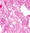
- First-trimester chorionic villi
- Composed of delicate mesh of central stroma surrounded by two discrete layers of epithelium:
1. Outer layer of syncytiotrophoblast (group of cells; two arrows) and
2. Inner layer of cytotrophoblast (single cells; arrow) - Smaller, more vascular, less trophoblast thickness as the pregnancy progresses
What is this?

- Third-trimester chorionic villi
- Composed of stroma with:
1. Dense network of dilated capillaries
2. Surrounded by markedly thinned-out syncytiotrophoblast and cytotrophoblast - REMEMBER:
1. Syncytio: think groups of cells
2. Cyto: think single cells
What kind of growth restriction do fetal abnormalities cause? Specific causes?
- SYMMETRIC: all organ systems are similarly affected
- Caused by chromosomal disorders, congenital anomalies, and congenital infections:
1. TORCH group of infections; usually transplacental -
a. Toxoplasmosis: usually 1st infection is the one that causes most problems (i.e., if mom has never been exposed before)
b. Rubella
c. Cytomegalovirus (CMV)
d. Herpesvirus (HSV)
2. Other viruses/bacteria, i.e., syphilis bacteria, listeria - NOTE: wear gloves if working in the garden while pregnant

What kind of growth restriction do placental abnormalities cause? Specific causes?
- ASYMMETRIC: spares brain (via a down-regulation of growth in latter 1/2 of gestation)
- Uteroplacental insufficiency may result from:
1. Umbilical-placental vascular anomalies (like single umbilical artery, abnormal cord insertion, placental hemangioma),
2. Placental abruption (detachment from uterus),
3. Placenta previa (covers opening of mom’s cervix),
4. Placental thrombosis and infarction,
5. Placental infection, or
6. Multiple gestations - NOTE: 3rd trimester loss is usually placental insufficiency
What kind of maternal abnormalities may cause fetal growth restriction?
- Those that result in DEC placental blood flow
1. Vascular diseases, like preeclampsia (toxemia of pregnancy) and
2. Chronic hypertension, are often the underlying cause - Usually some kind of vascular issue
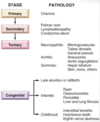
What is spontaneous abortion? Causes?
- MISCARRIAGE: pregnancy loss <20 wks gestation
1. Fetal chrom anomalies: approx 50% of early abortuses
2. Maternal endocrine factors: luteal-phase defect, poorly controlled diabetes, other uncorrected endocrine disorders
3. Physical defects of uterus: submucosal leiomyomas, uterine polyps or malformations, may prevent or disrupt implantation
4. Systemic disorders affecting maternal vasculature: antiphospholipid Ab syndrome, coagulopathies, and HTN - NOTE: families get very upset about losing the fetus early, but this may be a “good” thing in many cases, i.e., due to a chromosome abnormality
When might ascending infections cause pregnancy loss?
- 2nd trimester
- Losses due to fetal and maternal anatomic abnormalities are also common in the 2nd trimester
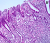
What is this?

- Listeria: necrotizing intervillositis
-
Non-pasteurized cheeses and milk (KNOW THIS: may get a question about it) -> if the infection is bad enough, you may lose this pregnancy
1. Outbreaks in some places where people eat a lot of unpasteurized cheeses (also cold cuts)
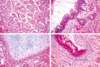
What is going on here?

- Chorioamnionitis: maternal inflammatory response to intervillositis
- These are ascending infections, so often maternal inflammatory response (not placenta)
- Lots of inflammation: would also call this deciduitis (this is acute inflammation -> mostly polys)
- NOTE: layers here are amnion, chorion, decidua
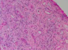
What do you see here?

- Necrotizing funisitis: due to Candida, in this case
- Punctate, 1-2mm yellow-white nodules on the cord (Candida always looks the same)
- Generally track the coils of the cord vessels, and are noted initially along the perimeter of the umbilical vein (3 vessels: 2 aa, 1 vein)
- Established, or long-standing infections can cause tissue necrosis and accumulation of cellular debris (necrotizing funisitis)
- False knot in the cord here
What is this?

- Subamniotic microabscesses typical of Candida funisitis
- Very characteristic, but not specific for this organism
- Inflammation of the umbilical vessels (vasculitis) and cord substance (funisitis) occurs in response to the infection -> FETAL INFLAMMATORY RESPONSE
-
Wharton jelly: supporting substance in umbilical cord that looks like jelly grossly, but pink ground substance in this micro
1. Right side of the image is the outside of the umbilical cord
2. Artery on the left
What is this?

- Candida pseudohyphae with GMS silver stain in subamniotic foci
- Funisitis: umbilical cord with subamniotic microabscesses
- Inflammation located at the periphery or surface of the umbilical cord may be the primary pattern seen (peripheral funisitis)
- Fungal ball
What is phlebitis?
- Inflammation of the umbilical vein
- Can be seen separately from arteritis (inflammation of umbilical arteries)
What do you see here?

- Chronic villitis with CMV: likes vascular endothelium
- Nuclear AND cytoplasmic inclusions (herpes would only have nuclear inclusions)
- This case is SEVERE
What is this?

Chronic villitis with CMV
What is going on here?

- Parvovirus B19: erythema infectiosum (slap cheek)
- Viral inclusions in early erythroid precursors
- Bone marrow sample b/c don’t see nucleated red cells in peripheral system, unless something is wrong
What do you see here? Arrow?

- Parvovirus B19: erythema infectiosum
- Erythroblasts in lumen of capillary vessels of the placental villi show eosinophilic nuclear inclusions
An image of fetal membranes are shown. Iron stain was negative. The histologic finding indicates what?

A. Meconium was present in the amnionic cavity
B. Placental abruption
C. Congestive heart failure in the fetus
D. Acute chorioamnionitis
A. Meconium was present in the amniotic cavity
- Passage of meconium in utero due to bowel peristalsis and relaxation of anal sphincter -> may be indicative of fetal distress
- Meconium components diffuse into placenta and cord, leading to vasoconstriction + hypoperfusion; damage to fetus INC with length of exposure
- Neonates are at risk for meconium aspiration
- Meconium is unlikely in fetuses <30 wks gestation
-
Meconiophages: brown-green pigment, usually right below amnion, in the chorion
1. Top a little bit of amnion layer (low, cuboidal epithelium), then chorion, then decidua (big, fluffy, pink cells)
2. Hemosiderin-laden macros would have been positive for iron
What is twin-twin transfusion syndrome?
- A complication of monochorionic twin pregnancy
- Monochorionic twin placentas have vascular anastomoses that connect twin circulations, and in some cases these connections include one or more INC blood flow to one twin at expense of the 2nd, one twin will be underperfused, while the second will be fluid overloaded
- If severe, this may result in the death of one or both fetuses
- Most twins Di/Di, so this syndrome is RARE

What may have happened here?

- Twin-twin transfusion syndrome: a complication of monochorionic twin pregnancy
- Monochorionic twin placentas have vascular anastomoses that connect twin circulations, and in some cases these connections include one or more INC blood flow to one twin at expense of the 2nd, one twin will be underperfused, while the second will be fluid overloaded
- If severe, this may result in the death of one or both fetuses

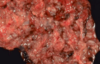
What is the most common site of ectopic pregnancy? Causes?

- Approximately 90% of cases in extrauterine fallopian tube
- Can rupture (gross image on front of this card), leading to hemoperitoneum, which is dangerous
- 50% are idiopathic, and the other 50% may be infection, leiomyoma causing obstruction, etc.
- Takes a “villous” to prove ectopic pregnancy; chorionic villi float on water

Describe each of these abnormalities of placental implantation (A-D).

- Fertilized ovum normally implants in uterine fundus
A. Normal placenta
B. Placenta previa: very low lying placenta, or one that covers the os -> severe hemorrhage can result, w/cervical dilation and passage of the baby through the birth canal
C. Placenta accreta: no formation of a normal decidual plate -> chorionic villi extend into myometrium, and placenta can’t separate normally post-delivery; severe hemorrhage
D. Abruptio placenta: premature separation of placenta prior to delivery, with formation of a retroplacental blood clot between decidua and uterine wall -> blood supply of O2 and nutrients to fetus compromised to greater degree with INC size of the abruption
What do you see here?

- Placenta accreta: NO formation of normal decidual plate -> chorionic villi extend into myometrium, and placenta can’t separate normally post-delivery, leading to severe hemorrhage
- Microscopically, the placental villi interdigitate directly with the uterine myometrium, without an intervening decidual plate
How is placental invasion into the myometrium classified (chart)?

The following gross and microscopic images show what pathology?
A. Placenta previa
B. Normal placenta
C. Abruptio placenta
D. Placenta accrete
E. Vasa previa

C. Abruptio placenta
- Retroplacental blood clot in gross image
What is this?

-
Amnion nodosum: seen in placentas affected by oligohydramnios, which may be associated with fetal renal agenesis and pulmonary hypoplasia
1. May be due to desquamated skin or membrane injury - GROSS: multiple yellow-tan superficial amniotic lesions (0.2 to 0.4 cm), usually near insertion of the umbilical cord
- Not really a bad thing, unless it is associated with oligohydramnios -> just a collection of squaffed off squamous cells, and maybe some fibroblasts
What is going on here?

-
Amnion nodosum: seen in placentas affected by oligohydramnios, which may be associated with fetal renal agenesis and pulmonary hypoplasia
1. May be due to desquamated skin or membrane injury - MICRO: nodules of protuberant eosinophilic fibrinous material with entrapped squamous cells
1. Associated w/stratified squamous metaplasia - Not really a bad thing, unless it is associated with oligohydramnios -> just a collection of squaffed off squamous cells, and maybe some fibroblasts
What happened to this little guy?

- Potter’s sequence: clubbed feet, pulmonary hypoplasia, and cranial anomalies
- Oligohydramnios experienced in utero
What may have caused the difference in size of these placentas?

- Preeclampsia: HTN and large amount of protein in the urine -> usually in 3rd trimester of pregnancy and gets worse over time (feared outcome is eclampsia = seizures)
- Most placentas are smaller than expected
- Infarcts and retroplacental hematomas are also more common
What do you see here? When are these more likely?

- Placental infarct: small infarct may be of no consequence to the fetus, but if >1/3 or 1/2 of the placental parenchyma is infarcted or lost in some fashion, blood supply to fetus can become severely compromised and fetal demise may occur (via utero-placental insufficiency w/multiple infarcts)
- Infarcts AND retroplacental hematomas are more common in PREECLAMPSIA
- Bread loafing for placenta section: maternal vessels on top, membrane on bottom, and pale area is an infarct
What is the difference between these two images?

- Preeclampsia
- LEFT: villi small, and syncytiotrophoblasts have thinned out (as they should)
- RIGHT: villous ischemia (increased syncytial knots) in the placenta
1. A little hemorrhage; don’t look as healthy
2. Only some blood vessels, and “nubbins” of syncytiotrophoblasts
What might be going on in this maternal vessel?

- Fibrinoid necrosis of the vessel wall: strong eosinophilic staining areas
- PREECLAMPSIA
- These are maternal vessels in the decidua
-
No cell nuclei, and decidua surrounding it (big, pink, fluffy cells)
1. Pink and amorphous (necrotic)
Why are hyatidiform moles important to recognize?
- Because they are associated with an increased risk of persistent trophoblastic disease (invasive mole) or choriocarcinoma
- Invasive: trophoblasts go into uterine wall
- Attached flow chart is important for pathology (not important for us to know yet) -> partial moles much more difficult than complete ones because often diagnosed very early

What is the origin of complete/partial hydatidiform moles? What hormone level could help you distinguish between the 2?
-
Complete: egg that has lost female chromosomes, so genetic material is completely paternally derived
1. NO fetal parts - Partial mole: fertilization of an egg with two sperm
-
hCG: compared to normal levels for gestation
1. Complete mole: very elevated
2. Partial mole: slightly elevated

What is this?

Gross image of a twin placenta
What is preeclampsia?
-
Vasculature problem: may be b/c trophoblasts don’t sufficiently invade/stimulate the spiral arteries in the uterus -> hypoxic state for the fetus, which is reflected in the gross and micro changes
1. Big, dilated, torturous blood vessels on fetal surface on left (attached image)
2. Right placenta much smaller: DEC blood supply, nutrients to fetus + more likely to get more infarcts and retroplacental hematomas

What do you see here?

-
Complete mole: no embryo, and no normal placenta
1. Marked villous enlargement, edema, and circumferential trophoblast proliferation - Villi get very large, and edematous, aka hydropic
- hCG very elevated (above what would be normal for that stage of the pregnancy) in these women because way too many syncytiotrophoblasts
What is this?

- Snowstorm appearance of complete mole on US
- Buzzword
- REMEMBER: no embryo, and no normal placenta
1. Elevated hCG for the corresponding stage of pregnancy
What do you see here?

-
Normal first trimester placenta: chorionic villi are large and covered by two layers of cells:
1. Cytotrophoblast and syncytiotrophoblast (syncytio make hCG) - Blood vessels in the villi are not prominent
What is this?

-
Partial mole: some villi appear normal; others are swollen, avascular, and grape-like (though not as large as a complete mole)
1. Minimal trophoblastic proliferation - hCG won’t be as high as with the complete mole
- Can be really hard to differentiate these from normal villi microscopically (b/c detected so early)
- Will karyotype if clinical suspicion is very high (looking for TRIPLOIDY)
What is this?

Complete mole: NO normal villi here
What is this?
- Partial mole: normal villi are pink, fluffy, light tan (vs. grape-like clusters of the molar villi)
What might this be?

-
Gestational choriocarcinoma: malignant neoplasm of trophoblastic cells from a previously normal or abnormal pregnancy, like an extrauterine ectopic pregnancy or complete mole
1. Complete moles more likely to progress to this than partial, but still only about 2-3% risk - Rapidly invasive and metastasizes widely, but once identified responds well to chemotherapy
1. Very hemorrhagic
2. >50% have metastasized by the time they are identified - NOTE: persistent trophoblastic disease does not metastasize, but pieces of it may embolize to lung or other parts of body
What is this?

- Choriocarcinoma: soft, fleshy, yellow-white tumor with large, pale areas of necrosis and extensive hemorrhage
- Histo: proliferating syncytio and cytotrophoblasts
1. Mitoses abundant and sometimes abnormal - Invades underlying myometrium, freq penetrates blood vessels, and in some cases extends out onto the uterine serosa and into adjacent structures
- Chorionic gonadotropins (hCG) very high!!!!
What are the histo and clinical features of complete hydatidiform mole?
- HISTO: large, avascular villi with trophoblastic proliferation
- CLINICAL: occurs when fertilized ovum contains only paternal chromosomes (usually 46, XX karyotype)
1. Marked uterine enlargement
2. “Snowstorm” effect w/no fetus on US
3. Some give rise to choriocarcinoma (2-3%)
What are the histo and clinical features of partial hydatidiform mole?
- HISTO: some villi enlarged, w/minimal trophoblast proliferation
- CLINICAL: typically triploid (69, XXX or 69, XXY or 69, XYY)
1. Malformed fetus present, but rarely goes to term
2. Rarely gives rise to choriocarcinoma
What are the histo and clinical features of choriocarcinoma?
- HISTO: malignant proliferation of syncytio-trophoblast with no villi
1. Often hemorrhagic
2. NO fetus is present - CLINICAL: hCG levels are often extremely high
1. Can metastasize
2. Many are sensitive to chemotherapy
What are the histo and clinical features of a placental site trophoblastic tumor?
- HISTO: rare, localized proliferation of intermediate trophoblast that can produce a grossly visible nodule
- CLINICAL: most are benign, with rare malignant cases
What are some of the hazards of prematurity?
- May give rise to:
1. Neonatal respiratory distress syndrome, also known as hyaline membrane disease
2. Necrotizing enterocolitis (attached image)
3. Sepsis
4. Intraventricular and germinal matrix hemorrhage

What is going on here?

- Necrotizing enterocolitis (NEC): potential hazard of prematurity
- LEFT: entire small bowel markedly distended, with a perilously thin wall (impending perforation)
- RIGHT: congested portion of ileum corresponds to areas of hemorrhagic infarction and transmural necrosis microscopically
1. Submucosal gas bubbles (pneumatosis intestinalis) can be seen in several areas (arrows on bottom left)
What is the difference between these two tissues?

-
Hyaline membrane disease (neonatal respiratory distress syndrome) in the image on the left
1. Alternating atelectasis and dilation of alveoli
2. Eosinophilic, thick hyaline membranes lining the dilated alveoli - Normal fetal lung on the right

What is fetal hydrops? What are some of the causes?
- Accumulation of edema fluid in the fetus during intrauterine growth
- CAUSES: CV, chromosomal, fetal anemia (immune, Parvo B19, homo alpha-thalassemia), twin-twin, etc.
1. Immune hydrops: major antigens known to induce clinically significant immuno rxns are certain Rh Ags and ABO blood groups
a. 2nd and subsequent pregnancies in Rh-(-) mom w/Rh-(+) dad
b. O- moms

What happened here?

- Fetal hydrops: generalized accumulation of fluid in the fetus
- RIGHT image: fluid accumulation is particularly prominent in the soft tissues of the neck, and this condition has been termed cystic hygroma
1. Characteristically seen, but not limited to, constitutional chromosomal anomalies such as 45,X karyotypes (Turner’s syndrome)
What is SIDS? Risk factors?
-
Sudden death of infant <1 year of age that remains unexplained after thorough case investigation, incl performance of a complete autopsy, examination of the death scene, and review of the clinical history
1. Aka, sudden unexpected infant death (SUID) - “Triple-risk” model that postulates the intersection of three overlapping factors:
1. Vulnerable infant,
2. Critical devo period in homeostatic control,
3. Exogenous stressor - NOTE: babies should sleep on their backs
Klinefelter syndrome
- XXY
- Eunucoid (indeterminate sex) features
- Loss of sertoli cells = loss of inhibin and INC FSH
1. FSH stimulates aromatase in gonads, INC estrogen
2. Female secondary sex characteristics at puberty
Androgen insensitivity syndrome
- XY phenotypic “female;” presents w/amenorrhea
- Normal testis: leydig cells make testosterone
1. Loss of testosterone receptor activity
2. No testosterone effect = NO development of male reproductive structures - No conversion of testosterone to DHT = no external developed male genitals
- XY means presence of MIF (sertoli cells), inhibits female reproductive organs, however, distal 2/3 forms due to estrogen (also normal breast)
Premature ovarian failure
- Female <40 with amenorrhea
- Loss of entire ovarian function
- DEC estrogen and inhibin feedback results in increased FSH and LH
- Progesterone withdrawal test is negative for bleeding
PCOS
- Female presents with amenorrhea and hirsutism
- INC LH with intact ovary -> INC testosterone (hirsutism)
- INC T converted to estrogen by aromatase, so INC LH (like LH surge) and INH FSH, leading to NO follicle development
1. Follicle becomes a cyst - Progesterone test is positive for bleeding
Turner syndrome
- 46, XO: monosomy
- Short, primary amenorrhea, lymphedema at birth
- Shield chest, wide nipples
- Webbed neck and low hair line
- Coarctation of the aorta
- Streak ovaries –> little estrogen
1. Elevated FSH and LH
Kallman syndrome
- Defect in hypothalamic production of GnRH
- Associated with anosmia: inability to perceive odor
- Delayed puberty
What is the progesterone withdrawal test?
- Give patient w/amenorrhea medroxyprogesterone acetate (Provera) 10 mg daily for 5-10 days or one IM injection of 100-200 mg of progesterone in oil
- If patient experiences bleeding after the progestin, she has estrogen present but is not ovulating (anovulation)
- If no withdrawal bleeding occurs, either patient has very low estrogen levels or there is a problem with the outflow tract such as uterine synechiae (adhesions) or cervical stenosis (scarring)
Appreciate this.

Good job!
What are the most common palpable breast lesions?
- Cysts
- Fibroadenomas
- Invasive carcinomas

Where are most carcinomas located in the breast? Do they metastasize?
- About 50% are located in the upper outer quadrant
- Some do metastasize, and the majority of those that do will have done so by the time they reach a size that can be palpated -> around 2-3cm
1. This is why mammography has become such a big deal -> helps us catch lesions earlier - Note: ductal and lobular carcinomas come from the same cell type, but appear different
What % of breast masses in women are malignant (2 age groups)?
- 10% in women younger than age 40 are malignant
- 60% are malignant in women over 50
- Breast cancer risk goes starts going back down after age 65 (see attached)

What are the 4 key symptoms of breast disease (image)?

What are the 4 main presentations of breast cancers?
- ABNORMAL MAMMOGRAM

What are fibrocystic changes? Categories and specific types (image)?
- Benign cyst formation and fibrosis
- Most common abnormality in premenopausal women
- Cyclic changes with menstrual cycle, especially fibroadenomas
1. Oral contraceptive pills (OCP) seem to DEC risk - Subdivided into proliferative (slightly more risk than non-proliferative to progress to something malignant, but still minimal) and non-proliferative
- IMAGE: UDH = usual ductal hyperplasia, ADH = atypical ductal H, ALH = atypical lobular H

What are non-proliferative fibrocystic changes?
- Cysts and fibrosis
- May have calcifications picked up on mammography
- Cysts may undergo apocrine metaplasia
What do you see here? How do you know?

- Gross image of fibrocystic changes
- Normal breast has a lot of yellow, fatty tissue, but fibrosis is white and springy (going to bounce back if you squish it in)
1. Cancer would be more gritty, and not have the same kind of bounce - Large cyst at the top left
- Blue dome cysts: no histologic correlation -> just a gross observation (middle-right of the image)
What is this?

- Benign fibrocystic changes: large, cystic structures
- Cystic and apocrine metaplasia near the bottom
- Some calcification
- Can do stains for myoepithelial cells because these are lost in breast cancer
What do you see here?

- Apocrine metaplasia: non-proliferative fibrocystic change
- Bright, bubblegum pink color to the cells
- Fibrosis instead of the typical breast fat and stroma
What do you see here?

- Usual ductal hyperplasia (UDH): proliferative fibro-cystic change
- Lumen filled by a heterogeneous, mixed population of luminal and myoepithelial cell types
1. Two cell layers: NOT DCIS (an invasive cancer that would necessitate lumpectomy or chemo) b/c myoepithelial layer present
2. Cells swarming, hanging out together: all on top of e/o, rather than individualied (as in atypia) - Irregular slit-like fenestrations prominent at periphery

What is this?

- Atypical ductal hyperplasia: proliferative fibrocystic change -> atypia b/c cells starting to dissociate from e/o rather than be all on top of e/o
- Histo resemblance to ductal carcinoma in situ (DCIS), but not classified as such due to its SIZE
1. >2mm or 2 ducts = DCIS (really ugly cytology or necrosis may also push patho to dx this as DCIS)
2. Still have myoepithelial cells - Relatively monomorphic proliferation of regularly spaced cells, sometimes with cribriform spaces
1. No slit-like spaces, but rather a cookie-cutter appearance (and Roman bridges; archways)
What do you see here?

- Atypical ductal hyperplasia: proliferative fibrocystic change -> atypia b/c cells starting to dissociate from e/o rather than be all on top of e/o
- Histo resemblance to ductal carcinoma in situ (DCIS), but not classified as such due to its SIZE
1. >2mm or 2 ducts = DCIS (really ugly cytology or necrosis may also push patho to dx this as DCIS)
2. Still have myoepithelial cells - Relatively monomorphic proliferation of regularly spaced cells, sometimes with cribriform spaces
1. No slit-like spaces, but rather a cookie-cutter appearance (and Roman bridges; archways)
What is this?

- Atypical lobular hyperplasia: cells identical to those of lobular carcinoma in situ, but the cells do not fill or distend more than 50% of the acini within a lobule
- Can become ductal or lobular carcinoma
- Don’t do anything with this right now per standard of care
What do you see here?

- Sclerosing adenosis: proliferative changes
- Involved terminal duct lobular unit enlarged, and acini compressed and distorted by dense stroma
1. Dilated glands - Calcifications present within some of the lumens
- Unlike carcinomas, acini are arranged in a swirling pattern, and the outer border is well circumscribed
- Myoepithelial marker (+) = “good to go” in calling this sclerosing adenosis rather than carcinoma
What is this?

- Radiograph: irregular central mass with long radiodense projections (needle-localized lumpectomy in this image)
- Grossly: solid and has irregular borders, but not as firm as an invasive carcinoma; tan/yellow instead of grey/gritty of cancer
-
Micro: central nidus of small tubules in a densely fibrotic stroma w/numerous projections containing epithelium with varying degrees of cyst formation and hyperplasia
1. Thick, keloid-like collagen with ducts radiating from central location
How do fibrocystic changes affect cancer risk?
-
Minimal to no INC risk:
1. Non-proliferative changes: cysts, fibrosis, apocrine metaplasia
2. Mild hyperplasia (UDH) -
Slightly INC (1.5-2-fold):
1. Moderate to florid hyperplasia (w/o atypia)
2. Ductal papilloma
3. Sclerosing adenosis - Significant risk (5-fold__): ADH, ALH, DCIS, LCIS
What inflammatory processes can affect the breast (5)?
- Acute mastitis: breastfeeding; staph aureus
- Fat necrosis: trauma, might calcify and appear abnormal on a mammography (attached image: shriveled and ghost-like, with lipid-laden macros coming in to clean up everything)
- Mammary duct ectasia: nonbacterial chronic inflammation
-
Lymphocytic mastitis: diabetes, possible autoimmune (not common; incidental finding)
1. Comes in and encircles the ducts - Granulomatous mastitis: TB, injections, breast implants, sarcoidosis

What are the 4 tumors of the breast?
- Fibroadenoma
- Phyllodes tumor
- Intraductal papilloma
- Carcinoma
What is a fibroadenoma?
- MOST COMMON benign neoplasm of F breast
- Usually relatively small, but can get pretty large
1. Juvenile FA: can rapidly enlarge in teens - Biphasic: glands AND stroma, but only stroma truly neoplastic
- 1-10cm fibroepithelial lesion
What is this?

- Fibroadenoma: usually very well circumscribed
- Not fixed (mobile); cancer tends to be more fixed because it can be invasive
- BIPHASIC: ducts and stroma (stroma squeezes the ducts closed/down)
What do you see here?

- Fibroadenoma: usually very well circumscribed
- Not fixed (mobile); cancer tends to be more fixed because it can be invasive
- BIPHASIC: ducts and stroma (stroma squeezes the ducts closed/down)
What is this?

- Phyllodes tumor (stromal): distinguished from FA’s on basis of higher cellularity & mitotic rate, nuclear pleomorphism, stromal overgrowth, and infiltrative borders -> leaf-life pattern
What is this?

- Phyllodes tumor: stromal
- Most of these are NOT malignant, but the ones that are will look kind of like a fibrosarcoma with lots of stromal overgrowth and mitotic figures
- Cigar-shaped cells
What do you see here?

- Malignant phyllodes tumor: most of these are NOT malignant, but this one is
- >10 mitotic figures per 10 high per fields AND stromal > ductal overgrowth
- Densely packed anaplastic stromal cells and infiltrative border
- Tumor necrosis, heterologous stromal elements, may have cellular stroma only around blood vessels
What is an intraductal papilloma? How does it present?
- Papillary projection inside the duct
- Can present with nipple discharge, and can rarely induce nipple retraction
- Double layer epithelium helps differentiate between papillary carcinoma, but not always so black and white
- Have to be really careful with diagnosing these because biopsy may alter the integrity of the sample
What is this?

- Intraductal papilloma: central fibrovascular core extends from the wall of a duct.
- The papillae arborize within the lumen and are lined by myoepithelial AND luminal cells (two cell layers distinguishes this from carcinoma)
- Fibrovascular stalk
- Location is one of the keys: sub-areolar
What is the lifetime risk of breast cancer for women?
- 12.3% (1 in 8 women)
- Most common cancer in women, and 3rd most common cause of death from cancer
What does this graph mean?

We have not made much progress in regards to limiting deaths from breast cancer
How does race affect breast cancer mortality?
- African-americans die at a much higher rate (then Caucasians)
What are the risk factors for breast cancer?
- Age: INC through life
- Geographic variations: environmental, higher in US and Northern Europe
- Race: AA
- Exogenous estrogens: shift to combined estrogen + progesterone created drop in new cases
-
Radiation
1. Genetics: ataxia telangiectasia -> likelihood of breast cancer and radiation - Others include obesity (estrogen), alcohol, and high fat diet
What are the most important factors in predicting breast cancer outcomes?
- Tumor size
- Lymph node status
What is the pathogenesis of breast cancer?
- Incompletely understood, but multifactorial:
1. Genetics
2. Hormonal influences
3. Environmental influences

What are the 4 molecular subtypes of breast cancer? How are they determined?
- Genetic profiling can separate the 4 molecular subtypes:
1. Luminal A: ER+, Her2(-) -> better molecular subtype for tx
2. Luminal B: ER+, HER2+ -> more high-grade, so more aggressive
3. Her2+: ER-, Her2 over-expressed -> NOT good (Her2 amplification = more aggressive tumors)
4. Basal type: ER-, Her2(-) -> really difficult to tx, esp. if also progesterone (-)
What % of breast cancer is linked to inherited muts? Characterize these cancers.
- 10%
- More likely to be bilateral and to have other cancers, ovarian
- More likely to have a family history
- To develop cancer before menopause
- And belong to certain ethnic groups
- 1/3 (of this 10%) have BRCA1 or BRCA2
- Other: Li-Fraumeni (p53), Cowden (PTEN: also endometrial cancer), ataxia-telangiectasia carriers
What are BRCA1/2?
- Tumor suppressor genes -> both alleles must be inactivated
- Most carriers devo cancer by age 70 (30-90%)
- 12% of women referred for genetic testing have BRCA1/2 deleterious muts; higher in Ashkenazi Jewish women
1. Pts really need to see a genetic counselor before being tested
What is the epi of BRCA1?
- Tumor suppressor gene at 17q21 that may interact with p53; autosomal dominant (DEC penetrance)
- BRCA1 muts present in 10% of women with:
(a) No family history
(b) Breast cancer onset by age 40 years, and
(c) High grade, triple negative tumors - Breast cancer usually by ages 40-59
- Most effective predictor is age of onset < 50 years, and triple negative status (greater incidence of medullary tumors)
What is the tx and prognosis for BRCA1?
- Carriers: close F/U or prophylactic mastectomy
- Tamoxifen may prevent bilateral breast cancers in ER(-) tumors
- INC risk of recurrence after breast conserving surgery
- BRCA1 status does not appear to affect death rates, but is associated with resistance to certain chemotherapy agents
What do you see here?

-
Medullary tumor of the breast: greater incidence in BRCA1+ pts
1. If you see this on patho report, test them for BRCA1, even if no family history - Usually high grade (basal-like phenotype)
- Abundant intra- and peri-tumoral lymphocytes
- Pretty well circumscribed, with pushing border rather than infiltrative
What is the typical micro appearance of BRCA1 breast tumors?
- Usually high grade (basal-like phenotype)
- Abundant intra- and peri-tumoral lymphocytes
- Greater incidence of medullary tumors
- High incidence of DCIS

What is the epi of BRCA2?
- Tumor suppressor gene at 13q12-13
- Usually get breast cancer by age 50
- Pts also have higher risk of cancers of the ovary (39-63%), bone, pharynx, prostate, pancreas, and other organs
1. More incidence of other tumors than with BRCA1 - Slightly more frequent in black (2.6%) versus white (2.1%) American patients
What is the typical micro of BRCA2 breast tumors?
- Usually invasive ductal carcinoma, no special type
- High grade features and pushing tumor margin
- High incidence of DCIS
- Don’t have same circumscription as BRCA1 tumors
How do hormones influence breast cancer?
- Estrogen/hormone imbalance play significant role
- Long duration of reproductive life, nulliparity, and late age at birth of first child
- All INC estrogen unopposed by progesterone
- Functional ovarian tumors
What are the noninvasive (2) vs. invasive (5) breast cancers?
-
Noninvasive: still have myoepithelial cells
1. Ductal carcinoma in-situ (DCIS)
2. Lobular carcinoma in-situ (LCIS) -
Invasive
1. Invasive Ductal Carcinoma (NOS)
2. Invasive lobular carcinoma
3. Medullary Carcinoma
4. Colloid/Mucinous Carcinoma
5. Tubular Carcinoma
What are the patterns, prognosis, and treatments of DCIS?
-
Many patterns: solid, comedo, cribriform, papillary, micropapillary
1. Comedo type: central necrosis (>50%) and high grade nuclei; more likely to progress to invasion (atypical cytology) - Calcifications frequent
- Good prognosis: can take a long time for these to progress to full-blown carcinoma
- Current tx options include lumpectomy, radiation and anti-estrogenic agents like Tamoxifen
What is this?

DCIS: cribriform pattern
What is this?

- High-grade DCIS: comedo type
- These cells are huge, prominent nuclei
- Easy to pick out mitotic figures
- Pyknotic
What do you see here? Morphological keys?

- Uniform, low grade monotonous cells
- Intracellular, “targetoid” mucin
- Treatment either chemotherapy OR close F/U
- Loss of e-cadherin: come from same cell as DCIS, but this loss of e-cadherin makes it different
-
Morphological keys: tend to be much simpler cytology than DCIS
1. Look kind of like lymphos
2. Round nuclei
What is this?

LCIS
What is this?

LCIS
What is invasive ductal carcinoma?
- 70-80% of tumors fall into this category
- Can be associated with DCIS
- NO myoepithelial cell layer
- Arises from terminal duct lobular unit (as does lobular carcinoma), not ductal epithelium, so nomenclature is not actually accurate
What do you see here?

- Invasive ductal carcinoma: atypical cells
- Some ducts
- Tumor cells infiltrating into and through the stroma and collagen
- NO myoepithelial cells here (single cell layer)
What is this?

Invasive ductal carcinoma
What is this?

- Invasive ductal carcinoma with desmoplastic response
- Lighter, purple kind of stroma in the background
- High N/C ratios
What do you see?

Invasive ductal carcinoma with desmoplastic response
What is lobular carcinoma?
- Loss of expression of CDH1, gene that encodes E-cadherin -> (-) for E-cadherin, while ductal will be (+)
- Males and females w/heterozygous germline muts in CDH1 also have a greatly INC risk of gastric signet ring cell carcinoma (image attached)
1. Lamina propria with cells with mucin pushing nuclei out to the edge
2. Form of adenocarcinoma that produces mucin - Start to fall apart because they have lost their E-cadherin
- She said this was IMPORTANT to remember
What is this?

- Lobular carcinoma: infiltrates with lines in the stroma -> caterpillars (or indian file)
What do you see here?

- Lobular carcinoma
- Targetoid mucin droplet pushing mucin to the side
What is this?

- Lobular carcinoma
- Indian file infiltration: cells line up (loss of adhesion due to loss of E-cadherin)
What do you see here?

- Medullary carcinoma: rare subtype w/strict criteria
- Unclear whether good or poor prognosis according to current research
- Minority of cases may be associated with germ line BRCA1 mutations
- Recurrences are very rare more than 5 years post detection
What is this? Describe the histo.

- Medullary breast carcinoma: indistinct cell borders (syncytial growth)
- Large pleomorphic tumor cells containing large nuclei, prominent nucleoli, numerous mitotic figures
- Prominent lymphoplasmacytic infiltrate at periphery
- Pushing borders, but well circumscribed
What do you see here? Prognosis?

- Colloid carcinoma: 10-yr survival for pure form >90% (much better than ductal carcinoma NOS)
- Slow growing
- Usually older women, around 70: good prognosis
- Tumor cells floating in huge pools of mucin
- *NOS = not otherwise specified
What is this? Prognosis?

- Tubular carcinoma: favorable prognosis
- Composed of:
1. Distinct, well-differentiated angular tubular structures (90%+ according to WHO) with
2. Open lumina,
3. Lined by a single layer of epithelial cells - Can be more difficult because can look like sclerosing adenosis
- Tadpole angles, gland looking
What is this?

Micropapillary variant carcinoma
What is this? Prognosis?

- Micropapillary variant carcinoma
- Very aggressive with poor prognosis
- 95% have lymph node metastases at presentation
- 70% recur, 50% die of disease
- Clinicians need to know this is what it is because it needs to be treated more aggressively
What do you see here?

- Paget disease of the nipple: extension up the lactiferous duct and into skin of nipple
- Produces crusted nipple and skin
- In almost all cases, have underlying carcinoma
- DDx: melanoma
What is inflammatory carcinoma?
- Clinically defined
- Enlarged swollen erythematous breast with no palpable mass (red, inflammatory)
- Underlying carcinoma usually poorly differentiated and diffusely infiltrative
- 5-year survival <50%, and even lower if metastatic
- Gone to dermal lymphatics under skin -> huge chunks of carcinoma in dermal lymphatics

What happened here?

- Peau d’orange: thickened skin due to lymphatic congestion
What is in this section from male breast?

- Gynecomastia: men do NOT have lobules
- INC ducts and prominent stroma: may be edematous with INC cellularity, and late phase may have fibrosis (pale areas around ducts)
What are epi and hypospadias?
- EPI: abnormal urethral orifice on dorsal aspect of penis
- HYPO: ventral aspect; can result in obstruction, and may be associated with other abnormalities, like inguinal hernia and undescended testes

What is this?

- Germ tube: outgrowth produced by spores of spore-releasing fungi during germination
- Think about Candida
What is this (male genitalia)?

-
Squamous cell carcinoma of the penis: associated with HPV infection (16, 18)
1. Cellular atypia, parakeratosis, and mitotic figures -> carcinoma in situ
2. Can also get condylomas (HPV 6 and 11) - Ulceration on the glans or shaft of the penis
- Can spread to inguinal nodes and rarely to distant sites
- Most cases occur in uncircumcised males: SCC of the scrotal skin one of the first that was linked to hygiene
What is cryptorchidism?
- Failure of testicular descent into the scrotum
- Diagnosis after 1 year of age (1%)
- Unknown cause
- Associated with infertility, and 3-5-fold risk of testicular cancer
-
Orchiopexy: surgery to remove the cryptorchid testicle -> reduces risk of sterility and cancer
1. Often infertile, even if they move the testicle down to where it is supposed to be; also still have a risk of cancer

What happened here?

-
Torsion: obstruction of testicular veinous drainage, but thick-walled and more resilient arteries remain patent
1. NEONATAL: in utero or shortly after birth; lacks any associated anatomic defect
2. ADULT: typically sudden onset of testicular pain; results from a bilateral anatomic defect whereby the testis has increased mobility - One of the few urologic emergencies – if fixed in 6 hours, there is a good chance that the testis will remain viable
- Histo: residual testicle on left and hemorrhage on right
What is this?

-
Seminoma: sheets of polygonal cells with lymphos in the stroma (20-30-y/o)
1. Delineated, large cells w/prominent nucleoli, lymphos in background, and clearing around the nucleus - Syncytiotrophoblasts in 15%: minimally elevated serum hCG concentrations (NO bearing on prognosis)
- Also looks like germinoma that happens in brain or mediastinum
What do you see here?

- Non-seminoma embryonal carcinoma: primary can be small, but metastatic
- Pure cases rare, typically mixed
- Sheets of undifferentiated cells and primitive gland-like structures
- Nuclei are large and hyperchromatic -> more high-grade than the seminoma, and much more anaplastic-looking
- With chemo, these cells can actually start to differentiate, and look more like a teratoma
What is this?

- Yolk sac tumor: children <3; good prognosis
- Often have eosinophilic hyaline globules with α-1-antitrypsin and alpha fetoprotein (AFP)
- Areas of loosely textured, microcystic tissue and papillary structures resembling a developing glomerulus (Schiller-Duval bodies: circled on attached image)

What do you see here?

- Yolk sac tumor (non-seminoma): children <3; good prognosis
- Often have eosinophilic hyaline globules with α-1-antitrypsin and alpha fetoprotein (AFP)
- Areas of loosely textured, microcystic tissue and papillary structures resembling a developing glomerulus (Schiller-Duval bodies: circled on attached image -> vessel surrounded by anaplastic cells and a space)

What is this? Arrows?

- Choriocarcinoma: cytotrophoblastic cells with central nuclei (arrowhead, upper right) and syncytiotrophoblastic cells with multiple dark nuclei embedded in eosinophilic cytoplasm (arrow, middle) are present
- Hemorrhage and necrosis are prominent
- hCG in syncytiotrophoblasts can be identified by IHC staining and is elevated in the serum
- Invasive: go to vessels quickly and metastasize -> may be small, and already have metastasized
What is this?
- Teratoma: mature cells from endodermal, mesodermal, and ectodermal lines
- Four different fields from same tumor specimen contain:
1. Neural (ectodermal; ganglion cells) (A),
2. Glandular (endodermal) (B),
3. Cartilaginous (mesodermal) (C), and
4. Squamous epithelial (D) elements
What 3 conditions can affect the prostate?
- Prostatitis
- BPH (nodular hyperplasia): TZ
1. Symptoms: hesitancy, dribbling - Carcinoma: PZ

Describe the normal prostate histo (image).
- Note the intraluminal secretions -> helpful for ID’ing this tissue
-
Two layers of cells: when basal layer is lost, this is malignancy
1. Glands also get more simple when they become carcinoma: nice and round, rather than tortuous

What is this?

- Bacterial prostatitis
- May be acute or chronic
- Responsible organism usually is E. coli or another gram-negative rod
- Can also be STI’s in younger men
- Can still see 2 cell layers, so not a neoplasm
What is BPH? Hormone? Symptoms?
- Proliferation of benign stromal and glandular elements -> DHT (androgen derived from T) is the major hormonal stimulus for proliferation
1. Normal proliferation rate, but NOT dying at a normal rate -> growth factors keep them alive - BPH nodules can compress the prostatic urethra
-
Clinical symptoms: hesitancy, urgency, nocturia, and poor urinary stream
1. Chronic obstruction predisposes to recurrent UTI’s (can cause pyelonephritis, but would likely come in before that) - NOT pre-malignant

What is this?

BPH
What do you see here?

- BPH
- Nodular, mostly in the central transition zone
- Still has the 2 cell layers (see attached image), so this is a benign process: a lot more stroma
- Really fibrous

What is this?

- Adenocarcinoma of the prostate
- Carcinomatous tissue on posterior aspect (lower left)
- Note the solid whiter tissue of cancer, in contrast w/the spongy appearance of the benign peripheral zone on the contralateral side
What is this?

- Prostate adenocarcinoma: normal gland at bottom left, but the rest are carcinoma glands -> small, simple, round, invading stroma
- This is low-grade: grade 3
What do you see here?

- Prostate adenocarcinoma -> loves to invade nerve tissue (perineural invasion shown here)
What is the Gleason system?
- Grading system of prostate cancer -> correlates with pathologic stage and prognosis
- Pathologists start reporting at 3
1. 4: cribriforming; glands start fusing together
2. 5: single cells infiltrating the stroma - Biopsy done in systematic way to get an idea of the volume of the tumor (volume + grade is what determines whether it stays or goes)
1. 2 grades: most commonly seen pattern + 2nd most commonly seen -> 4 + 3 in younger patient getting surgery; grade 5 definitely getting surgery

What is this?

- Prostate cancer
- Normal glands have more cytoplasm and 2 cell layers; carcinoma glands DO NOT
1. Both types of glands shown in these two images
What do you see here?

- PROSTATE CANCER
- IHC stains showing normal basal cells (brown) in a benign gland and NO basal cells in the malignant glands on the right side (no brown staining)
- Malignant glands show increased expression of racemase (AMCAR; red cytoplasmic stain) -> stains malignant glands in the prostate
- Perinueural invasion in the image on the left (this is a common feature of prostate cancer)
What is this?

-
Urothelium: composed of 5-6 layers of cells with oval nuclei, often with linear nuclear grooves
1. Surface layer of large, flattened, umbrella cells with abundant cytoplasm - Found in renal pelvis, ureters, bladder, and urethra (except terminal portion)
- Aka, transitional epithelium
- Think about this like a specialized squamous epithelium: mucosal surface
- NO mitotic figures or atypia
Briefly describe the histology of the urinary bladder (image).
- Defining whether or not there is invasion into the muscular layer is important -> determines prognosis

What is going on here?

- Double/bifid ureters: most are unilateral, and of NO CLINICAL SIGNIFICANCE
- Congenital
What happened here?

- Hydronephrosis (cystic spaces): ureteropelvic junction (UPJ) obstruction is the most common cause in infants and children
- Males, bilateral (20%); often associated with other congenital anomalies
- Abnormal organization of smooth muscle bundles at the UPJ, to excess stromal deposition of collagen between smooth muscle bundles, or rarely to congenitally extrinsic compression of the UPJ by renal vessels
- Congenital, and can be associated with other congenital abnormalities

What is this?

- Diverticula: saccular outpouchings of the ureteral wall
- Uncommon: may be congenital or acquired
- Typically asymptomatic, but urinary stasis with recurrent infections possible
What is this? Consequences?

-
Low-grade papillary urothelial carcinoma: 1o malignant tumors of the ureter resemble those arising in the renal pelvis, calyces, and bladder
1. Papillary lesion: fibrovascular core, too many cell layers (>6-7) - Majority are urothelial carcinomas -> this example is low grade
1. If there is any area of high-grade, the whole lesion is high-grade: more pleomorphism, hyperchromatic, mitotic figures - Occur most frequently in 6-7th decades of life and cause obstruction of the ureteral lumen
- Can be multifocal; commonly occur concurrently w/similar neoplasms in bladder or renal pelvis
1. Can be hard to get control of; like to come back, esp. in the bladder
What is this?
- Sclerosing retroperitoneal fibrosis: uncommon, and can cause ureteral narrowing or obstruction
- Middle to late age males
- Subset related to IgG4-related disease: associated w/elevated serum IgG4 and fibroinflammatory lesions rich in IgG4-secreting plasma cells
1. Reidel’s thyroiditis association (woody, fixed thyroid) - Tubulointerstitial with fibrous and prominent infiltrate of lymphocytes, plasma cells (frequently IgG4-positive), and eosinophils
What do you see here?

- Exstrophy of the bladder: developmental failure in the anterior wall of the abdomen and the bladder
- Bladder either communicates directly through a large defect with the surface of the body or lies as an opened sac
What is this?

- Cystitis of the bladder: malakoplakia (raised mucosal plaques)
- Filled with foamy macrophages, occasional multinucleate giant cells, and lymphocytes
- Macros have abundant granular cytoplasm due to phagosomes stuffed w/particulate & membranous debris of bacterial origin
- Mineralized concretions resulting from deposition of calcium = Michaelis-Gutmann bodies (circled in the attached image)

What is this? Characteristic histo?

- Cystitis of the bladder: malakoplakia (raised mucosal plaques)
- Filled with foamy macrophages, occasional multinucleate giant cells, and lymphocytes
- Macros have abundant granular cytoplasm due to phagosomes stuffed w/particulate & membranous debris of bacterial origin
- Mineralized concretions resulting from deposition of calcium = Michaelis-Gutmann bodies (circled in the attached image)
- Repeated bacterial infections: macros don’t eat up and destroy the bacterial particulate correctly
- Yellow-white, plaque-like lesions

What is this? Causes?

- Squamous cell carcinoma of the bladder
- CAUSES: cigarette smoking, various occupational carcinogens (hair dyes), and Schistosoma haematobium (blood drop; see attached image)
1. Schistosoma: Japonicum (Japanese flag), Mansoni (man); young, middle eastern men

What are the 4 bladder neoplasms? Recurrence? Invasion? Progression? Death (chart)?
- PUNLMP: papillary urothelial neoplasm of malignant potential
- LGUC: low-grade urothelial carcinoma
- HGUC: high-grade urothelial carcinoma

What is this?

- Bladder carcinoma in situ: umbrella cells gone or falling off -> can clue you in to carcinoma of the bladder
- Mitotic figure
- Whole urothelium is affected
- FLAT lesion (NOT papillary): these can be a little more difficult to see, and may progress faster than some of the papillary lesions
What do you see here?

- Low-grade papillary urothelial carcinoma (LGUC): >5-6 cell layers
- Mitotic figures
- Some atypia
What is this?

- High-grade urothelial carcinoma: HGUC
- Lots of atypia
- Cells falling off the top
- Very large cells
What do you see here?

- High-grade invasive urothelial carcinoma: FLAT lesion
- Not an INC # of cell layers, but hyperchromatic and have very high N/C ratios
- Can even have areas that undergo metaplasia to squamous cells, and keratinize
- A little bit of desmoplasia
What is this?

- High-grade invasive urothelial carcinoma
- BM-like division between the stroma and the papillary lesion
- Necrosis, apoptotic cells, large, prominent nucleoli, high N/C ratios, lots of mitotic figures
Syphilis stages and pathology? Congenital?
- Aka, lues: chronic venereal infection caused by the spirochete Treponema pallidum
- Like all other ulcerative genital diseases, syphilis promotes transmission of HIV, and HIV stimulates the progression of syphilis
What is this? Pathognomonic micro lesion?

-
Syphilis spirochetes are readily demonstrable in histologic sections of early lesions with the use of standard silver stains (e.g., Warthin-Starry stains)
1. T. pallidum - Pathognomonic micro lesion is a proliferative endarteritis with an accompanying inflammatory infiltrate rich in plasma cells
- Lg areas of parenchymal damage in 3o syphilis result in formation of gumma, an irregular, firm mass of necrotic tissue surrounded by resilient connective tissue (see attached image)

What do you see in these images?

- LEFT: syphilitic chancre of the scrotum
1. These lesions are typically are painless despite ulceration, and heal spontaneously - RIGHT: histologic features of the chancre include a diffuse plasma cell infiltrate beneath squamous epithelium of skin
1. Plasma cells: perinuclear hof, like to hover around the blood vessels
What is this?

- Syphilis gumma: central zone of coagulative necrosis (tertiary syphilis)
- Surrounded by a mixed inflammatory infiltrate of lymphocytes, plasma cells, activated macrophages (epithelioid cells), occasional giant cells
- Peripheral zone of dense fibrous tissue
What org is this?

- Trichomoniasis
- Pear-shaped organisms with flagella
What do you see here?

- Genital herpes simplex
- Margination of the chromatin, multi-nucleation, nuclear viral inclusions (with molding)
What is this?

- Urethral caruncle: inflammatory lesion that presents as a small, red, painful mass about the external urethral meatus
- Typically in OLDER FEMALES
- Inflamed granulation tissue covered by an intact, but extremely friable urothelial mucosa, which may ulcerate and bleed with the slightest trauma
- Surgical excision affords prompt relief and cure
What is going on here?

- High-grade urothelial carcinoma: invasion of the detrusor muscle
- Up to half of people that have this invasion can have occult metastatic disease
What pathologies affect the ovary?
- Non-neoplastic: 1) cysts: follicular and luteal, and 2) PCOS
-
Neoplastic:
1. Surface epithelial tumors: serous, mucinous, clear cell, Brenner, MMMT, metastatic
2. Sex cord stromal tumors: granulosa cell, fibromas/fibrothecomas, Sertoli-Leydig
3. Germ cell tumors: teratoma, dysgerminoma, yolk sac tumor, mixed

What is this?

- Follicular cyst: granulosa lining cells are present if the intraluminal pressure has not been so great as to cause their atrophy
- Outer theca cells may be conspicuous due to INC amounts of pale cytoplasm
- Mostly just see a space because granulosa and theca cells squeezed out to the edge
What do you see here?

- Corpus luteal cyst: present in normal ovaries of women of reproductive age
- Lined by a rim of bright yellow tissue containing luteinized granulosa cells -> occasionally rupture and cause a peritoneal reaction
- HISTO: luteinized granulosa cells (top arrow) bright yellow grossly and thecal cells (bottom arrow)
- Can hemorrhage and cause abdominal pain

Which surface epi tumors are sub-classified? How?
- Benign: cystadenoma
- Borderline: malignant, but low malignant potential; don’t see invasion, so don’t know exactly how it is going to behave
- MMMT and metastatic do NOT have this classification system

Describe the 2-pathway pathogenesis of epithelial ovarian tumors.
- STIC: serous tubal intraepithelial carcinoma; early malignant cells break off fimbriae, get into ovary somehow, and develop into high-grade serous
- Low-grade serous develop in the ovary proper
-
NOTE: endometrioid tumor looks like endometrial carcinoma, but arises in ovary
1. Can have an endometrioid tumor, and endometrial carcinoma at the same time: arise independently, not metastatically

What is this?

- Serous cystadenoma (left): stromal papillae with a columnar epithelium
- Not a lot of cytoplasm, and no atypia
- No architectural complexity
What do you see here?

- Serous cyst adenofibroma: like a cystadenoma, but with a fibrous component
- Not a lot of cytoplasm, and no atypia
- No architectural complexity
What is this?

- Serous borderline: INC architectural complexity and epithelial cell stratification
What do you see in these 2 images? How do these patients present?

- LEFT: low-grade serous carcinoma -> complex micropapillary growth (pseudo-papillary appearance)
- RIGHT: high-grade serous carcinoma -> invasion of underlying stroma
1. Distinguished from low-grade tumors by more complex growth patterns & widespread infiltration, or frank effacement of underlying stroma - PRESENTATION: usually really late with abdominal pain and INC abdominal girth: ascites because bits of tumor will seed abdominal cavity
What is this?

- Mucinous tumor of the ovary: mutation of KRAS oncogene a consistent genetic alteration in these
- Looks like colon adenocarcinoma, which likes to metastasize to the ovary
- Can get very large: tenacious, gross mucous
- MALIGNANT: serosal penetration and solid areas of growth -> architectural complexity, incl. areas of growth, cellular stratification, cytologic atypia, and stromal invasion
1. When you get one of these, you will tell the surgeon to check the appendix too (to make sure it is not a met) - BENIGN: nice and smooth a good indicator
What is this?

- Psudomyxoma peritonei: clinical condition with extensive mucinous ascites, cystic epithelial implants on the peritoneal surfaces, adhesions, and frequent involvement of the ovaries
- Recent evidence points to the source being, in almost all cases, extraovarian (usually appendiceal)
1. Mucinous neoplasms of the appendix can look pretty benign, and still cause this
2. Always tell surgeon to check the appendix - ASCITES
What is this?

- Endometrioid adenocarcinoma: 15-20% of cases coexist with endometriosis
- Some have PTEN mutation, similar to endometrial carcinoma
What is going on here?

- Brenner (transitional cell) tumor: neoplastic epithelial cells resembling urothelium
- Usually benign; characteristic epithelial nests (or islands of cells) in the ovarian stroma
- NOTE: also a cystic teratoma on the left in the gross image
Where do mets in the ovary come from?
-
Tumors of müllerian origin -> uterus, fallopian tube, contralateral ovary, or pelvic peritoneum
1. These are MOST COMMON -
Extra-müllerian tumors: breast and GI tract (see attached image)
1. Rare cases of pseudomyxoma peritonei, derived from appendiceal tumors

What is this?

- Krukenberg tumor: classic metastatic GI carcinoma involving the ovaries
- Characterized by bilateral metastases composed of mucin-producing, signet-ring cancer cells, most often of gastric origin (diffuse variant gastric carcinoma from the stomach)
- Signet rings: plump cells with lots of cytoplasm, and mucin pushing the nucleus to the side; see attached image (look for this on exam)

What do you see in these 2 images? Clinical importance?

- Granulosa cell tumor of the ovary: arranged in sheets punctuated by small, follicle-like structures (Call-Exner bodies; image attached)
- Right image shows strong IHC positivity for Inhibin
- CLINICAL IMPORTANCE: 1) may elaborate large amounts of estrogen, and 2) may behave like low-grade malignancies (not a high-grade behaving malignancy)
- Make little circles because they are trying to recapitulate the follicles
- Little grooves on cells -> look like coffee beans

What is this?

- Fibroma/fibrothecoma/thecoma in ovary: benign, white fibrous bands grossly
- Loose syncytium of stromal cells
- Meig’s syndrome: combo of findings -> ovarian tumor, hydrothorax, and ascites; genesis unknown
- Also associated with basal cell nevus syndrome
- REMEMBER: hydrothorax is a pleural effusion when pleural fluid collects in the pleural cavity
What do you see here?

- Sertoli-Leydig cell tumor in the ovary: often functional, and commonly produce masculinization or defeminization
- Few have estrogenic effects
- <5% metastasze
What is this?

- Teratoma in the ovary: most common
- 3 categories: 1) mature (benign), 2) immature (malignant), and 3) monodermal/highly specialized
- Teeth, skin, hair, nails
- Right image: brain, hair follicles, skin
- Key is to make sure there are no malignancies here, like SCC (most common) or melanoma
- Germ cell tumor
What do you see here?

- Immature tissue in teratoma: resemble embryonal and immature fetal tissue (neural most common)
- Found in pre-pubertal adolescents, young women
- Risk for subsequent extra-ovarian spread is histo grade of tumor (I through III), which is based on the proportion of tissue containing immature neuroepithelium
1. This determines how they behave: the more of this they have, the worse they behave - Always want to ask: Are there immature tissue variants inside? (when you find a teratoma)
What is this?

- Monodermal teratoma in ovary: thyroid tissue
- Struma ovarii
What is this?

- Dysgerminoma: ovarian counterpart of testicular seminoma
- Also present in: mediastinum, pineal gland in brain (germinoma), retroperitoneum
- Express receptor tyrosine kinase, KIT and about one third have activating mutations in the KIT gene (IHC staining attached)
- All malignant, but only 1/3rd aggressive and spread; all radiosensitive; 80% cure rate
- HISTO: fried egg appearance to the cells, with prominent nucleoli
1. Lymphocyte infiltration seals the deal

What do you see here?

- Yolk sac tumor: α-fetoprotein
- Schiller-Duval body: glomeruloid shape; vessel with malignant cells crowding around it
- Hyaline droplets
- Aka, endodermal sinus tumor

Appreciate this.

Good job!
What are the 2 layers of the endometrium?
- Functional: grows thicker in response to estrogen and is shed during menstruation
- Basal: forms the foundation from which the functional layer develops -> is NOT shed

What are the 3 “metriums” (image)?

What phase is this endometrium in? Arrow?

- Proliferative: mitotic figure at the arrow (can also see these in the stroma sometimes, but may be difficult to find)
What phase is this in?

- Proliferative: simple, columnar epithelium with tubular glands -> multiplying, but not tortuous yet
What phase is this in?

- Proliferative: nice, round glands
What phase is this endometrium in?

- Secretory: nice tubes have become squiggly glands with secretions inside them
- Vacuolization: from basilar to luminal side in early to late
- There will be a question about this on the exam
What phase is this?

- Early secretory: vacuoles on basilar side of the glands
What phase is this in?

- Late secretory: vacuoles on the apical side of the glands
What phase is this endometrium in?

- Menstruation/breakdown: clumps of cells with blood in between them
- Breakdown typically associated with menstruation
What phase is this in?

- Menstruation/breakdown: clumps of cells with blood in between them
- Breakdown typically associated with menstruation
What are some of the causes of dysfunctional uterine bleeding (by age group; chart)?
- Functional endometrial disorders

What is the most frequent cause of dysfunctional bleeding? Causes?
-
Anovulation (failure to ovulate): due to subtle hormonal imbalances, which are most common at menarche and in the perimenopausal period
1. Usually too much estrogen, and/or not enough progesterone -> proliferative phase, but no normal secretory phase, so glands big and dilated, encroach on stroma (normally a 1:1 ratio); disordered endometrium -> prone to breakdown and abnormal bleeding - Less commonly, anovulation is the result of:
1. Endocrine disorders: thyroid disease, adrenal disease, or pituitary tumors
2. Ovarian lesions: functioning ovarian tumor (granulosa cell tumors) or polycystic ovaries
3. Generalized metabolic disturbance: obesity, malnutrition, or o/chronic systemic diseases - Attached image: menstruation/breakdown stage of endometrium -> glands, stroma in clumps, and bleeding in between

What is this?

- Endometrial polyp: can cause anovulatory bleeding
- Cervix on the left and uterus on the right
What do you see here?

- Endometrial polyp: polypoid shape with dilated glands
- Cystic change, large thick walled blood vessels, fibrous stroma
What is this?

- Endometrial polyp: cystic change, large thick walled blood vessels, fibrous stroma

What are Tamoxifen polyps?
- Endometrial polyps in association w/admin of tamoxifen (anti-estrogenic activity on the breast)
- Tamoxifen has weak pro-estrogenic effects in the endometrium
- Link between estrogen and endometrial changes, including hyperplasia and carcinoma
- Not very common, but they do happen -> can look a little different than normal polyps (normal polyp attached)

What is this?
- Acute endometritis: not very common
- Usually retained products of conception (POC)
- Removal of the retained gestational fragments by curettage, accompanied by antibiotic therapy, promptly clears the infection
What do you see here? Arrows?

-
Chronic endometritis -> occurs in assoc with:
1. Pelvic inflammatory disease (PID)
2. Retained gestational tissue, postpartum or post-abortion
3. Intrauterine contraceptive devices
4. TB: from miliary spread or, more often, from drainage of tuberculous salpingitis -> both are rare in Western countries - Plasma cell is what you want to find: perinuclear hof and clock-face chromatin

What bug is associated with IUD’s and this image?

- Actinomyces -> sulfur granulomas
What is this? Cause?

- Disordered endometrium histo: due to ovulatory failure
- Regardless of cause, ovulatory failure results in excess of estrogen relative to progesterone
1. Endo goes through proliferative phase NOT followed by normal secretory phase
2. Endo glands may devo mild cystic changes or appear disorderly
3. Endo stroma, which requires progesterone for growth, may be scarce -> this combo of abnormalities makes endo prone to breakdown and abnormal bleeding
What is endometriosis?
- Defined by presence of “ectopic” endometrial tissue at a site outside of the uterus (see attached image -> brown-grey discoloration on surface)
- Almost always contains functioning endometrium, which undergoes cyclic bleeding (adenomyosis does NOT do this)

What is adenomyosis?
- Endometrial tissue in the myometrium
- Does NOT undergo cyclic bleeding like endometriosis typically does

What are the theories for the mechanism of endometriosis (image)?

What might this be?

- Endometriosis: endo glands, stroma, and hemosiderin
- Image of chocolate cyst attached: ovarian cyst rupture

Pt with abdominal pain and abnormal thickening of the sigmoid colon (histo attached). What is the diagnosis?

- Endometriosis of the sigmoid colon
What is this?

- Simple endometrial hyperplasia with no atypia: mild glandular crowding and cystic glandular dilation
- Loss of organization, but NO cytologic atypia in these glands
- Normal proliferative phase endometrium attached

What do you see here?

- Complex endo hyperplasia with no atypia: INC glandular crowding w/areas of back-to-back glands and cytologic features similar to proliferative endo
- Loss of stroma, but carcinoma would have NO stroma, and be a larger SIZE (needs to be a certain # of cm)
- Glands still look the same (no atypia), but they are more crowded
- Image of normal proliferative phase endometrium attached

What is this?

- Complex endo hyperplasia with atypia: very little intervening stroma
- Cancer would have NO STROMA
- Prominent nucleoli, cells bigger, less cytoplasm, likely to have necrosis
- Normal proliferative endo image attached

What is this?

- Complex endo hyperplasia with atypia: clumpy chromatin (arrow)
- Elongated cells with apoptotic debris
- High-power view
What are the characteristics of the two types of endometrial carcinoma (table)?
- Most common invasive cancer of F genital tract
-
Type I: micro-satellite instability (MSI) -> lynch syndrome association (HNPCC)
1. Depth of invasion usually <0.5 endometrium
2. Defects of DNA mismatch repair genes in about 20% of sporadic tumors (esp. prevalent in HNPCC pts) - Type II: serous -> think HIGH-GRADE (p53)

What do you see here? Differences b/t each image?

-
Endometrioid carcinoma (FIGO): grading based on pattern, and amount of glandular crowding
1. Grade 1 (top right): non-squamous solid growth pattern 5% or less
2. Grade 2 (bottom left): 6% to 50% non-squamous solid growth pattern
3. Grade 3 (bottom right): 50% non-squamous solid growth pattern - Will often have squamous differentiation (almost a metaplastic change within the cancer)
What is this?

- Serous endometrial carcinoma: grade 3, regardless of histologic pattern
- Despite relatively superficial endo involvement, may be assoc w/extensive peritoneal disease
1. Can be metastatic, even if it is a small lesion - p53(+) nuclear stain
- Pseudo-papillary projections with small balls of cells that break off
What are these?

- Malignant mixed mullerian tumors (MMMT’s): aka, carcinosarcomas -> mitotic figures, big cells (only epi components may metastasize)
- Epithelial AND stromal components appear to be derived from the same cell
- Tumors that have heterologous mesenchymal components (bone, cartilage) do worse
- Poor outcomes
What is this?

- Uterine leiomyoma: (commonly called fibroids)
- Perhaps the most common tumor in women
What do you see here?

- Leiomyosarcoma: DIFFERENT than leiomyomas (NOT a malignant transformation of them)
What are these lesions?

- Herpes: margination, molding (cells all being squished together like “play-doh”), multinucleation
- Glassy look; not clear chromatin because the virus has marginated the chromatin, “pushing it out”

What it this?

- Molluscum contagiosum: poxvirus
- Common in young children between 2-12 yrs of age -> transmitted through direct contact or shared articles (e.g., towels)
- Sexually transmitted in adults, but not always
- Always looks like this -> pink volcano exploding up towards the surface
- Bright pink, eosinophilic inclusions
What do you see here? Risk factors?

- Candida: extremely common
- Typically a result of a disturbance in the patient’s vaginal microbial ecosystem, i.e., via douching or lubricant
- RISK FACTORS: diabetes, antibiotics, pregnancy (due to immunocompromised state), and conditions resulting in a compromised immune system
- Semi-branching appearance w/pseudohyphae
- Tend to cluster: SHISH KEBAB’ING
What is this? Clinical presentation?

- Trichomonas vaginalis: protozoan
- Asymptomatic OR may complain of yellow, frothy vaginal discharge, vulvovaginal discomfort, dysuria (painful urination), dyspareunia (painful intercourse)
- Marked dilatation of cervical mucosal vessels results in characteristic colposcopic appearance of “strawberry cervix” (attached image)
- Arrow: little falling leaves or heart-like look with red granules

What is this?

- Gardnerella vaginalis: G(-) bacillus; main cause of bacterial vaginosis
- Microbial balance is off
- Patients typically present with thin, green-gray, malodorous (fishy) vaginal discharge
- Small nuclei and abundant cytoplasm
- CLUE CELLS
What do you see here?

- Chlamydia trachomatis: mainly cervicitis
- One of the causes of PID: ascending infection (can also cause tubo-ovarian abscesses and other peritoneal infections)
- Histology non-specific
- Diagnosis is based on molecular tests (NAAT)
What is going on here?

- Acute salpingitis: dilated lumen and edematous tubal plicae (folds) expanded by inflammatory cell infiltrates
- Pus fills the center of the fallopian tube
- Whole tube can be inflamed, and you can get abscesses between the fimbriae and the ovary
- Normal tubal histo attached here; showing fimbriae, delicated finger-like projections

What is this?

- Chronic salpingitis: scarring and fusion of plicae
- Scarring may cause infertility, or ectopic tubal pregnancy
- Whole tube can be inflamed, and you can get abscesses between the fimbriae and the ovary
- Normal tubal histo attached here; showing fimbriae, delicated finger-like projections

What are these? Associated syndrome?

- Fitz-Hugh-Curtis syndrome: perihepatitis due to fibrous ADHESIONS to the liver from PID
- Generally, presents with righ upper quandrant pain and minimal (or no) pelvic pain or cervical motion tenderness
What are Bartholin’s cysts?
- Obstruction of the Bartholin’s glands in the vulva
- Common
- Affects all ages, including little girls

What is the difference b/t these two images?

-
Lichen sclerosis: marked thinning of epidermis, sclerosis of superficial dermis, and chronic inflam cells deeper (normal VULVA histo on the right)
1. Normal ridges dipping into the dermis are gone, and the dermis is now pale - CLINICAL: white patch, atrophy of skin, vaginal stenosis -> squamous cell carcinoma can also look like this, so these often get biopsied
- Can get stenosis of the vagina too
What is this?

- Condyloma accuminatum: benign genital warts on VULVA via low oncogenic risk HPVs -> 6 and 11
- Very rarely, if ever, progress to carcinoma, so treated as BENIGN
- Velvety, undulating, pedunculated
- Hyper-granular squamous cells that aren’t completely normal: viropathic effect -> can have binucleation, halos (koilocytic change), and wrinkly nuclear membrane
What is this? Difference b/t the 2 images?

- HPV(+) VIN: whole entire surface looks the same
- Everything looks like a blue, basal cell -> SCC in situ on the left because has not crossed the BM
- Invasive on the right: pink/red is necrosis too
What do you see here?

- HPV(-) VIN: keratinizing SCC on the right -> most likely developed from lichen sclerosis
- In situ lesion on the left, but it looks like there may be some microscopic invasion
What is this? IHC?

- Paget disease: distinctive intraepithelial proliferation of malignant cells -> abundant clear cells in epi that are malignany, abnormal
- Typically NOT associated with underlying cancer, and is confined to the epidermis of vulvar skin
- Red, plaque-like clinical appearance
- Big differential going to be a melanoma bc they look the same, but clinical impression is important here -> can also do staining to help (keratin should not be + in a melanoma, but is + here -> K7)
- NOT in the dermis
What is this?

- Embryonal rhabdomyosarcoma: aka, sarcoma botryoides -> uncommon malignant vaginal tumor most frequently found in infants, children < 5-y/o
- Tend to invade locally and cause death by penetration into peritoneal cavity or by obstruction of the urinary tract
- Conservative surgery coupled with chemotherapy offer best hope, esp. in cases dx’d sufficiently early
- Cluster of grapes at the vaginal opening
1. Cambium layer: cluster of cells right underneath the epithelium
Appreciate this. Transformation zone.

Good job!
What is the major risk for death by cervical cancer?
- LACK OF SCREENING: accessibility of the cervix and slow progression make it amenable for screening, detection, and prevetive treatment
1. Most pts w/advanced cervical cancer die of the consequences of local tumor invasion (e.g., ureteral obstruction, pyelonephritis, and uremia), rather than distant metastases
2. Survival really high with localized cancer - REMEMBER: transformation zone is the area you want to look at, and sample

How is HPV implicated in cervical cancer (image)?
- E6: dysregulation of p53
- E7: promotes degradation of RB
- Also prevents cell cycle arrest, and INH p21 and p27, 2 important cyclin-dependent kinase inhibitors
- Removal of these controls:
1. Enhances cell cycle progression and
2. Impairs ability of cells to repair DNA damage - Something else has to happen for these to progress, i.e., weakened immune system, genetics, hormonal changes, etc.

What might this be?

- Cervical cancer: can be fungating (exophytic) or infiltrative masses
- Transition zone a little more granular in appearance
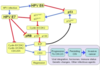
What is the terminology for squamous lesions of the cervix?
- All HSIL’s are considered at-risk for progression to carcinoma
- SIL terminology is current
- Severe dysplasia and carcinoma in situ are the same thing

What is this?

- Normal cervical epithelium
- BM and nice maturation of the cells
What do you see here?

- CIN I: koilocytic change, with halos and raisin-like nuclei, bi-nucleation in lower 1/3rd of the epithelium
- Highest viral load at top, maturing cells -> high proliferation at surface
What is this?

- CIN II: still have a little bit of maturation, with para-keratotic cells on top
- Higher mitotic figure (in middle) is a giveaway for these
- Highest viral load at top, maturing cells- high proliferation at surface
- 20% can develop without LSIL
What is this?

- CIN III: NO maturation, and may start looking for invasion
- Highest viral load at top, maturing cells -> high proliferation at surface
- 20% can develop without LSIL
What staining methods are highly correlated with HPV infection?
- Ki-67 and p53: can really help in older women because their cervix can become atrophic
A. LSIL
B. In-situ hybridization for HPV DNA -> dark granular staining (typically most abundant in the koilocytes)
C. Ki-67 (brown nuclear staining): abnormal expansion of the proliferating cells
D. Upregulation of CDK INH p16 (brown stain) characterizes high-risk HPV infections
- p16: has to be strong AND diffuse to be (+), i.e., staining every cell (will often see a block of brown cells next to “normal-appearing” cells)

What do you see here?

- Invasive SCC: this is what you are looking for in CIN III
- A little bit of keratinization in the bottom right
How likely are LSIL and HSIL to persist, regress, and progress (table)?

What is the outcome of most cervical HPV infections?
- Most women clear the HPV in 12-30 months

What are these two lesions? Difference?

- Cervical adenocarcinoma: AIS on the left and invasive adeno on the right
- Adeno in situ: have lost mucin-containing cells at the top (normal gland at the bottom)
- Invasivet: back to back glands with cribriform architecture, hyperchromatic, necrotic debris because turning over very fast, loss of maturation
- 2nd most common tumor type in the cervix: 15% of cervical cancer cases and devo from a precursor lesion called adenocarcinoma in situ
- Caused by high-risk HPV;s
- Note: adenosquamous and neuroendocrine tumors exist, very rare, and typically aggressive
What do you see in these cytology samples?

- Normal in top left
- LSIL: nuclear membranes are wrinkly (top right)
- HSIL: punctuated nuclear membrane and hyperchromatic (bottom)