Female Repro Flashcards

1
Q

What gnes promote ovarian development vs testerone?

A

DAX-1 gene promotes ovarian development and differentiation

SRY gene coding for TDF which upregulates Sox9 expression for testicular development

2
Q

Intersex

A

Intersex is a general, nonspecific term meaning that ambiguous genitalia are present, but does not indicate the nature or etiology of the abnormality

3
Q

Sexual development disorders are categorized as:

A

Abnormalities of chromosomal sex

Abnormalities of gonadal sex

Abnormalities of phenotypic sex

4
Q

Abnormalities of chromosomal sex

A

Animals with these disorders have an abnormality in the number or structure of the sex chromosomes

◦ XXY → Klinefelter
◦ XXX
◦ XO → Turner
◦ XX/XY (Chimeras and mosaics)

 In general, animals with trisomy or monosomy have underdeveloped genitalia and are sterile

5
Q
A

An example of these chromosomal sex disorders are male tortoiseshell or calico cats; they have testicular hypoplasia and are almost always infertile ( some may be XXY)

6
Q

Chimeras

A

individuals composed of two or more cell populations each arising from different individual

7
Q

Mosaics

A

individuals composed of two or more cell populations, but the cells originate within the same individual

8
Q

What is the most common example of a chimera?

A

Genetic female born co-twinwithamale

 Pathogenesis → vascular anastomoses between placentas allow male hormones (incl Mullerian Inhibitory Substance) and cells to cross and suppress development of the female genital system

Macroscopically, freemartins have small ovaries, blind- ended uterus, poorly developed vagina, enlarged clitoris and seminal vesicles

 Maletwinisminimallyaffected

9
Q

Abnormalities of gonadal sex

True hermaphrodites

How do you define them?

A

 Ovary and testis present in the same individual

 Lateral → testis one side, ovary the other

 Bilateral → ovotestes both sides

 Unilateral → ovotestis one side, ovary or testis on other

 Ambiguous external genitalia
 Rare, seen more in dogs, goats and pigs
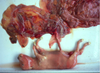
10
Q
A

True hermaphrodite

mix of male and female

11
Q
A

Gilt, lateral hermaphrodite (testis one side, ovary the other)

12
Q
A

True hermaphrodite → Ovotestis

Bilateral

13
Q

Sex reversal

A

Sex reversal; animal in which gonadal sex does not follow chromosomal sex

 Gonad is not the type corresponding to the XX or XY makeup of the individual

◦ American Cocker Spaniel
 Dogs with XX sexual reversal may be XX true

hermaphrodites or XX males

◦ Polled goats (gene with Y effect close to gene for hornlessness)

14
Q

pseudohermaphroditism

A

Abnormalities in phenotypic sex (pseudohermaphroditism)

 Occur when chromosomal and gonadal sex agree, but the internal or external genitalia are ambiguous

Female pseudohermaphrodites

 Often the result of iatrogenic administration of androgens or progestagens during gestation

15
Q

Male pseudohermaphrodites

A

Due to failure of Mullerian duct regression

 Persistent Mullerian Duct Syndrome in the Miniature Schnauzer

16
Q
A

XY dog with testes- Clitoral enlargement The clitoris protrudes between the labia and is visible on the ventral floor of the vulva

17
Q

Segmental aplasia of the paramesonephric ducts

A

DEVELOPMENTAL ANOMALIES of Phenotype Sex

 Segmental aplasia of the paramesonephric ducts  Failure of short or long segments of the uterine horn to

develop
 Complete absence of an entire horn → uterus unicornis

 Commonly found in white Shorthorn cattle → “white heifer disease” → associated with the recessive gene for white coat color

18
Q
A

Uterus unicornis

19
Q
A

Uterus unicornis; ovaries on both sides

20
Q

Imperfect fusion of the paramesonephric ducts

A

Results in double vagina, double cervix, and uterus

didelphys

21
Q
A

Uterus didelphys

22
Q
A

double cervix

23
Q

Failure of fusion of the paramesonephric ducts with the urogenital sinus

A

Persistence of a tissue band running across the vagina just cranial to the opening of the urethra (imperforate hymen)

24
Q
A

Imperforate hymen

25
Q
A

imperferate hymen

26
Q
A
27
Q

Duplication of an ovary

A

incidental

28
Q

Ovarian hypoplasia

A

Results in small ovaries without follicles

 Seen in Swedish Highland cattle and in other cattle and mares with chromosomal abnormalities (XXX or XO)

29
Q
A

Bilateral ovarian hypoplasia (reproductive tract may remain infantile)

30
Q
A

Paraovarian cysts

31
Q

Cystic Graafian follicle

A

Cystic Graafian follicle → commonest type of cystic change

◦ Occur as a result of insufficient release of luteinizing hormone

◦ Cysts may be simple or multiple, and if they persist, can cause changes associated with prolonged estrogen stimulation

32
Q
A

Cow→ cystic Graafian follicle

33
Q

Cystic subsurface epithelial structures (SES) of the bitch

A

Frequently give rise to single or multiple cysts extending along ovarian surface

◦ Occasionally undergo neoplastic transformation (adenomas, and adenocarcinomas)

34
Q
A

Ovarian carcinoma

35
Q

oophoritis

A

Inflammation (oophoritis)
◦ Rare → tuberculosis of the peritoneal cavity ◦ In poultry → Salmonella pullorum
◦ IBR,BVD

36
Q
A

Inflammation (oophoritis

37
Q

Intrafollicular hemorrhage

A

◦ In mares can be quite severe
◦ In cows → manual enucleation of the corpus luteum ◦ May lead to reduced fertility

38
Q
A

Mare → Intrafollicular hemorrhage

39
Q
A

Cow → Periovarian hemorrhage

40
Q

Epithelial tumors

A

Ovarian tumors

 Epithelialtumors

 Develop from the surface epithelium of the ovary

 Frequent in dogs (40–50% of all ovarian neoplasms)

 Benign and malignant forms are difficult to differentiate

 Papillary cystadenomas in bitches may contribute to the appearance of ascites

 Affected bitches may have cystic hyperplasia of the endometrium

41
Q
A

Ovarian tumors

 Epithelial tumors

42
Q

Dysgerminoma

A

Dysgerminoma
Tumors of primordial germ cells of the embryonic

gonad (female counterpart of testicular seminoma)  All considered malignant; up to 20% metastasize

43
Q

Teratoma

A

Uncommon neoplasm composed of abnormal tissue derived from at least two of the three germ cell layers (endoderm, mesoderm, ectoderm)

44
Q
A

Ovary- dysgerminoma

45
Q
A

Ovary → Teratoma

Younger animals

features= really odd structures growing them

46
Q

Granulosa cell tumor (granulosa-theca cell tumors)

A

Most common in horses

Most common ovarian tumor in cows and mares

May be steroidally active (estrogens or androgens)

Generally unilateral and large; may be solid, cystic or polycystic with abundant hemorrhage and necrosis

 Microscopically tumor cells resemble normal granulosa cells

 Call-Exner bodies are diagnostic (rosettes of granulosa cells surrounding pink proteinaceous fluid)

 Malignant forms will also metastasize to other organs (especially in cats)

47
Q
A

Granulosa cell tumor (granulosa-theca cell tumors)

48
Q
A

granulosa cell tumors

Call-Exner bodies

49
Q

Hydrosalpinx

A

The uterine tube is distended and filled with clear watery mucus; usually secondary to obstruction (congenital or inflammatory)

50
Q

Salpingitis

A

Usually secondary to endometritis; may lead to

pyosalpinx and interfere with fertility

 Commonly seen with Mycoplasma and Ureaplasma infections

INFLAMMATION

51
Q

Pyosalpinx

A

Accumulation of pus in the tube following obstruction of the lumen

52
Q
A

Ewe → Hydrosalpinx

53
Q
A

Hydrosalpinx

54
Q

Cystic Gartner’s ducts

A

vestigial remnants of Wolffian ducts

55
Q

cystic Bartholin’s gland

A

vestibular glands

56
Q
A

Cow → Cystic Gartner’s ducts

57
Q

Vulval tumefaction

A

SWOLLEN VULVA

Physiological response to estrogens; also due to persistent hyperestrogenism (endogenous or exogenous)

In sows, often due to the estrogenic effect of zearalenone (Fusarium) in moldy grains

 May lead to vaginal prolapse  +/- mammary enlargement

58
Q
A

Vulval tumefaction

59
Q
A

Mare → rectovaginal fistula

60
Q

Inflammation of vagina and vulva

A

Associated with trauma, likely post partum

 Granular vaginitis → nodular appearance of the vaginal mucosa associated to lymphoid follicle proliferation in cattle (some cases associated with Mycoplasma or Ureaplasma infection)

61
Q
A

Chronic vaginitis

62
Q

Infectious pustular vulvovaginitis

A

Infectious pustular vulvovaginitis (IPV) disease of cattle caused by BHV-1 → disease is venereally transmitted and causes epithelial necrosis

63
Q
A

Small mucosal pustules lead to erosions overlying the submucosal lymphoid follicles

Infectious pustular vulvovaginitis (IPV) disease of cattle caused by BHV-1 → disease is venereally transmitted and causes epithelial necrosis

64
Q
A

Herpesvirus in horses (coital exanthema EHV-3) and Trypanosoma equiperdum causing “Dourine”

65
Q
A

Leiomyoma (fibroids)

 Can occur in the uterus, cervix or vagina

In dogs appear to be estrogen dependent (almost always occur in entire bitches)

begin as tumors of muscle

smooth under mucosal surface

66
Q
A

Transmissible venereal tumor (TVT)

67
Q

Transmissible venereal tumor (TVT)

A

 Contagiousneoplasm;IHCsuggestshistiocyticorigin  Tumor cells → 59 chromosomes (normal 78

chromosomes)

 Macroscopically → solitary or multiple, papillary to pedunculated or multi-lobulated masses often ulcerated, inflamed and friable

68
Q
A

Transmissible venereal tumor (TVT)

 Histologically → solid sheets of large round to ovoid cells; moderate to scant pale eosinophilic finely granular, often vacuolated or clear cytoplasm

THESE CAN BE ANYWHERE- ORAL CAVITY, SQ

69
Q
A

Squamous cell carcinoma (SCC)

 Occurs mostly in farm animals, especially those lacking pigment on vulvar skin

70
Q
A

Melanoma and melanocytoma

 Particular in white mares, under the form of large ulcerated nodules, located in the vulva or the perineum

71
Q

Torsion

A

 Most commonly occurs in enlarged uterus (pregnancy, pyometra or mucometra)

 May result in circulatory embarrassment, death of the fetus, and/or uterine rupture

Accounts for 5-10% of serious cases of dystocia in mares

72
Q
A

Unilateral uterine torsion

73
Q

Uterine prolapse

A

Common in ruminants

 Causes →

 Uterine hypotony  Prolonged dystocia  Retained placenta  Hypocalcemia
 Hyperestrogenism

May be followed by congestion, edema, hemorrhage, necrosis, gangrene and sepsis

74
Q
A

Uterine prolapse

75
Q

who is uterine prolapse common in?

A

Large animal

76
Q

Uterine rupture

A

From trauma at parturition, iatrogenic or spontaneous; can lead to fatal hemorrhage, perimetritis and peritonitis

77
Q
A

Uterine rupture

78
Q

Rupture of the uterine artery

A

Occurs in mares and results in death from exsanguination

79
Q
A

Ruptured uterine artery

80
Q

Endometrial hyperplasia

A

 Most common in the bitch and involves cystic distention of endometrial glands

 Ifendometrialsecretionsaccumulate,infectionmayfollow (cystic endometrial hyperplasia - pyometra syndrome)

Often due to prolonged hyperestrogenism (farm animals) or excess progesterone (from persistent CL) with estrogen priming (dogs, cats)

 Estrogen sources include → ◦ Cystic ovarian follicles
◦ Granulosa cell tumors
◦ Estrogenic pastures

◦ Zearalenone (and other mycotoxins)

 Lesions can become cystic and may lead to pyometra

Estrogen binds to estrogen receptors in endometrium  synthesis of intracellular progesterone receptors  progesterone immunosuppresses, providing a suitable environment for bacteria to grow and cause pyometra

81
Q
A

Endometrial hyperplasia . Cystic endometrial hyperplasia

82
Q
A

Cystic endometrial hyperplasia

83
Q

Adenomyosis

A

Presence of endometrial glands and stroma between the muscle bundles of the myometrium

84
Q
A

Adenomycosis

85
Q

Endometriosis (ectopic endometrial glands)

A

Actively growing endometrial tissues are explanted to aberrant sites within and outside the uterus

86
Q
A

Endometriosis in a non-human primate

87
Q

Subinvolution of placental sites

A

 Occur in the bitch
 Manifested by prolonged bloody vaginal discharge

postpartum

 Placental sites appear as multiple segmental thickenings of uterine horns

88
Q
A

Subinvolution of placental sites

89
Q
A

Subinvolution of placental sites

 Multiple segmental thickenings visible from the serosal surface Endometrium is hemorrhagic and thickened
 Placental sites are raised, rough, gray-brown plaques
 Uterine lumen contains small amounts of serosanguinous fluid Endometrium between sites is normal

90
Q

Hydrometra and mucometra

A

Accumulation of thin or viscid fluid in the uterus ( 2nd to endometrial hyperplasia or congenital obstruction)

91
Q
A

Hydrometra

92
Q

Endometritis

A

inflammation of endometrium only

93
Q

Metritis:

A

inflammation of all layers of uterine wall

94
Q

Perimetritis

A

inflammation extending to tissues surrounding uterus

95
Q

Pyometria

A

accumulations of pus in the lumen of the uterus

96
Q

Metritis, Endometritis, Perimetritis

A

The normal nonpregnant uterus is highly resistant to infection; resistance is greatly reduced during pregnancy and under the influence of progesterone

 Resistance is low postpartum, especially following dystocia, retained placenta, or slow involution

 Most uterine infections start as endometritis and may or may not progress to metritis, perimetritis or pyometra, all of which impact fertility

97
Q
A

Uterus: diffuse fibrinosuppurative metritis

98
Q

Contagious equine metritis (CEM)

A

Venereal disease of mares caused by Taylorella equigenitalis (G-)

 Clinical disease is manifested by temporary infertility and mild to moderate inflammation of the endometrium; stallions transmit the organism but don’t develop clinical disease

REPORTABLE

99
Q
A

Contagious equine metritis (CEM)

100
Q

Pyometra

A

Acute or chronic suppurative inflammation characterized by accumulation of pus in the uterine lumen; usually secondary to endometritis

101
Q
A

Pyometra

102
Q

Pyometra in bitches

A

Characteristically affects older animals (especially those that are not bred)

 Condition most often develops a few weeks after estrus; animals are depressed and anorexic, frequently vomit and have polyuria and polydipsia, usually accompanied by a vaginal discharge

103
Q
A

Pyometra

104
Q

What impacts the pus in a pyometra?

A

 The amount of pus varies with the duration and type of bacteria involved

 Extragenital lesions in dogs Bone marrow depression and anemia Marked EMH in spleen and liver Marked leucocytosis

Immune complex glomerulonephritis

105
Q

What species are endometrial biopsy?

A

Most commonly used in equine theriogenology-ONLY
 The evaluation system is based on identification and

scoring of 4 microscopic lesions →

Inflammation

Dilation of lymphatics

 Loss of gland

s Fibrosis

106
Q
A

Rabbit. Uterine adenocarcinoma with pulmonary metastases

107
Q
A

Lymphosarcoma in cows is the most common encountered metastatic neoplasm (BLV-positive cows)

Uterine lymphosarcoma

108
Q
A

Leiomyomas in bitches

109
Q

Mammary gland- natural defense mechanisms

A

 Mechanica lbarrier(teatorificeandcanal)

 Presence of bactericidal compounds on the surface of

the teat canal

 Humoral factors in milk (lactoferrin, opsonins, immunoglobulins)

 Cellularfactors(neutrophilsandmacrophages)

110
Q

Mastitis

A

 Most costly disease in dairy herds

 Inflammation of the mammary gland is usually due to infectious agents (mainly bacteria, some fungi) that gain access through the teat canal

 Hematogenous and percutaneous routes rarely occur

 Peak occurrence is shortly after calving and in the drying-off period when milk stagnates in the udder

111
Q

Types of mastitis

A

Contagious
◦ Streptococcus agalactiae ◦ Staphylococcus aureus
◦ Mycoplasma spp

 Environmental
◦ Coliforms (E. coli, etc)
◦ Environmental streptococci

 Others
◦ Other bacteria, fungi, viruses

112
Q

Streptococcus agalactiae

A

 Specific, contagious mastitis of ruminants; entry is through the teat canal

 Healing is characteristically by fibrosis; sequel → atrophy of quarters

Not a systemic sickness

113
Q

Staphylococcus aureus

A

 Acute inflammation due to α-toxin; may be peracute and fulminating or milder or more chronic

 Abscesses, necrosis, gangrene in cattle

SYSTEMIC SICKNESS

114
Q

Mycoplasma bovis

A

 Cattle and sheep

 Galactogenic and hematogenous routes

 Sudden onset of agalactia, involvement of all 4 quarters, a precipitous drop in milk production, severe swelling of the udder, poor response to therapy and lack of obvious signs of systemic illness

not systemic

115
Q
A

Staphylococcus aureus

Some strains elaborate an α-toxin that causes gangrenous staphylococcal mastitis. The toxin causes vasoconstriction and ischemia to the udder resulting in gangrene

116
Q
A

Mycoplasmal mastitis

117
Q
A

Head tilt in a calf with Mycoplasma otitis

118
Q

Coliform mastitis

A

Poor hygeine

 Organisms are ubiquitous (“environmental mastitis”) → infection is through the teat canal (may be associated with metritis and septicemia)

 E.coli may cause an acute and often fatal disease → potent endotoxin causes vascular damage leading to fluid exudates, hemorrhage, thrombosis and necrosis

 The disease is most severe in newly calved cows transiently immunosuppressed

 Peracute cases show systemic signs of severe toxemia

119
Q
A

Severe coliform mastitis

120
Q
A

Gangrenous mastitis

121
Q

Granulomatous mastitis

A

 Mycobacterium bovis, Nocardia asteroides, Cryptococcus neoformans, other Mycobacterium spp., Candida spp.

 Mycobacterium bovis is likely spread hematogenously, while the others are often reported to be iatrogenic

 Typical granulomas are formed and the organisms can usually be demonstrated in tissues by special stains or cultured from milk

 Nocardial mastitis has been associated with neomycin infusions in dry cows

122
Q
A

Granulomatous mastitis

Chronic inflammation of the lactiferous ducts and adjacent mammary gland has resulted in replacement of most of the gland by pyogranulomas and abscesses

Can feel the nodules in the gland

123
Q

Mastitis in other species

A

Mastitis in the ewe and goat is usually caused by Staphylococcus aureus or Mannheimia haemolytica

Contagious agalactia

 Primarily a disease of goats, sheep being slightly less susceptible, caused by Mycoplasma agalactiae

 Lactating females and kids are particularly susceptible to the infection and likely to succumb in the septicemic phase of the disease

124
Q

Other causes of mastitis

A
  • Viral diseases such as
  • Ovine progressive pneumonia (Maedi-Visna) • Caprine arthritis encephalitis (CAE)
  • They cause diffuse interstitial mastitis that is not recognizable grossly
  • Foot and Mouth disease may also cause mastitis
125
Q
A

Mastitis associated with FMD virus is assumed to be due to secondary bacterial infections.

126
Q

Mammary gland neoplasia

A

 Mammary tumors are seen most commonly in bitches and queens and are rare in other species

 Mammary fibroadenomas are common in rats

127
Q
A

Rat Mammary fibroadenoma

128
Q

What is the most common diagnosed tumor in female dogs?

A

 One of the most commonly diagnosed tumors in female dogs

 Epithelial tumors are most commonly diagnosed (>90% of all tumors), the remainder consisting of sarcomas and mixed tumors

 Approximately 50% of all tumors are malignant and of these, 50% express the metastatic phenotype

cats- these are usually malignant

dogs-benign

129
Q

If a female dog has mixed mammary tumors will spaying them will or will not stop the tumors?

A

WILL NOT

130
Q

Benign mammary tumors

A

 Adenoma

Simple → Proliferation of well-differentiated luminal epithelial cells

Complex → Mixed proliferation of secretory epithelial cells myoepithelial cells

 Fibroadenoma
◦ Mixture of epithelial and stromal cells

 Benignmixedtumor
◦ Proliferation of glandular and mesenchymal elements

 Ductpapilloma
◦ Papillomatous projections in distended ducts

131
Q
A

benign mixed tumor

132
Q

Malignant mammary tumors

A

 Carcinoma
 Osteosarcoma

◦ Mammary gland is the most common site of extraskeletal soft tissue osteosarcoma in dogs

 Carcinosarcoma:
◦ Also called malignant mixed tumor; malignant epithelial and

mesenchymal components  Fibrosarcoma

133
Q
A

metastatic spread very quickly; fibrosarcoma

134
Q

Prognostic factors for canine mammary tumors

A

Histological type

 Degree of invasion (very

important!!!)

 Degree of nuclear differentiation

 Lymphoidcellularreactivity

 Tumor size (>3 cm)

 Ulceration

 Inflammation/inflammatory carcinoma

 Estrogen or progesterone receptor status

135
Q

Clinical features associated with prognosis of canine mammary tumors

A

Clinical presentation

 Approximately 70% of dogs have more than one mammary tumor*

 Smaller tumors are more likely to be benign than larger ones

 Dogs with malignant tumors are more likely to develop new tumors in other mammary glands

 Tumors > 5cm in size are more likely to be malignant and more likely to show lymph node metastasis*

 Lymphatic invasion and lymph node metastasis are significant prognostic factors

*sampling errors may lead to incorrect diagnosis

136
Q

Feline mammary neoplasia

A

Third most common neoplasm in domestic cats- VERY LIKELY MALGIANANT

 Classification is similar to the canine classification;

most neoplasms in cats are malignant

 Most tumors are adenocarcinomas → grow rapidly and metastases are common

 Staging and grading of tumors is done similarly as in dogs

 Tumor size is the single most important prognostic factor in cats with mammary gland neoplasia (tumors < 2 cm in size have a better prognosis)

137
Q
A

Feline mammary tumor

138
Q

Mammary fibroadenomatous hyperplasia

A

“Juvenile fibroadenoma, feline mammary hypertrophy”

 Benign, nonneoplastic proliferation of mammary ducts and connective tissue of young intact cats, pregnant cats, or older neutered male and female cats on prolonged progesterone therapy (megestrol acetate)

139
Q
A

Mammary fibroadenomatous hyperplasia

140
Q
A

Mammary fibroadenomatous hyperplasia

 Histologically → lobules of branching ductal structures lined by epithelial cells surrounded by edematous myoepithelial stroma

141
Q
A

Amniotic plaques - Foci of squamous epithelium on the internal surface of the amnion; they are commonly present on the bovine amnion during the middle trimester of gestation

INCIDENTAL!

142
Q
A

Mineralized foci- incidential

143
Q

EMBRYONIC MORTALITY AND FOETAL DEATH

A

RARELY SEE EMBROYO

Early embryonic losses/ mortalities occur during the early stage of gestation (delayed return to estrus)

 Except for some infectious causes of infertility, it

appears that chromosomal abnormalities account for

many of these losses
 Eg. Campylobacter fetus venerealis

 Foeta ldeath later in development will lead to→  Abortion
 Stillbirth
 Mummification

 Maceration

144
Q

Abortion

A

Abortion is defined as the expulsion of a foetus prior to the time of expected viability

145
Q

Stillbirth

A

Stillbirth is the death of the foetus in the last part of gestation during the period where it is independently viable

146
Q

Mummification

A

The fetus is retained indefinitely and becomes dehydrated

 Typically no bacterial infection to promote tissue lysis or putrefaction

Dehydration of a fetus in utero usually takes longer than 1 week to occur

147
Q
A

Dehydration of a fetus in utero usually takes longer than 1 week to occur

148
Q

Maceration

A

The dead fetus is retained and infected by bacteria

 May be associated with dystocia or incomplete

abortion

 May lead to pyometra and maternal death from peritonitis and toxemia

149
Q
A

Uterus. Macerated fetus

150
Q

Placental insufficiency

A

 Important in the mare; may lead to fetal death, mummification or abortion

 Seen in cases of:
Twinning
Endometrial fibrosis Premature placental separation Uterine body pregnancy Torsion of the umbilical cord

151
Q
A

Placental insufficiency

152
Q
A

Torsion-umblicial cord

153
Q

Adventitial Placentation

A

Development of intercotyledonary placentation in cattle as a mechanism of compensation for inadequate development of placentomes

154
Q
A

Adventitial placentation -right

normal-left

155
Q

Hydramnios

A

Hydramnios(hydropsoftheamnion)
◦ Usually associated with malformation of the foetus (ie. facial

anomalies)

156
Q

Hydroallantois

A

Associated with uterine disease with inadequate numbers of caruncles and the development of adventitial placentation in cattle

157
Q

Incidental findings

A

 Amniotic Plaques and Placental Mineralization  Hippomanes

 Present in virtually all equine placentae and are proteinaceous soft calculi, aggregated mineral and organic allantoic concrements

 Yolk sac remnants and allantoic pouches are also commonly observed in mares

158
Q
A

Hippomane

159
Q
A

Yolk sac remnants

160
Q

Infectious causes of abortion

A

There are a group of abortogenic agents that occur in a variety of species and include →

Brucella spp

. Chlamydophila abortus

Coxiella burnetii
Leptospira spp.
Listeria monocytogenes Salmonella spp. Mycoplasma and Ureaplasma Neospora caninum Toxoplasma gondii Herpesviruses Pestiviruses

161
Q

Basic concepts concerning abortion in cattle

A

 Criteria for submission of aborted bovine foetuses

for diagnostic evaluation →

When the abortion rate exceeds 3% (maximum tolerable abortion rate)

When a number of animals abort over a short period of time

 Consider regional differences in the most common causes of abortion

 Be familiar with common causes of abortion in your area!

162
Q

What is the most common cause of pregnancy failure in cattle?

A

Infectious cause- usually bacteria/salmonella

163
Q

Several lesions may be found in foetuses that may carry diagnostic significance →

A

Dermatitis → Mycotic abortions

Amnionitis → Ureaplasma sp

Focal necrosis in the liver or other tissues → herpesviral infections/Campylobacter

Myocarditis and myocardial necrosis → BVD, Neospora caninum, nutritional myopathy

Brain microscopic lesions- Protozoal

Placental lesions-fungal/Chlamydiae/Protozoal/Bact.

164
Q

Campylobacter foetus subsp. venerealis

A

 True genital infection and is an important cause of infertility

 Early embryonic deaths at 3-4 weeks (rarely late term

abortions)

 Gross placental lesions are those of intercotyledonary placentitis and necrosis of the cotyledons

165
Q

Tritrichomonas foetus

A

 Embryonic death or overt abortion can occur; pyometra can be a complication

 Protozoa can be found in preputial washes, vaginal mucus and in cases of abortion, in the stomach contents of the foetus

166
Q

Brucella abortus

A

Abortions are more common at 7-9 months

Gross lesions in the placenta are characteristic but not pathognomonic (necrotising placentitis)

Most foetuses aborted in the last half of pregnancy will show evidence of pneumonia
•Easy to culture organisms from fluid or tissues

zoonotic

167
Q
A

Chronic placentitis. Placental lesions are not uniform; some cotyledons may appear more-or-less normal and others will be extensively necrotic

zoonotic

Brucella abortus

168
Q

Salmonellosis

A

Salmonella dublin

Salmonella typhimurium

ZOONOTIC Antibiotic Resistance

169
Q

Listeriosis

A

Abortions in both cattle and sheep due to L. monocytogenes → last trimester of pregnancy

 Gross lesions → exudative intercotyledonary placentitis and in the foetus there may be tiny pinpoint yellow foci of necrosis in the liver the lung, myocardium, kidney, adrenal, spleen and brain.

170
Q

Leptospiral abortion

A

Most of the abortions → last trimester of pregnancy

 No placental or foetal characteristic lesions so diagnosis is dependent on

demonstrating raising antibody titres in paired sera from the dam

 IFA on tissues also diagnostic

Leptospirres invade and kill the fetus which rapidly becomes autolysed

171
Q
A

Leptospiral abortion

172
Q

Ureaplasma diversum

A

Abortion usually occurs in the last trimester of gestation

 The amnion is the most severely affected portion of the placenta

173
Q
A

Aminoitis. There is patchy thickening with fibrosis and multifocal areas of necrosis, heamorrhage, and fibrin exudation.

174
Q
A

Mycotic abortion in cattle

 Variety of fungal species
Aspergillus fumigatus
Zygomycetes (Absidia, Mortierella, Rhizomucor, Rhizopus)

 Dermatitis in the aborted foetus is often associated with mycotic abortions

175
Q
A

Mycotic placentitis

176
Q

Neospora caninum

A

Most abortions occur at 5-6 months of gestation (distinctive from other kinds of abortion in cattle)

 No useful identifying gross lesions; bovine foetuses may be fresh, autolysed, or in early stages of mummification

 N. caninum is also a cause of abortion in sheep and possibly in goats

 Brainneededfordiagnosis

177
Q
A

Organisms may be identified in focal brain lesions (focal non-suppurative encephalitis)

178
Q
A

Lesions which are multifocal are pathogonomic for either neospora or toxoplasma.
Immunohistochemistry for definitive diagnosis

THIS IS A PATHOPNEUMONIC LESION!

179
Q

What are the most important viral pathogens for viral abortion in cattle?

A

Bovine viral diarrhea virus and Akabane virus (AKAV) are probably the most important viral pathogens; less commonly BoHV-1 and Bluetongue virus and more recently Schmallenberg (in Europe)

180
Q

What is the major cause of preg. failure in sheep?

A

Most are infectious

181
Q

Campylobacter spp.

A

Campylobacter foetus subsp. foetus is an important

cause of abortion in sheep

 Lesions are characterized by intercotyledonary placentitis and multifocal necrotizing hepatitis

182
Q
A

Campylobacter spp.

183
Q

Chlamydophila abortus

A

“Ovine enzootic abortion” and “enzootic abortion of ewes”

 Important cause of in utero infections in sheep and goats resulting in abortion, stillbirth, and the birth of weak offspring

 If a ewe is infected early in gestation → will abort in the same gestation; if infection occurs in late gestation → will abort during the next pregnancy

 Placentallesionsacutesuppurativetochronicplacentitis

 Diagnosis → organism should be demonstrated in a smear (placenta) stained by a modified ZN, Gimenez, or Giemsa method

184
Q
A

Transmission occurs upon exposure to infected birthing fluids

“Ovine enzootic abortion”

185
Q
A

Foci of intercotyledonary necrosis

“Ovine enzootic abortion”

186
Q

Toxo

A

 Major roleinovineabortion;thesourceofinfectionis usually infected cats

 Naive ewes will develop a protective immunity but if they are pregnant, infection of the cotyledon will result in abortion

 Gross placental lesions → cotyledons are bright to dark red and have numerous small soft white nodules (“strawberry-like lesions”)

187
Q
A

Toxo

188
Q
A

Toxo

189
Q
A

Toxo lesions in the brain

190
Q

Border disease

A

Border disease (hairy shaker disease)

 Infection of border disease virus can result in embryonic or foetal death, abortion, mummification, dysmorphogenesis, early postnatal death, and birth of weak lambs

191
Q

What are common preg. failures in goats?

A

PREGNANCY FAILURE IN GOATS

 Similar to abortion in sheep, except that Coxiella burnetti, trauma and stress are more common causes

 Other causes include Brucella melitensis, Salmonella spp. and Iodine deficiency

192
Q

Coxiella burnetii (Q fever)

A

Very important disease, of goats and sheep; poses a

significant zoonotic risk !
 Abortion tends to occur late in gestation and weak

lambs and kids may be born during an outbreak

 Organisms can be demonstrated using modified Ziehl- Neelsen or Macchiavello’s stain

Gross lesions are confined to the placenta → acute

diffuse suppurative placentitis

193
Q
A

Acute diffuse suppurative placentitis

Coxiella burnetii (Q fever)

194
Q
A

The placenta is thickened and leathery, with multifocal to confluent areas of mineralization; exudate is copious, off-white and most obvious in the intercotyledonary region

Coxiella burnetii (Q fever)

195
Q

What is the most common cause of preg. failure in mares?

A

Infectious and non-infectious

YOU HAVE TO SUBMIT THE PLACENTA!

196
Q

Umbilical cord abnormalities

A

 Cordshouldbebetween36-83cmlong

 Longer cords are associated with foal strangulation and

necrosis of the cervical pole of the placenta

 A cord that is less than 36 cm may be associated with premature separation of the placenta at the site of attachment

197
Q
A

Umbilical cord torsion

198
Q

Mare reproductive loss syndrome and late- term abortions (MRLS)

A

 Abortion syndrome that reached epidemic proportions (Kentucky 2001-2002)

 The setae (hairs) of the eastern tent caterpillar embed into the submucosa of the alimentary tract of the mare create microgranulomatous lesions → bacteria from the alimentary tract invades the circulatory system → infections are established in tissues where the immune surveillance is reduced (fetus and placenta)

199
Q

Equine herpesvirus abortion in horses

A

EHV-1 is widespread throughout the world and causes respiratory, neurologic and generalized neonatal disease, as well as abortion

 Majority of infections occur during the last 3 months of gestation and there may be prominent changes in the foetus →

◦ Fibrin cast in the trachea
◦ Interstitial pneumonia
◦ Focal necrosis in the liver
◦ Prominent lymphoid follicles in the spleen

200
Q
A

Equine herpes virus

 Foals infected with this virus in utero may be born alive at, or near, term; many of them die in the first few days with severe interstitial pneumonia and secondary bacterial septicemia

Equine herpesvirus 4 and equine viral arteritis virus may also induce abortion in mares
Diagnosis based on PCR and/or observation of inclusion bodies in lung and liver tissues

201
Q

What is the cause of most of the of preg. failure in pigs?

A

Infectious

It is suggested that some 30-40 % of porcine abortions are primarily infectious

 Opportunistic infections associated with ubiquitous microorganisms → Actinomyces pyogenes, Escherichia coli, Staphylococcus aureus and Aspergillus, Erysipelothrix, Pasteurella, Salmonella and Streptococcus sp.

202
Q

Brucella suis

A

Abortion occurs between the 2-3 month of pregnancy; there is also a high incidence of stillborn and weak piglets born at term

203
Q

SMEDI

A

Stillbirths, mummification, embryonic death and infertility complex (SMEDI)

 SMEDI-type problems are caused by porcine parvovirus; less frequently enteroviruses

 Classical sign of SMEDI → full-term litter consisting of small mummified foetuses ranging up to full-grown stillborn or live weakly piglets, due to progressive infection in utero; abortion rarely occurs

204
Q
A

Brucella canis

Pregnant bitches may abort after 30 days, but most abortions occur after 50 days; there is often prolonged vaginal discharge after abortion

205
Q

Protocals of abortion

A

Access status of foetus
 Culture (stomach fluid typically)

 Histopathology- best for protozoals
 Serology of pleural fluid
 Leptospira FAT
 Tissue culture
 Dam Serology- Neosporia, Salmonella

Sheep- chlmyadia and toxo- PLACENTA AND BRAIN

206
Q
A

Bovine amorphus globosus

207
Q
A