Respiratory system Flashcards
Main purpose of the respiratory system
Supply itself with oxygenRemove waste products
4 main functions of the respiratory system
To Extract oxygen from atmosphere
To excrete water vapour and carbon dioxide
To maintain the normal acid base balance of the blood
To ventilate the lungs
Inspired Air The components are
Oxygen=20%Nitrogen=79%Inert gases=1%Carbon dioxide=0.04%Water vapour = variable
Expired AirThe components are of expired air
Oxygen 16%Nitrogen 79%Inert gases 1%Carbon dioxide 4%Water vapour to saturation
Function of the nose 5
To warm the airTo filter the airTo moisten the airTo assist in resonating soundOrgan of the sense of smell
Function of the larynx
To provide a passageway for air from the pharynx to the trachea
To continue to moisten
,To warmTo filter the air
To produce sounds
To protect the airway during the act of swallowing food

Structure of the pharynx

Pharynx is funnel shaped tube 13cm long composed of skeletal muscleCan be divided into three sectionsNaso-pharynxOro-PharynxLaryngo-Pharynx

Function of the Pharynx
Respiratory functionDigestive functionAssist with the sense of tasteTonsils assist in the fight against infection

what is this

Larynx
Pic lungs label

1 epiglottis
2 thyroid cartilage
3 cricoid cartilage
4 trachea
5 left lung ( superior lobe/apex)
6 bronchiole
7 left primary bronchus
8 secondary bronchus
9 right primary bronchus
10 tertiary bronchus
11/17 mediastinum
12 parietal pleura
13pleural cavity
14 visceral pleura
15 left lung (inferior lobe)
16 diaphragm
18 mediastinal surface
19 cardiac notch
20 diaphragmatic surface
21 right lung (superior lobe )
22 right lung (middle lobe )
23 right lung inferior lobe
Structure of the trachea
16-20 c-shaped incomplete rings of cartilageApproximately 12cm long
Functions of the trachea
Maintain airway normal and forced respirationAllowed distension of oesophagus during the act of swallowingTo remove dust by the secretions of the goblet Reflex centre for coughing at the bifurcation of the trachea carina
Bronchi what are they
The two bronchi are formed the trachea divides at the carina

The right bronchus is
The right bronchus is wider and shorter than the left after entering the right lung it divides into three branches on to each lobe

The left bronchus is
The left bronchus is longer and narrower than the right 5cm after it enters the lung divides into two branches one to each lobe

Smaller air passages 4
Terminal bronchiolesRespiratory bronchiolesAlveolarAlveoli

Structure of the lungs
Right side three lobesLeft side two lobes
The pleura is
The pleura is a serous membrane that surrounds each lung Has two layers
Chemical control of respiration Normal drive is driven
In normal healthy people the basic drive to breathe is high levels of C02Basic rhythm of respiration is controlled by parts of the CNS
Hypoxic Drive
Patients with copd the C02 chemoreceptors become worn out, The stimulus for this type of patient becomes low concentrations of O2 and is termed the hypoxic drive
Mechanism of external respiration
Inspiratory phase 2sec active stageExpiratory phase 3 Sec passive stageRespiratory pause phase 0.25
Factors that can decrease breathing rates
Head injuryCvaDrugs heroin opiates methadone barbiturates
what is this

pharynx
what is this

trachea
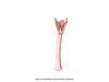
what is this

larynx
Oxygen is taken to the lungs and exchanged at which point in the lungs
Alveoli

Pulmonary volumes Alveoli volume Dead air Tidal

Picture

Lung capacity Inspiratory reserve forced inhalation Tidal Expiratory forced expiration Residual air
Picture

The exchange of oxygen for carbon dioxide is termed what
Gas exchange

external and internal respiration
label
where does external respiration take place

external

Respiratory tract
The respiratory tract is made up of ten main parts that follow one after the other in a line leading from the nose (or nasal cavity) to the pleura membrane in the lungs
1 nose larynx lungs bronchi trachea alveoi hilum
pharynx bronchioles
10 pleura

what happens on expiration

what happens on inspiration
