Path Flashcards
histology of PBC
- florid duct lesion
- granulomatous inflammation
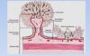
- Histology of PBC: the pt has high antimitochondrial antibodies. He has a granulomatous inflammation [top right image – black arrow points to the granuloma]. You have a destruction of the bile duct – the bile duct is no longer intact in the top left image [black arrow].
- In the bottom image you can see the jigsaw puzzle cirrhosis – very unique for PBC.

risk for cholesterol gallstones
older
female (estrogens) - OCPs
obesity and metabolic syndromes
rapid weight loss
gallbladder stasis

inflammatory polyps histo
reactive/regenerative
epithelial changes with inflammatory infiltrates in lamina propria
non neoplastic

intragastric balloon
restrictive
restricts food intake for 60 months - 20-40 lbs lost


hemorrhoids
secondary to elevated venous pressures
straining at defecation or pregnancy or portal htn
thin walled dilated submucosal vessels beneath anal or rectal mucosa
Types of bile duct epithelial lsions
bile duct adenoma - benign
cholangiocarcinoma - malignant
invasive adenocarcinoma
if lesion penetrates muscularis mucosa
metastatic potential

anal condyloma
squamous papilloma caused by HPV
papillary gorwth
enlarged keratinocytes w central hyperchromatic wrinkled nucleus

aflatoxins
in food can cause damage
- The primary food contaminants are aflatoxins, which are especially seen in developing countries
- If peanuts in particular go bad, it can cause a certain fungal infestation that can produce aflatoxins and AFB1
- This aflatoxin can directly cause a mutation in the p53 tumor suppressor gene
- The 249ser gene mutation is very unique for aflatoxic damage
- This aflatoxin toxin can react synergistically with HBV infection
- Aflatoxin in the liver, in human cells, can induce much more damage related to HBV infection
- In another sense, this can also mean that aflatoxin prevalence parallels that of HBV infection
- In the area that has high HBV infection, you have high incidence of the toxin
stellate cells
in space of disse

- Stellate cells, under normal conditions, are very quiet; they store some fat and minerals
- When the activate, they become fibroblasts and produce collagen, which can eventually cause fibrosis leading to cirrhosis, which we will discuss later
carcinoid tumors
neuroendocrine
from endocrine stem cell in crypt
more indolent than carcinoma
can make many bioactive things

hereditary non-polyposis colon cancer
i.e. lynch syndrome
increased risk of many cancers
colorectal cancers often multiple at young age in right colon
inherited germline mutations in DNA repair caretaker
most common syndromic form of colon cancer
sessile polyps
tumoral masses or nodules which project into the lumen, usually refers to epithelial lesions
sessile polyps have a broad pase


PBC
- Histology of PBC: the pt has high antimitochondrial antibodies. He has a granulomatous inflammation [top right image – black arrow points to the granuloma]. You have a destruction of the bile duct – the bile duct is no longer intact in the top left image [black arrow].
- In the bottom image you can see the jigsaw puzzle cirrhosis – very unique for PBC.
hyperplastic polyps etiology and location
non neoplastic!
age 60-70, asymptomatic
*left colon and rectum
adenoma
precursor of colorectal adenocarcinoma
tubular, villous, tubulovillous
risk of malignancy with size, architecture, dysplasia
familial, higher chance with age
pathogenesis of hepatocellular adenoma
idiopathic
female hormones (contraceptoves)
acute cholecysitis
acute inflammation of the gallbladder
90% from obstruction of the neck of the cystic duct by stones (calculus cholecystitis)
10% from ischemia of systic aretey
sepsis, immunosuppression, trauma, diabetes, nfection
budd chiari syndrome
hepatic venous outflow obstruction
blockage of 2 major hepatic veins
passive congestion and centrilobular necrosis
- This is a typical presentation for Budd-Chiari Syndrome.
- [top left image] Here is a thrombus. If the vessel is blocked, you cause congestion of blood. The blood spills over from the sinusoids and damages the hepatocytes.
- [bottom left image] This is partial. You can see the thrombosis [black arrow]. If you block the left hepatic vein, you cause damage to the left lobe [the darker left portion of the liver shown].
- Histologically, you can see the ischemia in the liver parenchyma [right images]. The hepatocytes are gone b/c the oxygen is depleted. There are no nutrients, causing damage.

juvenile polyp
hamartomatous non-neoplastic polyps
30-50% of patients develop AC by age 45
usually sporadic in kids under 5
usually in rectum
in adults: “retention polyp”
can mean there is a rare polyposis syndrome
colon polyp:

tubular adenoma
neoplastic/premalignant
epithelial cells fail to mature as migrate to crypt surface
crowded disorganized rounded glands, numerous goblet cells and enlarged hyperchromatic nuclei
dysplastic change
before hepatocellular carcinoma

- In 10 to 30 years, you can have a clear preneoplastic change (pre-neoplasia)
- It does not necessarily have to go through an adenomatous change; the adenoma is a different animal
- That is called a dysplastic change
- You will see high- or low-grade dysplasia before HCC
- There may be another 3-5 years before the hepatocytes become dysplastic
- Most of the time, the process will stop here à the patient will not develop cancer
- However, a certain percentage of patients pass that boundary over another 5-10 years and progress on to hepatocellular carcinoma (neoplasia)
neoplastic lesion
- If the proliferation goes out of control without a boundary or limits, you get neoplastic disease
- Benign disease
- Adenoma
- Hemangioma
- Malignant disease
- Metastasis
- Primary hepatocytic carcinoma, ductal carcinoma, cholangiocarcinoma
histo in cronkhite-canada syndrome
mortality in 50-60%
cystically dilated crypts w marked inflammation
mucosa adjacent to polyps also shows cystic dilation

Histo in cowden syndrome
stroma rich polyp with cystically dilated crypts
risk of colon cancer = gen pop


chronic pancreatitis
pancreas is hard w extremely dilated ducts and visible calcifications

hematochromatosis
- You do a biopsy on this pt. This is what you see in the liver biopsy.
- This is the brown colored deposit [black arrow in left image]. It is iron.
- If you are not sure, do an iron stain [right image].
angiosarcoma risk factors
- Some major risk factors in the United States include exposure to vinyl chloride
- This used to be used in the plastic industry, but no longer
- If there is contamination in the water, this can potentially cause development of angiosarcoma
- Exposure to thorium dioxide, which was previously used as contrast for radiology, is also associated with angiosarcoma
- Now we know this is associated with this disease, so it has been banned
- Arsenic and arsenite can also cause angiosarcoma, particularly in developing countries where these can contaminate food
nodular regernative hyperplasia pathogenesi
similar to FNH - adaptive parenchymal hyperplasia due to heterogenous distribution of blood flow
- Top left: wedge resection with a multiple nodular appearance
- The yellowish tissue is liver parenchyma
- There is some bile staining (green)
- Bottom left: the trichrome stain shows a nodule almost separated by incomplete septa
- It is not like cirrhosis
- Cirrhosis, by definition, is a completely separate nodule
- Top right: high power view shows a nodular appearance
- There is a central area; a central vein
- Around the area are the proliferative hepatocytes
- There is still a portal tract with a bile duct à it is hyperplastic, not neoplastic
- Bottom right: reticulin stain highlights the hepatocytes
- This is classic for nodular regenerative hyperplasia (NRH)
- This is not a neoplastic process

nodular regenerative hyperplasia
- Top left: wedge resection with a multiple nodular appearance
- The yellowish tissue is liver parenchyma
- There is some bile staining (green)
- Bottom left: the trichrome stain shows a nodule almost separated by incomplete septa
- It is not like cirrhosis
- Cirrhosis, by definition, is a completely separate nodule
- Top right: high power view shows a nodular appearance
- There is a central area; a central vein
- Around the area are the proliferative hepatocytes
- There is still a portal tract with a bile duct à it is hyperplastic, not neoplastic
- Bottom right: reticulin stain highlights the hepatocytes
- This is classic for nodular regenerative hyperplasia (NRH)
- This is not a neoplastic process

- See the pink globules [black arrow in left image] – protein structures within the hepatocytes.
- If you do a special stain PASD, you would see the pink proteins [black arrow in right image], which are glycoproteins, stuck in ER and cannot be transported outside of the ER for further processing.
- Another hereditary disease is a1-antitrypsin deficiency. It is another autosomal recessive disorder. The gene is on chromosome 14, encoding a protease inhibitor.
- The abnormal protein that is produced cannot be folded properly. Proteins are synthesized in the ER, but this mutated protein cannot be processed well. It is stuck in the ER and cannot get out. It forms a globule.
- Wild type genotype is normal; most common mutant is PiZZ.
- Histologically, will see round globules depositing in hepatocytes.
•
adenocarcinoma on left side of colon symptoms
occult blood in stool
change in powel habits
“napkin ring” tumors
obstruction uncommon
endoscopic gastroplasty
endoscopy with stitch device that stitches stomach closed to make it smaller
safe


micronodular cirrhosis

intraductal papillary mucinous neoplasm
pancreas
chronic cholecystitis
persistant inflammation of the gallbladder wall
almost always associated w gallstones


metastatic carcinoma

portal fibrosis stage 1
- , there’s a significant amount of portal fibrosis (collagen) here
- It’s stage 1 because you don’t see any fibrosis above the portal tract
pathology and degrees of HCC
- The degree of cellular differentiation is very important for patient prognosis
- If the tumor cells have very similar cytology to the hepatocytes, they are well-differentiated
- The cells can be recognized as hepatocytic in origin
- The other extreme is poorly differentiated—you cannot tell that the tumor cells came from liver; you cannot recognize the liver at all
- In the middle is moderately differentiated
- This is very important for the hepatologist to know when the patient is diagnosed with HCC
- Other important pathologic features for this disease include vascular invasion
- All hepatic surgeons know that if the patient has vascular invasion, the patient has a very poor prognosis
- The lesion may contain bile
- Top left: well-differentiated hepatocellular carcinoma
- This is obviously from the liver; it looks like hepatocytes
- Bottom: poorly-differentiated HCC
- You cannot tell that this tumor is derived from the hepatocytes
- Top right: moderately differentiated
- This looks like a tumor, but you can still recognize that this is hepatocellular in origin
- There is cribriforming, a very high N/C ratio
- Well-differentiated carcinoma has a very good prognosis
- After resection, you probably do not need chemotherapy
- For a poorly-differentiated carcinoma, the patient needs adjuvant therapy (post-surgical treatment); this may be chemotherapy or radiation

mucinous cystic neoplasm
pancreas
slow growing mass in the tail of the pancreas
cystic cavities villed w mucin
cysts lined by columnar mucin-producing epithelium associated w dense stroma
no commnication w pancreatic ducts
can progress to invasive adenocarcinoma

histology of polyps in juvenile polyposis
dilated crypts filled w mucin and inflammatory debris
lamina propria expansion by mixed inflammatory infiltrate

angiosarcoma
- Angiosarcoma is a malignant type of hemangioma
- This is a very uncommon disease
- It is a malignant tumor arising from the endothelial lining
- Grossly, it is usually a grey-white tumor with hemorrhagic areas
- It affects the vessel
multicentric with both lobes involved 70% of the time
grey white tumor with hemorrhagic areas


mucinous adenocarcinoma
syndrome criteria for juvenile polyposis
more than five juvenile polyps in the colon or erectum
Types of vascular lesions
- Hemangioma: people think of this as a hamartomatous change, but most people think of this as proliferation of the endothelial lining
- Angiosarcoma is a malignant type of hemangioma

HCV
lymphoid aggregate (sometimes seen in hep B)
bile duct epithelial cell proliferation
•You have a lymphoid aggregate [black arrow in left image], which is clinically a very important hint for the diagnosis of Hep C infection. If you see this plus bile duct epithelial cell proliferation [green arrows in right image] – normally you have one, but here you have multiple – this is very typical for Hep C.
•

villous adenoma
pathogenesis of focal nodular hyperplasia
parenchymal hyperplasia
- Parenchymal hyperplasia resulting from abnormal blood flow
- In certain areas, there is an arterial abnormality with a thicker wall; more blood comes to the area with more oxygen and nutrients
This particular area has a high proliferative potential and can form very large lesions
- This is associated with female hormone stimulation: estrogen
- This is due to high estrogen receptor protein expression in the affected area
- These receptors respond to hormone stimulation

budd chiari syndrome and veno-occlusve disease
- This is a typical presentation for Budd-Chiari Syndrome.
- [top left image] Here is a thrombus. If the vessel is blocked, you cause congestion of blood. The blood spills over from the sinusoids and damages the hepatocytes.
- [bottom left image] This is partial. You can see the thrombosis [black arrow]. If you block the left hepatic vein, you cause damage to the left lobe [the darker left portion of the liver shown].
- Histologically, you can see the ischemia in the liver parenchyma [right images]. The hepatocytes are gone b/c the oxygen is depleted. There are no nutrients, causing damage.
cholangitis
bacterial infection in the bile ducts

•portal fibrosis with septal formation
In stage 2, in addition to the fibrosis in the portal tract, you see some fibrosis penetrating into the liver parenchyma
•We call this septal formation
pathogenesis of hemangioma
- It is a congenital disease that is sometimes characterized by hormone-promoted growth
- As a result, you need to closely monitor pregnant women with hemangiomas
- The hemangioma can grow to be very large and compress the fetus, particularly late in pregnancy

HBV
- There is a specific histology associated with Hep B.
- For Hep A, it is not chronic so we don’t usually do biopsy. If you do a biopsy, you see acute hepatitis, which has no specific features.
- Hep B has a unique feature: ground glass cytoplasm. This [black arrow in left image] is ground glass cytoplasm, which contains lots of viral hepatitis surface antigens. If you do an immunostain for the surface antigens, they will show up like this [black arrow in right image].
•
pathology of metastatic carcinoma
mult nodular metastases causing hepatomegaly
central necrosis (outgrow blood supply)
cells usually resemble primary (not hepatic) tissue
histology of polyps in peutz-jeghers
large, pedunculatd
arborizing network of CT, SM, glands with normal epithelium


autoimmune hepatitis
prominent plasma cell infiltrate
central lobular necrosis/bridging necrosis
increase in serum auto ab titers
- Autoimmune hepatitis is unique clinically b/c it normally occurs in young women and postmenopausal women. Men can have it but at a much smaller percentage.
- Histologically, it shows prominent plasma cell infiltrate [left image]. Plasma cells produce antibodies – that’s why it is autoimmune. They produce antibodies against the human antigen.
- Another feature is the central lobular necrosis or bridging necrosis [right image]. This is the central vein [see label]. There are tissue and cells surrounding the central vein which is damaged and collapsed.
- To make a diagnosis, you have to have an autoantibody increase in the serum, ANA, SMA and some other autoantibodies.
- [Inaudible student question: “What is the difference…”] Answer: No, these are plasma cells [points to left image]. Plasma cells are mature lymphocytes. If an antigen stimulates the lymphocytes, they turn into plasma cells which produce antibodies. This is why it is autoimmune, different from Hep B or Hep C or drug toxicity. [Student: “They still look the same to me.”] Plasma cells are morphologically different from lymphocytes, which have small nuclei and very limited amount of cytoplasm. Plasma cells have a lot of cytoplasm and contain antibodies. Pathologists can look at these and immediately tell they are plasma cells.

angiosarcoma
- Histologically, the tumor is extensively infiltrating, anaplastic-like, with spindle cells derived from blood vessels
- The tumor is growing around the sinusoids
- You cannot recognize any hepatocytic architecture; the hepatocytes are all damaged, or have been replaced by the malignant cells
- This can sometimes form a solid mass, infarct, atrophy and fibrosis
intramucosal carcinoma in adenoma
lamina propria invasion
little or no metastatic potential
cholelithiasis
gallstones
within lumen of gallbladder or in extrahepatic billiary tree
most are non symptomatic
made of cholesterol or pigmented (Ca-bilirubin)
Attenuated FAP
fewer polyps (average 30)
50% lifetime risk

sessile serrated adenoma location and etiology
right colon!
50-60, asymptomatic
neoplastic
etiology of chronic pancreatitis
chronic alcoholism
long standing obstruction
autoimmune disease
idiopathic

- Bottom: the high power view shows a poorly differentiated hepatocellular carcinoma
- Compare to normal hepatocytes in the hyperplastic lesion; these hepatocytes have lost their normal, recognizable architecture
- It has a clustered, infiltrative pattern
- There is a single artery at bottom left, but with no bile duct
high grade dysplasia - carcinoma in situ (in adenoma)
does not metastasize, clinically benign

hepatocellular adenoma
- This is also well-demarcated, but there is no central scar
- capsulated!! unlike FNH
neoplastic!!
- One very important feature of this disease is that there is no normal portal triad
- Because this is a neoplastic disease, this disease has an unpaired artery without a bile duct companion
- No bile duct, only an artery
We have a prominent vessel and draining vein

adenoma polyps histo
epithelial dysplasia
nuclear hyperchromasia
elongation and stratification
sessile or pedunculated
tubular, tubulovillous, villous
neoplastic

most common primary sites of metastatic carcinoma of the liver
colon, breast, lung
any cancer in any site except leukemia and lymphoma
intraductal papillary mucinous neoplasm
pancreas
mucin-producing epithelial neoplsm from major pancreatic ducts or branches
more common in the head of the pancrease and in men
can progress to invasive carcinoma

ballooning degeneration
hepatocyte swelling by failed osmoregulation
rupturing of hepatocyte

microsattelite instability
caretaker patheway
DNA mismatch repair pwathway
HNPCC = germline MLH1, MSH2
can be acquired
- germline or somatic mutations of mismatch repair genes
- alteration of 2nd allele
- microsatellite instability/mutator phenotype


stage 3
portal to portal bridging fibrosis
2 portals connected by thin fibrous bands
- In bridging fibrosis, you can see the fibrosis trying to form nodules but they’re not completely separated yet.
- This is stage 3
How does alcoholism lead to HCC?
- Alcoholism causes damage directly and indirectly
- Indirectly, alcohol can be metabolized by CYP450 (2E1) and produce intermediate components called reactive oxygen species (ROS)
- These ROS are very active and can damage the proteins and DNA, causing DNA instability
- Side note: this is important in lipid metabolism, but causes damage in this context
- There is also production of an intermediate factor called acetaldehyde, which can also cause protein and DNA damage
- DNA damage can cause cellular transformation, which can persist and eventually cause the mutation that leads to oncogenesis

anal intraepithelial neoplasia
AID
HPV
neoplastic proliferation of squamous cells confied to anal mucosa
high N/C ratio
desaturation
mitotic figures above basal layer (more immature cells in high grade)


cirrhosis = stage 4 of fibrosis
- In stage 4, there’s going to be cirrhosis
- As comparison, this is normal (left)
- The surface is very smooth with normal contour. Histologically, it has normal architecture.
- In cirrhosis, you can see the formation of nodules
- If you did a biopsy, you’ll see these nodules surrounded by fibrotic bands

serous cystadenoma
pancreas

tubular adenoma
colon
hyperplastic polyps histo
serration in the upper third
mature goblet cells and absorptive cells
non neoplastic

histo of drug toxicity
central necrosis or diffuse necrosis (need transplant or die)
lobular inflammation
parenchymal necrosis
bile duct damage and prolf

tubular adenoma
most in colon (90%)
low grade dysplasia = adenoma
smaller - sessile and smooth
larger - pedunculated, lobulated
stalk = fibroconnective tissue and vessels covered by non neoplastic mucosa


PSC
peri-ductal or onion skinning
histology of fulminant hepatitis
- This is what you see from the biopsy
- The liver parenchyma is totally gone; this is near total necrosis with the liver parenchyma totally destroyed
- You may have a few remaining hepatocytes, but they aren’t functioning and the patient isn’t going to survive
- The patient is on life support and needs a liver transplant
•

colon polyp?

hyperplastic polyp
non neoplastic
delayed shedding of epithelial cells result in crowding and tufting toward surface of the crypt
elongated crypts with tufting serrated surface lined y mature goblet cells and absoroptive cells

sqamous cell carcinoma
increasing incidence
HPV
lymph node metastasis
cholelithiasis factors and mech
cholestrol supersaturation
- high Ch output
- decreased bile acid synthesis
- gallbladder hypomobility
- excessive mucus


ascending colon polyp
smooth, rounded protrusion above the surrounding mucosa
kuppfer cells
- Kupffer cells are also very important; kind of act as macrophages, which engulf damaged cells, particularly hepatocytes (such as in Hepatitis)
- The can also engulf certain things like ions and small mineral compounds
•
•

acinar cell carcinoma
pancreas
like normal acinar cells, form zympgen granules and produce exocrine enzymes (trypsin and lipase)
metastatic fat necrosis caused by the releae of lipase into the circulation

pedunculated polyps
tumoral masses or nodules which project into the lumen
have a stalk
epithelial lesions

mallory hyaline
collapsed, dense condensation of cytoskeletal proteins in the cytoplasm of hepatocytes

histology of ductal adenocarcinoma
tubular structures in abundant desmoplastic stroma
solid firm infiltrative tumors w ill defined borders

acidophil body
a single apoptotic hepatocyte
in acute hepatitis

focal nodular hyperplasia
well-circumscribed lesion with a central scar
central grey-white depressed stellate scar
lympocytic infiltrates
normal hepatocytes!!
no association w cirrhosis

pancreatic neuroendocrine tumors (PEN)
clinically - attacks of hypoglycemia, CNS system manivestation
90% benign
functional - hormone production (insulin, glucagon, somatostatin)
nonfunctional - larger lesions at diagnosis
malignancy - mitotic activity

Nodular Regernerative Hyperplasia
•Hepatocellular nodules distributed throughout the liver in the absence of fibrous septa between the nodules
Diffuse nodular lesions throughout the liver
- The hepatocytes are plump and very enlarged
- The reticulin stain highlights changes in the hepatocellular architecture, which takes on a nodular appearance


- These are a few examples of hepatocellular carcinoma
- Top left: single nodule, appears well-demarcated
- Top right: another nodule
- Two nodules are separated by a thin septa
- This is not well-demarcated à it has a very irregular border
- Bottom left: several nodules, diffuse changes
- Bottom right: large nodule with satellite lesions
- This is a sign of intrahepatic metastasis
- There are different types, shapes and demarcations
risk for pigment gallstomes
biliary infection
ileal disease (crohn, resection, CF)

Classic FAP
100-2500 tubular adenomas
100% risk of carcinoma
early detection, prophylactic colectomy

carcinoma of the gallbladder
rare, poor survival (1%)
major risk factors: gallstones, crhonic infection
adenocarcinoma

sessile serated adenoma histo
serration throughout the full length of the crypt
lined by goblet cells wihtout cytologic features of dysplasia
neoplastic


acute cholecystitis
serosal congestion, fibrinous exudates, edema
red purple mucosa edema necrosiss

serous cystadenoma
multicystic neoplasms that usually occur in the tail of the pancreas
cysts are small, lined by glycogen rich cuboidal cells with clear, thin, straw colored fluid
amost always benign
VHL

chromosomal instability
gatekeeper pathway
CIN
FAP, germline APC inactivation
can have multigene acquaired in activation
- “first hit” = germline or somatic mutations of cancer suppressor genes
- methylation abnormalities - inactivation of normal alleles
- protoncogene mutations

chronic cholecystitis
chronic inflam and fibrosis of the wall

bile duct adenoma
- Top right: if you take one slice from the tissue at left, you see clusters of bile ducts
- This is benign bile duct proliferation
- Bottom: high power view
- This is benign
- The proliferative index is very low; there are no mitotic figures
- This is a benign lesion
Histology of PSC
periductal or onion skinning fibrosis


mucinous cystic neoplasm
pancreas
choledochal cyst
gallbladder
congenital dilations
can lead to obstructive jaundice, pain, abdominal mass


•focal nodular hyperplasia
If you section one piece of tissue and look at it histologically for microscopic evaluation, what do you see?
- Top left: well-circumscribed lesion
- Top right: a central scar composed of fibrous tissue à by definition, a scar is fibrotic tissue that replaces normal parenchyma
- In the middle, there are some thicker-walled blood vessels
- This is a vein or artery à it is a thicker-walled vessel
- Bottom left: on the edge of focal nodular hyperplasia, you see some inflammation and bile duct proliferation
- Bottom right: if you do a special stain, you can highlight the fibrous tissue

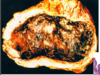
- This hemangioma was resected
- This is typical of a hemangioma à there is very hemorrhagic, sponge-like tissue
- The tissue shows a lot of vasculature filled with blood
- Bottom: the vasculature becomes large, sclerotic and fills with blood
- Macroscopic:
- These are usually solitary lesions, but there may be multiple lesions
- Typically these are less than 3 cm in size, but can become very large à these are called giant hemangiomas
- Microscopic:
- Virtually all consist of vascular spaces
- These are mostly venous-type

pancreas:

chronic pancreatitis
fibrosis
reduced number of acini
chronic inflam infiltrate
enlarged persistent islets
dilated ducts w proteinaceous material

wilson’s disease
copper depsition in liver
- This is what you see if you do a biopsy. Slightly different from hematochromatosis.
- You see darker, brownish deposits in the left image.
- If you use a copper stain [right image], you will see the very brown, granular copper deposits.

•Grossly:
- Cholangiocarcinoma usually occurs in non-cirrhotic liver, which distinguishes it from HCC
- HCC usually occurs in a cirrhotic liver except in the case of HBV, which has oncoproteins that can integrate into the human host and can induce HCC without cirrhosis
- The etiology of HCC is usually related to cirrhosis
- The tumor is composed of a tree-like tumorous mass and is firm; it has a gritty, tumor surface
•Histology:
- This is a type of adenocarcinoma with a marked desmoplastic reaction
- Demoplasia is a fibroblastic reaction to tumor invasion
- Early, you can identify lymphatic and vascular invasion
- This is rarely bile stained because it does not produce bile
villous adenoma
larger and more ominous than tubular adenoma
rectum and rectosigmoid
sessile
finger like extension
all degrees of dysplasia
carcinoma will directly invade the bowel wall

duodenal sleeve
malabsorption procedure
duodeno-jejunal bypass sleeve
open at both ends
food can pass but sleeve prevents contact w duodenum
delays digestions

histology of acute hepatitis
- portal and interface inflammation (portal tract filled w lymphocytes)
- lobular inflammation
- bile duct damage
- no significant fibrosis

serum/infiltrate in autoimmune chronic pancreatitis
IgG4 infiltrate and in serum
ANA

autoimmune chronic pancreatitis
lymphoplasmacytic sclerosing pancreatitis
mass like lesions and irregular beading of the pancreatic duct
associated w autoimmune conditions


macronodular cirrhosis

- This is a hepatocellular adenoma
- Adenomas are usually asymptomatic; normally we do not remove an adenoma surgically unless it is enlarged, the patient wants to have it taken out, or wants to get pregnant
- This particular lesion also responds to hormonal stimulation
- Left image: in contrast, to FNH, this lesion is capsulated
- It is an enucleated tumor with a capsular surface
- Right image: if you cut it in cross-section, you can see the lesion is very well circumscribed and encapsulated
- The central area has some kind of hemorrhage
histo of acinar carcinoma
solid nests, acini, scant stroma
large solid well circomscribed w extensive necrosis


cholangeocarcinoma

- If you do a resection, you will see a large, irregular lesion that is not well-demarcated with a smaller satellite lesion
- There is intrahepatic metastasis
- Right image: there is a very proliferative bile duct-like structure
- Left: the high-power view shows an infiltrative pattern and a desmoplastic reaction
- This is a fibroblastic reaction of the glands
- Bottom right: desmoplastic reaction
- Top right: intravascular invasion
- This is typical for cholangiocarcinoma