68 Facial Trauma Flashcards

1
Q

What important elements of the physical exam are considered in facial trauma?

A

What important elements of the physical exam are considered in facial trauma?

  1. Airway, breathing, and circulation (ATLS)
  2. Disability: cervical spine and brain injury (ATLS)
  3. Cranial nerves: motor (CN VII) and sensory (CN V1, V2, V3)
  4. Eyes: vision (CN II), pupils, movement (CN IV, VI) fields, pressure, globe injury, globe position
  5. Ears: hearing, hemotympanum, ear canal fracture, temporal bone fracture (CN VII, VIII)
  6. Bones: calvarium, midface, and mandible for deformity and dysfunction
  7. Throat: occlusion, TMJ function, bleeding, hematoma, airway, speech and swallow (CN IX, X)
2
Q

What type of imaging should be ordered to evaluate facial trauma?

A

What type of imaging should be ordered to evaluate facial trauma?

High-resolution (fine cut) axial computed tomography (CT) with coronal and sagittal reconstruction is ideal. Cervical spine imaging should be included in facial fractures caused by high-energy impacts such as motor vehicle accidents (MVA). Coronal and sagittal reconstructions are helpful in evaluating the orbital floor, frontal sinus outflow tracts, and mandibular condyles. Three-dimensional CT scans are very helpful in surgical planning when multiple fractures are present. Direct radiographic signs of facial fractures are nonanatomic linear lucency, cortical defects or suture diastasis, overlapping bone fragments causing “double density,” and facial asymmetry. Indirect radiographic signs include soft tissue swelling, periorbital or intracranial air and fluid in the paranasal sinus.

3
Q

What characteristics of the mechanism of trauma are considered important?

A

What characteristics of the mechanism of trauma are considered important?

Facial fracture results when the tolerance of facial bone is overcome by the kinetic energy (KE = ½mv2) transfer from blunt or penetrating trauma. Mechanisms have variable energy from low (fall from standing) to high (MVA). Understanding the mechanism of injury can help predict the extent of facial injury and the risk of associated cervical or brain injuries. High-impact and low-impact forces are defined as greater or lesser than 50 times the force of gravity (g). Facial bones differ in ability to withstand force: nasal bones can resist 30 g, zygoma 50 g, mandible angle 70 g, frontal-glabella 80 g, midline maxilla and mandible 100 g, and supraorbital rim 200 g. The most common facial fracture is the nasal bone.

4
Q

How is the facial trauma patient evaluated?

A

How is the facial trauma patient evaluated?

Each patient must be evaluated and treated according to the ATLS guidelines. Once the patient is medically stable, definitive facial fracture assessment and management can proceed. Facial trauma can range from a minimally displaced nasal fracture to a highly comminuted compound panfacial fracture involving the orbit, brain, and cervical spine. Facial trauma evaluation is best done by dividing the face anatomically into three sections as each has its own unique characteristics. The upper third assesses frontal bone, frontal sinus, and frontal lobe injury. The middle third or “midface” contains nasal, nasal-orbital-ethmoid (NOE), orbit, zygomaticomaxillary complex (ZMC), and maxillary structures. The lower third includes the mandible and temporomandibular joint.

5
Q

How would you evaluate a suspected frontal sinus injury?

A

How would you evaluate a suspected frontal sinus injury?

High-resolution thin cut computed tomography (CT) is best to evaluate anterior and posterior table fractures and outflow tract injury. In addition to the standard axial and coronal images, sagittal reconstructions of the paranasal sinuses can enhance visualization of the frontal outflow tract. Additional findings such as NOE complex fractures and anterior skull base injury near the junction of the posterior table and the cribiform plate strongly suggest injury to the frontal outflow tract.

6
Q

What are the treatment goals of frontal sinus repair?

A

What are the treatment goals of frontal sinus repair?

  • Protection of intracranial structures
  • Stopping CSF leak
  • Prevention of posttraumatic infection or mucocele (late complications)
  • Restoration of facial aesthetics
7
Q

What are the surgical indications for anterior and posterior table frontal sinus fractures?

A

What are the surgical indications for anterior and posterior table frontal sinus fractures?

Surgical indications for anterior table fractures include bony displacement causing a deformity or frontal sinus outflow tract impairment. Surgical indications for posterior table fracture include displacement of the posterior table greater than one table width, dural injury, CSF rhinorrhea, or frontal sinus outflow impairment.

8
Q

How do you treat a frontal sinus fracture of the anterior table?

A

How do you treat a frontal sinus fracture of the anterior table?

Isolated, nondisplaced, or minimally displaced anterior table fractures are not treated. Displaced fractures are treated by open reduction and internal fixation. Management options include osteoplastic flap with open reduction and internal fixation of anterior table fracture with or without obliteration, or an attempt at outflow tract reconstruction. Observation and medical management with future endoscopic surgery if needed is also an option.

9
Q

How would you treat a posterior table frontal sinus fracture?

A

How would you treat a posterior table frontal sinus fracture?

Uncomplicated nondisplaced posterior table fractures are generally not treated, but nondisplaced posterior table fractures with continued CSF leakage despite initial conservative measures require repair. Surgery is generally recommended for displaced posterior table fractures greater than one posterior table width, or severely comminuted fractures. The risk of dural injury in these cases is high and consultation with a neurosurgeon is recommended for possible dural repair. Mucosal removal and obliteration with abdominal fat or cranialization of the frontal sinus may be considered.

10
Q

What is the “osteoplastic flap with frontal sinus obliteration” procedure?

A

What is the “osteoplastic flap with frontal sinus obliteration” procedure?

The osteoplastic bone flap is created by a frontal sinus outline marked on the cranium classically using a template from a 6 ft. Caldwell radiograph. Osteotomies are performed and the sinus is opened. The mucosa of the sinus is completely removed, the frontal recess is occluded with temporalis fascia or muscle, abdominal fat is used to fill the sinus, and the bony flap is replaced. Postoperative CT/MR surveillance imaging is used for detection of postoperative mucocele formation; however, imaging is often difficult to interpret.

11
Q

What is “frontal sinus cranialization”?

A

What is “frontal sinus cranialization”?

The posterior wall of the frontal sinus is removed and the sinus mucosa is stripped away from the remaining bone. The brain and dura are evaluated by a neurosurgeon for possible debridement and dural closure. A previously mobilized anterior pericranial flap is inserted beneath the brain to separate it from the paranasal sinuses. The brain and dura are permitted to rest against the repaired anterior wall in the area originally occupied by the frontal sinus, which no longer exists.

12
Q

What are the complications of frontal sinus fractures?

A

What are the complications of frontal sinus fractures?

Early complications include wound infection, CSF leak, meningitis, acute sinusitis, deformity, pain, hypesthesia, and brain abscess. Late complications include mucocele, mucopyocele, osteomyelitis, cosmetic defect, brain abscess, and headache.

13
Q

What are the surgical approaches to repair the frontal sinus?

A

What are the surgical approaches to repair the frontal sinus?

  1. Frontal sinus trephination and elevation of the anterior wall with limited exposure
  2. Frontoethmoidectomy using a Lynch incision or endoscopic repair of the outflow tract
  3. Open reduction and internal fixation through the laceration or by coronal flap
  4. Frontal sinus obliteration
  5. Frontal sinus cranialization
  6. Frontal sinus ablation (Reidel) with removal of the anterior wall (rarely used today)
  7. Endoscopic frontal sinus surgery (delayed)
14
Q

What are the dangers of raising a bicoronal flap for facial fracture repair and how are they avoided?

A

What are the dangers of raising a bicoronal flap for facial fracture repair and how are they avoided?

  1. Injury to the frontal branch of the facial nerve can be avoided by incising the superficial layer of the deep temporal fascia at the temporal line of fusion so elevation can be deep to this layer.
  2. Injury to the supraorbital and supratrochlear nerves at the supraorbital rims is prevented by removing the inferior lip of the nerve foramen with an osteotome to allow the nerve to move inferiorly.
  3. Laxity of the midface soft tissues occurs if the fascia is not resuspended at the time of closure.
15
Q

What endoscopic procedure is used to treat severe chronic frontonasal outflow obstruction?

A

What endoscopic procedure is used to treat severe chronic frontonasal outflow obstruction?

The modified Lothrop procedure (Draf III procedure) may be used to restore severely obstructed frontal outflow pathways after trauma.

16
Q

What key features are evaluated in nasal trauma?

A

What key features are evaluated in nasal trauma?

Nasal fractures are commonly identified by epistaxis and bony nasal deformity. Often the patient complains of nasal obstruction. The external exam should include evaluation of deformity, mobility, step-offs, and telecanthus. The internal exam should examine for septal deviation, mucosal tears, or septal hematoma. Clear rhinorrhea may indicate cerebrospinal fluid (CSF) leak. Epistaxis in severe facial trauma may be life threatening and require surgery or embolization of the feeding arteries if packing fails.

17
Q

What are the dangers of a nasal septal hematoma and how is it treated?

A

What are the dangers of a nasal septal hematoma and how is it treated?

A septal hematoma is a collection of blood under the nasal septal perichondrium following trauma. The lack of blood supply to the cartilage can lead to cartilage necrosis or septal abscess and can produce a saddle nose deformity. Urgent treatment is required to evacuate the clot or purulence.

18
Q

How is a nasal fracture treated?

A

How is a nasal fracture treated?

Timing is critical. Acute nasal fractures are treated best by closed reduction immediately following the fracture (1 to 2 hours) or after swelling has subsided (5 to 10 days). The bones are repositioned and splinted for 7 to 14 days. Chronic nasal fractures (>10 days) may be more difficult to treat and often require complete healing (3 to 6 months) followed by formal septorhinoplasty.

19
Q

What is an NOE fracture?

A

What is an NOE fracture?

The nasal-orbital-ethmoid (NOE) complex is the confluence of the frontal sinus, ethmoid sinuses, anterior cranial fossa, orbits, frontal bone, and nasal bones. An NOE fracture is a telescoping fracture of the nasal, lacrimal, and ethmoid bones, which occurs from blunt trauma at the nasal bridge. Injury to the bony septal attachment at the cribiform plate can produce a CSF leak and anosmia. NOE fractures involve the attachment of the medial canthal tendons (MCT) and can produce telecanthus. Failure to diagnose and repair an MCT can lead to functional and cosmetic complications that are difficult to repair secondarily. Long-term sequelae of NOE fractures include blindness, telecanthus, enophthalmos, midface retrusion, cerebrospinal fluid (CSF) fistula, anosmia, epiphora, sinusitis, and nasal deformity.

20
Q

How are medial canthal tendon injuries classified?

A

How are medial canthal tendon injuries classified?

Markowitz classified NOE fractures based on the status of the MCT and the degree of comminution of the “central fragment” of bone to which it remains attached. In Type I fractures the fracture lines leave a single noncomminuted central fragment with MCT attached. In Type II the central fragment gets comminuted but the MCT stays attached to its fragments. In Type III fractures there is severe central fragment comminution and the MCT is detached. Type II and III are the most difficult to repair and require transnasal wiring in a posterior superior direction to keep medial orbit deformity to a minimum.

21
Q

What is a blowout fracture?

A

What is a blowout fracture?

An orbital blowout fracture results from hydraulic compression of the orbital contents into the paranasal sinuses through the weakest portions of the orbit. This usually occurs through the thin portion of the orbital floor (0.5 mm) and less frequently through the thin lamina papyracea (0.25 mm), which is supported by the honeycomb structure of the ethmoid sinuses. The pure form is purely hydraulic without rim injury; the impure form is caused by rim deformation and fracture extending posteriorly to create the blowout fracture.

22
Q

What are the indications for surgery of the blowout fracture of the orbital floor?

A

What are the indications for surgery of the blowout fracture of the orbital floor?

Surgery is indicated for: (1) enophthalmos greater than 2 mm, (2) double vision on primary or inferior gaze, (3) entrapment of extraocular muscles on forced duction testing, or (4) fracture greater than 50% of the orbital floor on CT imaging.

23
Q

What is a “white-eyed blowout fracture”?

Why is it treated emergently?

A

What is a “white-eyed blowout fracture”? Why is it treated emergently?

This is a trapdoor or greenstick fracture of the orbital floor, most commonly in children. The orbital floor opens under hydraulic pressure from the compressed globe, forcing the orbital fat and muscle into the maxillary sinus. The elastic bony floor will immediately close on these contents and trap them tightly. Under these circumstances the sclera remains white without hemorrhage, and the child is often nauseated and in severe pain. Careful examination of the irritated child may be difficult and CT scanning of the orbit should be considered for the diagnosis. Urgent surgery is required to preserve the entrapped ischemic inferior rectus muscle.

24
Q

What are the surgical approaches to repair orbit wall fractures?

A

What are the surgical approaches to repair orbit wall fractures?

The orbital floor is approached through the lower lid, which includes infraorbital, subciliary, and transconjunctival (preseptal or postseptal) routes. The medial wall is accessed through a transcaruncular incision, an endonasal endoscopic approach through the ethmoid sinuses, or by an external ethmoidectomy (Lynch) incision. The lateral wall is approached through an infrabrow incision, an upper lid skin crease (blepharoplasty) incision or through an extended lower lid transconjunctival incision with a lateral canthotomy. Orbital roof approaches include the external ethmoidectomy (Lynch), transbrow, or coronal incisions.

25
Q

What structures can be damaged using a Lynch approach to the orbit?

A

What structures can be damaged using a Lynch approach to the orbit?

The Lynch orbitotomy is used to access the medial wall using a curved skin incision half-way between the medial canthus and the bridge of the nose. Disadvantages of the Lynch incision are skin scarring, disinsertion of the medial canthal tendon, damage to the lacrimal sac, diplopia caused by trauma to the trochlea of the superior oblique muscle, and scarring of adjacent structures.

26
Q

Describe the transcaruncular approach to the medial orbit. Why is it used?

A

Describe the transcaruncular approach to the medial orbit. Why is it used?

The caruncle is divided to access a plane between Horner’s muscle and the medial orbital septum to expose the medial extraperiosteal space. Advantages include rapid entry into the orbit, less damage to skin and muscle layers, better cosmetic result, and less manipulation of the medial canthal tendon and lacrimal sac.

27
Q

What are common complications of orbital fracture repair?

A

What are common complications of orbital fracture repair?

  • Diplopia: double vision from paresis of an extraocular muscle (usually due to the initial injury) or fibrosis of an extraocular muscle causing restriction
  • Enophthalmos: posterior displacement of the eye within the orbit from changes in orbit volume in the setting of fat atrophy or wall malposition
  • Entropion: inversion of the eyelid toward the globe
  • Ectropion: eversion of the lid margin away from the globe
  • Proptosis: forward displacement of the eye from overcorrection of a blowout fracture
  • Hypoglobus: downward displacement of the eye in the orbit
  • Telecanthus: intercanthal distance is bigger than the width of the eye
  • Dacryocystitis: inflammation of the lacrimal sac related to nasolacrimal duct obstruction
  • Orbital cellulitis: infections in the orbit or from orbital implants
28
Q

What is the quickest way to decompress the eye with increased intraocular pressure?

A

What is the quickest way to decompress the eye with increased intraocular pressure?

Orbital compartment syndrome (OCS) is an ocular emergency requiring prompt diagnosis and treatment to prevent blindness from ischemia of the optic nerve and retina. Orbital pressure can be relieved with an emergent lateral canthotomy with inferior cantholysis. Absolute indications for lateral canthotomy include retrobulbar hemorrhage resulting in acute loss of visual acuity, increased IOP, and proptosis. In the unconscious or uncooperative patient, an IOP greater than 40 mm Hg is an indication for lateral canthotomy (normal IOP is 10 to 21 mm Hg).

29
Q

What is the first indication that the optic nerve is injured following orbital trauma?

A

What is the first indication that the optic nerve is injured following orbital trauma?

Traumatic optic neuropathy (TON) is a condition of acute injury to the optic nerve from direct or indirect trauma. TON is thought to result from shearing injury to the intracanalicular portion of optic nerve, which can cause axonal injury or disturb the blood supply of the optic nerve. The optic nerve may swell in the optic canal after trauma, resulting in increased luminal pressure and secondary ischemic injury. Patients with TON may have decreased central visual acuity, decreased color vision, an afferent pupillary defect or visual field deficits. An early clinical finding of optic nerve injury in the traumatized eye is loss of red color vision. Treatment is with high-dose corticosteroids or surgical decompression.

30
Q

What is the difference between a forced duction test and a traction test?

A

What is the difference between a forced duction test and a traction test?

The forced duction test is an upward tug on the anesthetized sclera to test for inferior rectus muscular entrapment after blowout fracture. The traction test is the grasping of the lower eyelid and pulling laterally against its medial attachment to determine if there is abnormal laxity indicating a disruption of the medial canthal tendon following NOE fracture.

31
Q

How is an open globe injury treated?

A

How is an open globe injury treated?

A ruptured globe should be protected from any pressure or contact by placing a rigid eye shield without an eye patch on the patient. Foreign bodies should be left undisturbed. Medications should include antiemetics, sedation, analgesics, and prophylactic antibiotics to prevent endophthalmitis. Tetanus immunity should be updated, as open globe lacerations are considered tetanus prone. The patient should be kept NPO and definitive surgical repair by an ophthalmologist should be expedited.

32
Q

What is a zygomatic arch fracture and how is it treated?

A

What is a zygomatic arch fracture and how is it treated?

A zygomatic arch fracture is usually a medially displaced deformity in the zygomatic arch from an external blow. The defect can be seen and palpated; the indented bone can impinge on the coronoid process of the mandible and cause pain with jaw movement. Treatment is via fracture reduction often without fixation through a (1) direct cutaneous approach using a hook or suture, (2) a Gilles approach—an incision behind the hairline over the temporalis muscle to reach the fracture, or by (3) a transoral approach through a gingivobuccal sulcus incision. Comminuted arch fractures may require a coronal flap and ORIF.

33
Q

What is a tripod fracture?

A

What is a tripod fracture?

A tripod or malar fracture is officially known as the zygomaticomaxillary complex (ZMC) fracture. The zygoma is separated from the face at the (1) zygomaticomaxillary (ZM) suture (infraorbital rim); (2) zygomatic arch, and (3) zygomaticofrontal (ZF) suture (lateral orbital rim). A fourth sphenozygomatic (SZ) suture is at the lateral orbital wall, which when counted makes the ZMC fracture technically a “tetrapod” rather than a tripod fracture.

34
Q

What are the midfacial buttresses and why are they important in fracture treatment?

A

What are the midfacial buttresses and why are they important in fracture treatment?

The midface is reinforced by strong vertical and weaker horizontal buttresses. Three vertical buttresses resist the forces of mastication; the medial (nasomaxillary) buttress extends from the nasomaxillary region to the frontal bone. The lateral (zygomaticomaxillary) buttress extends from the molar region superiorly to the zygomaticomaxillary complex along the lateral orbital rim to the frontal bone. The posterior (pterygomaxillary) buttress is from the pterygoid plates to the skull base. The medial and lateral vertical buttresses are accessible for repair while the pterygoid sites are not. Four horizontal buttresses are bridging supports between the vertical buttresses and consist of the palate, a central facial buttress from malar to malar interrupted by the piriform aperture, the frontal bar, and the anterior-posterior zygomatic arch.

35
Q

How are middle third (midface) facial fractures classified?

A

How are middle third (midface) facial fractures classified?

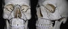
In 1901, French military surgeon René Le Fort published a classification of midface fractures that is still in use today. All three fracture types traverse the pterygomaxillary fissure to interrupt the pterygoid plates (Figure 68-1). Le Fort I fracture is a horizontal fracture above the maxillary alveolus producing a floating palate. The fracture usually involves the nasal aperture and extends above the apices of the teeth, causing the maxilla and hard palate to move separately. Le Fort II fracture is a pyramidal fracture that usually involves the inferior orbital rim. It extends from the nasion through the lacrimal bones and inferior orbital floor and rim through or near the inferior orbital foramen, and inferiorly through the anterior wall of the maxillary sinus. The Le Fort III fracture is a transverse fracture that separates the face from the skull, which is known as craniofacial dissociation. It includes fractures through the zygomatic bone, nasofrontal and frontomaxillary sutures, and orbit. The thick greater wing of the sphenoid bone usually prevents the continuation of the fracture into the optic canal thus preserving vision. Le Fort fractures often present in “mixed combinations” and should be reported as such (Figure 68-2).

Figure front: Le Fort fractures. Le Fort I fracture (1) is a horizontal fracture above the maxillary alveolus. Lefort II fracture (2) is pyramidal and usually includes the infraorbital rim. Le Fort III fracture (3) includes the zygoma and orbit and is considered a craniofacial dissociation when present bilaterally.

FIgure back: A, Left photo is a preoperative 3DCT of a patient with a right Le Fort II, left Le Fort I-II-III, and a palate fracture and left coronoid fracture. B, Right is a postoperative 3DCT showing the nose and zygoma repositioned and fixed to the skull base. The maxillary buttresses were repaired in relation to both the upper stabilized segments and the mandibular occlusion. Note the untreated coronoid fracture in correct position following zygoma repositioning.

36
Q

How are midface fractures treated?

A

How are midface fractures treated?

The goal of midface fracture repair is to restore form (cosmesis) and function. It must be done in concert with the nose, orbit, zygoma, and maxilla in relation to the mandible. In treating the patient in Figure 68-2, the upper portions of the midface were treated by stabilizing the nose and ZF suture fractures first with titanium plates. The maxilla was then aligned with the mandible and the remaining right Le Fort II and left Le Fort I-II-III fractures could be repaired in an accurate and functional way to restore the patient’s occlusion and facial cosmesis. Facial lacerations and minimally invasive soft tissue access techniques aid in the final cosmetic result.

37
Q

What is a panfacial fracture and how are panfacial fractures treated?

A

What is a panfacial fracture and how are panfacial fractures treated?

Panfacial fractures are defined as fractures involving the lower, middle, and upper face. Treatment is challenging and requires an individualized treatment plan utilizing treatment principles for each individual fracture. Reconstruction should be performed from the stable to the unstable (see Figure 68-2). The mobile zygomatic bone and nasal bones are secured in their correct anatomic position to solid cranial bone. Occlusion and facial height are reestablished first by reconstruction of the mandible to the maxilla, which is then secured to the nasal and zygomatic bones and cranium.

38
Q

What are surgical complications associated with midface fracture repair?

A

What are surgical complications associated with midface fracture repair?

  1. Inadequate reduction: malocclusion (maxilla) and facial deformity (zygoma)
  2. Imprecise reconstruction of the orbit: globe malposition
  3. Diplopia: from globe malposition, residual entrapment, muscle or nerve injury
  4. Eyelid malposition: eyelid incision/dissection trauma, orbital septum injury
  5. Reduced vision and blindness: rare, preoperative vision evaluation required
  6. Scars and hair loss: irregular coronal incisions, avoid with careful design and preoperative counseling
  7. Numbness: traumatic versus surgical
  8. Nonunion: chronic implant infection/extrusion, rare in midface fractures
  9. Dental injury: avoid tooth roots when placing screws, tooth and gum care with arch bar use
  10. CSF leaks: recognize early and treat to keep intracranial cavity separate from nose/sinuses
39
Q

What are the clinical signs of mandibular fracture and how is the mandible assessed in a patient with facial trauma?

A

What are the clinical signs of mandibular fracture and how is the mandible assessed in a patient with facial trauma?

The head and neck exam may show lacerations, swelling, and hematoma in the area of the fracture. Bimanual palpation of the inferior border may identify swelling, step-off deformity or tenderness. Lip numbness occurs in mandibular fractures distal to the mandibular foramen. Oral exam may show deviation of the mouth on opening, limited opening (trismus), TMJ pain, coronoid impingement, occlusal changes, and floor of mouth ecchymosis from periosteal or gingival tearing. Occlusal evaluation may show obvious or subtle malocclusion. Imaging studies are necessary, as mandibular fractures usually occur in pairs—parasymphyseal and condyle fractures often occur together.

40
Q

What are the best imaging studies for mandibular trauma?

A

What are the best imaging studies for mandibular trauma?

An axial computed tomography (CT) with coronal and sagittal reconstruction is ideal when visualization is difficult and is the preferred method of imaging for multiple mandibular fractures. Three-dimensional CT is very helpful for treatment planning of complex mandibular fractures. A Panorex film of the mandible is an excellent screening study involving low cost, low radiation, and excellent for follow-up evaluation. Other studies include mandible series, occlusal films, and periapical films.

41
Q

How are mandibular fractures classified?

A

How are mandibular fractures classified?

Mandibular fractures are classified by anatomic region and by an additional descriptor. Each anatomic region has unique characteristics that require specialized treatment considerations. The additional descriptors describe severity (greenstick, simple, compound, comminuted), displacement by muscle pull (favorable or unfavorable), and malocclusion (open bite, cross bite). Each of these considerations will factor into the treatment plan.

42
Q

What are the anatomic regions of the mandible?

A

What are the anatomic regions of the mandible?

The mandible is divided into horizontal and vertical parts. The horizontal mandible has four anatomic regions: the dense basal bone consisting of the symphysis, parasymphysis, body, and less dense alveolar bone which holds the dentition. The vertical mandible has four anatomic regions: the angle, ramus, condyle, and coronoid. Fractures can occur in any of these regions but more frequently in the angle and condyle regions (Figure).

Figure: Common mandibular fracture sites: (1) condylar head, (2) condylar neck, (3) subcondylar, (4) coronoid, (5) ramus, (6) angle, (7) body, (8) symphysis (symphysis and parasymphysis), (9) and alveolar.

43
Q

How are condylar fractures classified and why are they considered difficult to treat?

A

How are condylar fractures classified and why are they considered difficult to treat?

Condyle fractures are classified by three sites: head, neck, and subcondylar (see Figure 68-3). Condyle fractures are difficult to treat because (1) condyle fractures occur under the facial nerve (CN VII), which can be injured in the approach; (2) the condyle is often malpositioned by pull of the lateral pterygoid muscle or by traumatic dislocation and are difficult to reduce; (3) the bone quality is often inadequate to support hardware. Subcondylar fractures are generally the only site considered for ORIF, while the other regions are often treated without operative intervention. The malocclusion that occurs from condyle fractures creates an open bite deformity.

44
Q

What are the indications for open reduction and internal fixation (ORIF) of a condyle fracture?

A

What are the indications for open reduction and internal fixation (ORIF) of a condyle fracture?

Absolute and relative indications for surgery are discussed by Zide and Kent (Table 68-1). Unfortunately there is no consensus on the treatment of condylar fractures in adults. The type of treatment must be chosen on a case by case basis and by professional experience. Functional therapy (early jaw mobilization) is essential to avoid ankylosis of the TMJ. Three treatments advocated for adults with condylar process fractures include: (1) a period of maxillomandibular fixation (MMF) followed by functional therapy, (2) functional therapy without a period of MMF and, (3) open reduction with or without internal fixation. ORIF in children is becoming more accepted due to technical experience and improvements in rigid fixation.

45
Q

Why is the mandibular angle subject to high fracture rates?

A

Why is the mandibular angle subject to high fracture rates?

First, the angle has a thinner cross-sectional area relative to the neighboring segments of the mandible and second is the presence of third molars which weaken the region. The thin bone and tooth socket creates a pathologic fracture site by weakening the junction between the vertical and horizontal segments. Unfavorable angle fractures are subject to displacement by pull from the masseter and medial pterygoid muscles. Mandibular angle fractures pose a unique challenge for surgeons because they have the highest reported postoperative complication rate of any mandibular region.

46
Q

How is tension and compression related to mandibular healing?

A

How is tension and compression related to mandibular healing?

During chewing, a functional load creates tension which separates the superior border of the mandible (Figure). This opens the fracture site and will allow bacteria and food to enter and produce a poor result. Compression occurs on the inferior border at the same time and closes the fracture. During repair it is important to place a tension plate on the superior border to reduce separation. This fixes the fracture and reduces risks of nonunion and infection.

Figure: Under chewing load a mandibular angle fracture opens at the superior border (open arrows). This is considered the “tension” or distracting site, which can be held together with a lightweight mini-plate. “Compression” or closure occurs on the inferior border during loading and needs no plate to keep the fracture reduced. Some surgeons feel additional help on the inferior border may be necessary.

47
Q

What do fractures of the mandibular body and parasymphyseal region have in common?

A

What do fractures of the mandibular body and parasymphyseal region have in common?

Both are regions of the horizontal mandible that bear teeth and require adequate occlusal reconstruction to prevent malocclusion. Patients who sustain bilateral parasymphyseal fractures can have a “flail mandible,” which can result in the fragment and tongue moving posteriorly and producing airway obstruction.

48
Q

Explain the angle classification of occlusion.

A

Explain the angle classification of occlusion.

Class I is considered normal and the mesiobuccal cusp (MBC) of the permanent maxillary first molar occludes in the buccal groove (BG) of the permanent mandibular first molar. Class II (retrognathia) is a posterior mandible, so the upper MBC is now in front of (mesial) the lower BG. Class III (prognathia) is an anterior mandible in which the upper MBC is behind (distal) the lower BG.

Figure: Angle classification of occlusion. A, Class I, normal occlusion. B, Class II, malocclusion. C, Class III, malocclusion.

49
Q

What are the indications for closed verses open reduction of mandible fractures?

A

What are the indications for closed verses open reduction of mandible fractures?

Closed reduction and fixation is considered for nondisplaced favorable fractures, pediatric fractures, grossly comminuted fractures, coronoid fractures, and adult condyle fractures. This is accomplished by maxillomandibular fixation (MMF) using Arch Bars, Ivy loops, Risdon wires, dental splints, and dentures. ORIF is considered for displaced unfavorable fractures, atrophic edentulous mandible fractures, complex facial fractures, and condylar fractures that can’t be treated with closed techniques. This is done by exposing and reducing the fracture, and fixating with wires, lag screws, or plates and screws.

50
Q

How should the edentulous mandible fracture be treated?

A

How should the edentulous mandible fracture be treated?

Mandibular atrophy occurs with the loss of teeth, resulting in a smaller fragile bone. This remaining “basal bone” is dense cortical bone that has decreased osteogenesis, reduced blood supply, and depends on the periosteum for nourishment. Closed treatment using Gunning splints, dentures, and external pin fixation is done to preserve the blood supply in a noncontaminated environment to promote fracture healing. Open treatment requires the placement of heavy load-bearing reconstructive plates using bicortical screws. Edentulous bone has no cross-sectional stability and is too weak to “load-share” the fracture with a small bone plate and monocortical screws. Failure to recognize this important concept when treating with ORIF can lead to serious complications.

51
Q

How are pediatric mandibular fractures different from adult fractures?

A

How are pediatric mandibular fractures different from adult fractures?

Pediatric mandible fractures are more difficult to treat than adult fractures. The teeth are conical in shape and have short roots that are not amenable to MMF. Tooth buds and growth centers can be damaged during the fracture treatment. Children 6 years of age and younger are generally treated with closed reduction techniques to avoid injury to developing teeth. Children 12 and older should have their permanent teeth in place and can be treated with ORIF using mini-plates. Mandible growth occurs because of elongation in the condylar region and remodeling and growth in the ramus and body. Injuries in the condylar region during fracture repair may lead to facial asymmetry.

52
Q

Posterior table fracture management.

A

Posterior table fracture management.

Controversy

Management of posterior wall fractures is most controversial of all fracture sites. The issue is assessing whether the fragments are displaced. Fine cut CT scans are helpful in determining if the fracture is linear or displaced. Linear fractures require no treatment. Nondisplaced fractures with CSF leak may be observed for 5 to 7 days. According to the current treatment algorithm, if the wall is displaced, frontal sinus exploration is indicated. The result of the exploration can result in doing nothing, doing a simple posterior table repair, doing a complete mucosal drill out and abdominal fat obliteration, or doing a frontal sinus cranialization procedure.

53
Q

Outflow obstruction and frontal sinus obliteration verses sinus sparing techniques.

A

Outflow obstruction and frontal sinus obliteration verses sinus sparing techniques.

Controversy

The question as to whether the frontal outflow tract obstruction can be successfully preserved in selected patient populations (sinus preserving) in contrast to sinus obliteration procedures is a controversial topic. There are no head-to-head comparisons of these two techniques. All agree that the consequences of nasofrontal outflow tract obstruction require treatment.

54
Q

Use of prophylactic antibiotics in the management of facial fracture.

A

Use of prophylactic antibiotics in the management of facial fracture.

Controversy

There is considerable variability in the management of patients with frontal sinus fractures in the use of prophylactic antibiotics. Prophylactic antibiotics are used to prevent infections; however, they also may inadvertently increase the risk of postoperative infections with opportunistic pathogens. Antibiotics may also alter the physiologic local flora and eliminate the reliability of microbiological analysis of CSF samples when meningitis is suspected. The efficacy of antibiotics is difficult to study given the heterogeneity in injury pattern, patient characteristics, and study designs.