Dermatology Flashcards

Karposi’s sarcoma
Principles of skin examination
Inspect, describe, palpate, systemic check
Inspect
General observations Site and number of lesions If multiple - pattern of distribution and configuration
Describe
S.C.A.M Size, Shape Colour Associated secondary change Morphology, Margin (border)
Pigmented lesion
A.B.C.D Asymmetry Irregular border two or more Colours within the lesion Diameter >6mm
Palpate
Surface Consistency Mobility Tenderness Temperature
Systematic check
Examine the nails, scalp, hair, mucous membranes and general examination of all systems

Concentric rings - erythema multiforme
What is shown and list the risk factors for this type of lesion?

Venous Ulcer
Risks for venous ulcers
Varicose veins.
Previous deep vein thrombosis in the affected leg.
Phlebitis in the affected leg.
Previous fracture, trauma, or surgery.
Family history of venous disease.
Symptoms of venous insufficiency: leg pain, heavy legs, aching, itching, swelling, skin breakdown, pigmentation and eczema.
What is shown and list the risk factors for this type of lesion?

Arterial Ulcer
Risks for arterial ulcers
Coronary heart disease.
History of stroke or transient ischaemic attack.
Diabetes mellitus.
Peripheral arterial disease including intermittent claudication.
Obesity and immobility.
What is shown? Name some differential diagnoses

Erythema Nodosum
Streptococcal infection.
Sarcoidosis.
Tuberculosis (TB).
Other infections. Infections such as chlamydia, Mycoplasma pneumoniae, Yersinia enterocolitica
Certain medicines.
Inflammatory bowel disease.
Pregnancy. Occasionally, pregnancy can trigger erythema nodosum.
Certain cancers, including lymphoma and leukaemia

Atopic eczema
Shortly after starting a medication this person developed…

Stevens Johnson syndrome
This is a form of toxic epidermal necrolysis, is a life-threatening skin condition, in which cell death causes the epidermis to separate from the dermis. The syndrome is thought to be a hypersensitivity complex that affects the skin and the mucous membranes. The best known causes are certain medications (such as lamotrigine), but it can also be due to infections, or more rarely, cancers

Candida Albicans

Urticaria

Melanoma

Pitting

Henoch Schonlein

Herpes Zoster

Thrombophlebitis

Keloid Scar

Seborrheic Keratosis

Excoriation eczema

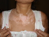
Naevus flammus - Vascular malformation

Pyogenic granuloma

Normal mole

Phlebitis

Keloid scar

Basal cell carcinoma

Angioedema

Livedo reticularis

Tinea corporis

Melasma

Venous ulcer

Mucosal desquamation - Stevens Johnson

Melanoma

Squamous cell

Normal moles

Superficial phlebitis

Necroytic migratory erythema

Lichenification eczema

Vitiligo

Candida

Squamous cell

Varilla - chicken pox

Basal cell carcinoma

Squamous cell carcinoma

Hypertrophic scar

Sebhorreic Keratosis

Thrombophlebitis

Melanoma

Necrolytic migratory erythema

Candida - mouth

Venostatis ulcer

Serbhorreic keratoses

Senile purpura

Cystic acne

Chronic arterial insufficiency

Erythema nodosum

Basal cell carcinoma

Normal moles

Eczema herpeticum

Dermatitis herpetiformis -
associated with coeliac disease and gluten sensitivity

Psoriaris

Neuropathic ucler

Herpes zoster

Eczema herpeticum

Psoriasis

Koilonychia
Vitiligo

Pyogenic granuloma

Lichenefication

Phlebitis

Hypertrophic scar

Varicella zoster

Acute hand eczema

Onycholysis

Urticaria

Eczema

Acne

Palmer erythema

Varicella zoster

Senile purpura

Henoch purpura

Hidrardenitis suppurativa

Acanthosis nigricans

Ab igne

Dermatitis herpetiformis

Bullous pemphigoid disease

Hidradenitis suppurativa

Erythema ab igne

Acanthosis nigrans

Keratoderma blenorrhagica (seen in reactive arthritis)

Keratoacanthoma
In the picture shown the central keratotic area, and the lack of the typically raised pearly edge would favour keratoacanthoma over BCC
Keratoacanthomas arise from a single hair follicle as they are only seen on hair-bearing skin. Untreated, a true keratoacanthoma will go on growing for several months, reach a maximum size then self-destruct over several more months.
Sometimes surgical excision is necessary and occasionally biopsy will remove the lesion entirely.
Risk factors for keratoacanthoma include those for skin cancer and so high levels of UV exposure are often present. Skin trauma is also often noted in the patient’s history and research has shown a link with human papilloma virus (HPV).

keratoacanthoma
A 64-year-old asylum seeker has arrived in the United Kingdom with this painless lesion on his forearm.
It started as a small, red, itchy lesion, which then blistered, bursting to form the lesion shown. He was employed as a sheep herder.

Cutaneous anthrax is an infection of the skin caused by direct contact with the bacterium
Bacillus anthracis.
It is a differential in all skin lesions in subjects who may have had contact with infected animals, mainly hoofed animals (for example, sheep, goats).
Cutaneous anthrax accounts for 95% of cases of anthrax worldwide.
There is potential for anthrax spores to be utilised as a means of bioterrorism. In case of this event, stockpiles of ciprofloxacin have been set aside to treat the affected population. It can also be treated with penicillin and doxycycline
A 30-year-old man presents with a five day history of chills, sore throat and some dyspnoea
What is the rash called? What is most likely cause of his symptoms and rash? List drugs which can preciptate it.

Erythema multiforme
Mycoplasma pneumonia
Penicillins, sulphonamides, phenytoin, barbiturates, carbamazepine, and vaccinations