Mid semester exam Flashcards

1
Q

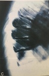
1 - Describe the major radiographic findings.

2 - What is the name of this condition and what is its aetiology?
A

1 -
Loss of height of the anterior portions of the vertebrae over multiple levels.

Trapezoidal shaped vertebrae. Irregular end plates.

2 -

Scheuermann’s

Avascular necrosis of the secondary ring of the vertebral body. Could be hereditary, endocrine or osteoporosis

2
Q

What is the site and age of Legg-calve-perthes?

A

Femoral capital epiphysis

3-12 (peak 5-7)

3
Q

What is the site and age of Friebergs?

A

Metatarsals heads of 2nd and 3rd digits of the feet

13-18 year old girls

4
Q

What is the site and age of Kienbocks?

A

Lunate

20-40 year olds, predominately males

5
Q

What is the site and age of Kohlers?

A

Navicular

5 year olds, predominantley males

6
Q

What is the site of osgood schlatters?

A

Tibial tuberosity (tibial tubercle apophysis)

7
Q

What is the aetiology of osgoods?

A

Traction apophysitis/ tendonitis caused by trauma or repetitive microtrauma.

8
Q

What are the radiological features of Osgood schlatters?

A

Soft tissue swelling

Patella tendon thickening

Bones appear with irregular isolated ossicles towards anterior margin of tuberosity.

Possible chondro osseous avulsion

9
Q

What is the best image to view radiographic findings?

A

T2 or Fat sat sagittal

10
Q

1 - Describe the radiographic findings?

2 - Give 3 different differentials.

3 - Choose one and outline your reasoning behind your diagnosis and radiographic findings.

A

1 -

Osteopaenia (widespread) enlargement of bones and epiphysis regions.

Eggshell thin cortices

Reactive sclerosis

Increased soft tissue density

Pseudocysts

Erosive changes - anteroinferior and posterosuperior calcaneus = interruption to cortices

2 -

Charcots joint

JRA

Osteogenesis imperfecta

Haemophilia

3 -

Juvenile RA - Causes enlargement of the epiphyseal regions. Also results in osteopaenia due to disuse or medications. Cortical thinning

Haemophilia - Juxtaarticular osteopaenia, haemarthrosis could cause the osteopaenia, joint disorganisation, cortical thinning, osteophytes.

11
Q

What are the films in a complete ankle series?

A

Dorsoplantar, lateral, medial oblique

12
Q

How would you go cobbs on this diagram?

A

:)

13
Q

What are the 3 areas of the body used to determine skeletal maturity?

A

Non-dominant hand - distal radial epiphysis

Vertebral ring epiphysis

Iliac epiphysis (risser sign)

14
Q

Provide some complications that may arise from the image seen above right

A

Degeneration (levels just above and below hardware), # around hardware, sensitivity to weather, infections from surgery, DJD, pain, Supp OM

15
Q

What age bracket and sex do we need to be concerned about with scoliosis?

A

Females, 12-16, convex left

16
Q

Outline the method to interpret chest films.

A

1 - Request details

2 - Technical

3 - Trachea

4 - Heart and mediastinum

5 - Diaphragm

6 - Pleura

7 - Lung fields

8- Hidden areas

9 - Hila

10 - Below diaphragm

11 - Soft tissue density

12 - Bone

17
Q

What are the major radiographical findings?

What is the diagnosis

A

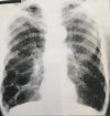
Pancake/vertical heart

Hyperlucent lung fields

Barrel chest

Depressed diaphragm

Blebs

Hyperinflated

Diagnosis = emphysema

18
Q

1 - What are the black arrow heads pointing at?

2 - What are 2 possible reasons for the radiographic findings?

A

1 - Apophysis of the calcaneus

2 - Avulsion

Normal finding

Severs

19
Q

1 - What is the diagnosis?

2 - How might the patient present in the clinic?

A

1 - Hiatal hernia

2 - heart burn, chest pain, dysphagia, loss of weight, anorexia, reflux

20
Q
  1. What is the diagnosis
  2. What is the arrow pointing at?
  3. What is the common age bracket for this patients condition?
  4. Any other radiographic findings?
A
  1. Perthes or Dwarfism
  2. Sagging rope sign - intertrochanteric line
  3. 3-12
  4. Buttressing of femoral head, mushroom cap deformity, coxa magna, coxa vara
21
Q
  1. What is this congenital malformation called?
  2. Is it clinically significant?
    3.
A
  1. Pectus carinatum
  2. Yes, the restriction of the ribs could alter lung capacity - low tidal volume predisposes to pneumonia
22
Q
  1. Describe 4 radiographic findings
  2. What is the condition?
A
  1. Flattened pelvis (champagne glass pelvis), mushroom cap femoral head, coxa magna, enlargement of lesser trochanter, diverticuli
  2. Dwarfism
23
Q
  1. What type of study is this?
  2. Outline 8 muscles attaching to the scapula
A

CT bone window axial and coronal

Rotator cuff

Teres major

Levator scapulae

Latissimus dorsi

Trapezius

Deltoid

Pec minor

Omohyoid

24
Q

This is the L4/5 disc space

  1. Which nerve root is displaced?
  2. What reflexes would be affected
  3. What muscles are affected by this nerve root?
  4. What is A, B, C and D
A
  1. L5
  2. Achilles
    • gluteus maximus muscle mainly S1.
    • gluteus medius muscle.
    • gluteus minimus muscle.
    • tensor fasciae latae.
    • tibialis anterior.
    • tibialis posterior.
    • extensor digitorum brevis.
    • extensor hallucis longus.
  3. A = left L4 nerve root, B = right common iliac artery, C = Colon, D = facet
25
Q
  1. What is the arrow pointing at?
  2. Is this clinically significant?
  3. Provide 1 other radiographic finding?
  4. What is the type of study?
A
  1. Bakers cyst
  2. Can be painful and cause decreased ROM, and can also burst and cause a lot of pain. Can also cause pain with movement.
  3. Increased fluid in the lateral femoral condyle
  4. MRI Axial Fat sat
26
Q
  1. What is the major radiographic finding?
  2. How does it happen
  3. What is the diagnosis?
  4. Which ethnicity is this condition common in?
A
  1. ‘hair on end’
  2. Due to marrow hyperplasia and widening of the diploe.
  3. Sickle cell anaemia or thalassemia
  4. Thalassemia = mediterranean, sickle cell anaemia = african american
27
Q
  1. What is the radiographic finding?
  2. What are 3 conditions that can cause this?
A
  1. Splenomegaly
  2. Sickle cell anaemia, rheumatoid arthritis, AIDs, osteoporosis, thalassaemia, haemophilia (blood transfusions)
28
Q
  1. What is the arrow pointing to?
A
  1. Renal calculi, calcified lymph node, malignant mass
29
Q
  1. What is the type of image?
  2. Which structure can be seen?
  3. What are the contraindications to this study?
A
  1. Intravenous pyelogram
  2. Renal pelvis and part of the ureter
  3. Allergy to contrast media
30
Q

What type of image is this?

A

T1 MRI

31
Q

What type of image is this

A

MRI T2

32
Q

What type of image is this?

A

Proton density

33
Q

What is the type of image?

What lines of alignment would be most appropriate?

A

CT soft tissue window

Cervical curve angle, cervical line of gravity, ALL, PLL

34
Q

What type of image is this?

What lines of alignment would be most appropriate?

A

CT bone window

Cervical curve angle, cervical line of gravity, ALL, PLL

35
Q
A