14. GI Radiology Flashcards
Briefly describe X-rays. What are the 3 MC views?
Describe fluroscopy and CT.
Label A-G of the anatomy on this abdominal XR.

Static tube + camera. Images usually taken on inspiration with XR source behind pt (PA). MC views: supine, erect, L lateral decubitus.
Fluoroscopy: mobile camera + tube, provides living, moving images, use of XR
CT: rotating camera + tube, use of XR to give ‘slices’. 3D view of body but radiation can be 100x XR
A) Liver
B) Ascending colon
C) Rectal gas shadow
D) Stomach bubble
E) Kidney
F) Psoas muscle
G) Descending/sigmoid colon
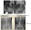
Small bowel = central; colon = frame

How would you distinguish between colon and small bowel on an XR?
Why is fluoroscopy not used much now, what has it been replaced by, and when might it still be used?
Label A-F in this CT.

Colon: haustra - folds DON’T cross entire length of bowel width
Small bowel: valvulae conniventes (plicae circulares) folds CROSS entire length of bowel width [Pic]
Replaced with MRI small bowel b/c of radiation. Only use if pt has MRI contraindication e.g. pacemaker or claustrophobia
A) Liver
B) IVC
C) Abdominal aorta
D) Kidney
E) Splenic flexure
F) Stomach

Label A-F of this CT.
What are the 4 cardinal elements of the XR (opacities)?
What would you see on an XR of ascites?

A) Liver
B) IVC
C) Kidneys
D) Aorta
E) Colon loops
F) SMA
G) SMV (always on RHS of SMA)
Bone (white = opacity), air (black = clarity), fat, water/soft tissue
Too white, bowel loops more central and float like balloon, air shadow replaced by soft tissue shadow, NO FAT STRIPE! [Pic]

Label A-C of this CT pelvis
What is the FREE ABDO approach to interpreting an abdo XR?
What density is free fluid the same as?
What is an indication of fluid buildup in the abdomen?

A) Small bowel loops
B) Bladder
C) Rectum
FREE: free fluid; A: air (inside, outside); B: bowel wall; D: density; O: organs
All same density as soft tissue.
Distance from the colon to the flank = fat stripe [Pic]. Should be close to descending colon loop. If there’s fluid = decreased width of fat stripe

It is normal to see intraluminal air in the small bowel on XR, however it it is abnormal to see intra-peritoneal air. What might this suggest?
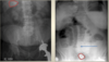
What 3 signs are indicated in the following 3 XRs?
Perforation of duodenum - urgent! Peptic ulcer. Diverticulitis. Cancer. Obstruction. Look for air under diaphragm in RUQ (liver diaphragm) on erect AXR/CXR, and air in R lateral flank in lateral decubitus AXR [Pic - L = normal, middle = erect abdo XR, see RUQ air, R = L. lateral decubitis, air fluid level]
L: Rigler’s sign - outer and inner aspect of bowel wall. Intra-peritoneal air, free air inside and outside lumen. Pneumoperitoneum may be due to perforation or recent surgery
Middle: Falciform sign - enough free gas to outline falciform ligament running to umbilicus from liver, seen with pneumoperitoneum
R: Football sign - free gas, abdominal cavity outlined by gas from perforated viscus, massive pneumoperitoneum

How long should it take post-op air to be resorbed?
What could a sudden increase indicate?
What causes air in the retroperitoneal space?
- *10 days.**
- Perforation.** [Pic - Day 1 post-op on top L, can see free air under RUQ, Day 5 post-op on top R, volume of air has increased = perforation of bowel during surgery?*]
Part of duodenum, ascending/descending colon = retroperitoneal.
If perforate = air seeps into retroperitoneal space. Look for air outlining the retroperitoneal structures (kidneys, psoas muscles etc.) - will be clear if perforated [Pic - bottom L = kidney and psoas clearly outlined, bottom R = kidney]

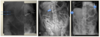
Describe the 2 different instances of air in the liver in an XR, and explain why this may have happened.
How would you distinguish an abscess in an abdo XR?
How would you distinguish pneumatosis in an abdo XR?
- *1. Pneumobilia = central hilar.** Due to manipulation of bile ducts (sphincterotomy)
- *2. Air in portal vein = central and peripheral.** Devastating event e.g. massive bowel infarction and necrosis
Black air collection in strange location, doesn’t move on sequential AXRs, no haustra/valvulae conniventes. [Pic - top, abnormal air shadow on liver = liver abscess. Can’t see diaphragm so not free intraperitoneal air]
Air in bowel wall. Sign of bowel suffering from ischaemia and impending perforation. Look for linear black streaks in bowel wall. [Pic - bottom]

What should you look for when checking for abnormal intra-luminal air?
What classifies small and large bowel as obstructed?
An ileus is usually secondary to an obstruction. What are the 2 types?
What could a secondary ilius from an obstruction be caused by?
Signs of ileus (paralyzed bowel loops) or obstruction (cancer): dilatation, air-fluid levels (horizontal line with air above and fluid below)
Measurements: >3cm for small bowel, >6cm for colon, >9cm for caecum
1. Localised: caused by near inflammatory process
2. Generalised: drugs (e.g. opiates), surgery, pain… e.g. Meckel’s diverticulum
[Pic - top = ileus]
Cancer, foreign bodies e.g. stone, loop on itself etc. [Pic - bottom = small bowel obstruction, >3cm, can see air-fluid level on right (arrow)]
Aside from ileus and obstruction, what is the third cause of abnormal intra-lumian air? What should you look for?
What is bowel wall thickening a sign of?
What would you look at when examining the bowel wall on an AXR?
Volvulus: bowel twists on itself and obstructs. Commonest places = sigmoid, caecum.
Look at sigmoid (dilated loops - coffee bean or inverted U in pelvis) and caecum (second stomach bubble in middle of abdomen) [Pic - top]
Ischaemia or IBD
Narrowing of lumen, thickening of folds (thumbprinting) [Pic - bottom]
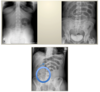
What should you look at when covering the ‘densities’ section of FREE ABDO?
What desities can you see on the 2 AXRs?
Most organs are surrounded by fat so are visible on AXRs. What would you specifically look for in the ‘organ’ section of FREE ABDO?

Bones, appendicolith (calcified deposit in appendix), stones, pancreatic calcifications, abdominal aortic calcifications.
Top: pancreatic calcifications, mid-line to LUQ
Bottom: linear calcifications, sign of vessel calcification on L side of spine - aortic aneurysm has dilated + calcified
Hepatosplenomegaly [Pic]

Case 1
30yo pt presents with acute abdominal pain, N+V.
Lab = increased inflammatory markers.
Exam = marked peritonism in all 4 quadrants.
AXR below
What can you see on the AXR?

Free intraperitoneal air in RUQ. Had a perforated peptic ulcer from e.g. NSAIDs, cocaine addiction (most likely causes in young person). Sent to OR.
Case 2
65yo pt presents with acute abdominal pain, N+V
Lab: increased imflammatory markers
Exam: marked distension
AXR below
What can you see?
What would the next steps be?

Colonic obstruction with distended pelvic loops of colon (coffee bean) likely due to sigmoid volvulus.
Next step = urgent CT (to check no perforation) [Pic - can see ‘whirl’ shape of sigmoid volvulus].
Then surgery

Case 3
65yo pt presents with acute abdominal pain, PMH of psychiatric disorders
Lab: normal
Exam: diffusely distended with metallic sounds
AXRs below (R = L lateral decubitis)
What can you see?
Dilated small bowel loop (can see plicae circulares) on L AXR.
R AXR - small bowel obstruction with multiple air-fluid levels.
Radio-opaque material R iliac fossa.
Pt ingested stone… -> surgery!
Case 4
85yo pt presents with 1 week of acute abdominal pain
Lab: inflammatory markers
Exam: diffusely peritonitic (can’t touch)
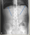
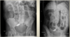
AXR and CT below
What can you see?

Top L = more translucency on L side. Lateral decubitis = Free intra-peritoneal air. Upright = air under diaphragm.
CT: blue arrow = dilated aorta - linear calcification. Red arrow = free air.
At surgery: perforated duodenal ulcer and incidental aortic aneurysm
Case 5
70yo female presents to A&E with RUQ pain
Lab: inflammatory markers
Exam: RUQ pain
24hrs after hospitalisation, RUQ pain decreases but develops diffuse abdominal pain.
Exam: marked distension
Supine and erect AXRs below
What can you see?

NB: RUQ = liver and gallbladder.
L AXR: RUQ opacity in keeping with gallstone and cholecystitis
R AXR 24hrs later: small bowel dilatation and air-fluid levels in keeping with small bowel obstruction. Radio-opaque stone now visualised in pelvis. This is gallstone ileus (and choleduodenal fistula - pt had lots of gallbladder inflammation - fistulated into duodenum and now in distal ileum)